Project 2
PROJECT(A)
SELECTED SYSTEM:- GASTROLOGY SYSTEM
GASTROINTESTINAL SYSTEM
- The gastrointestinal system is also known as the digestive system. It is a collective name of the alimentary canal and accessory organs.
- The alimentary canal forms a continuous tube that is open to the outside environment at both ends.
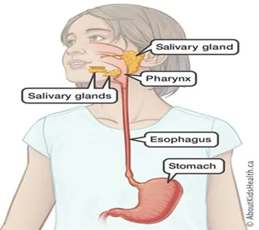
- The organs of the alimentary canal are the mouth, pharynx, esophagus, stomach, small intestine, and large intestine.
- The overall process of digestion and absorption occurs in the alimentary canal.
- Accessory structures include the teeth, tongue (in the oral cavity), salivary glands, liver, gallbladder, and pancreas.
UPPER GI TRACT
- The upper GI tract is responsible for starting digestion and preparing it for the lower GI tract to process and absorb nutrients.
- The upper GI consists of the following organs: Mouth, Pharynx, Oesophagus, Stomach, Duodenum.
LOWER GI TRACT
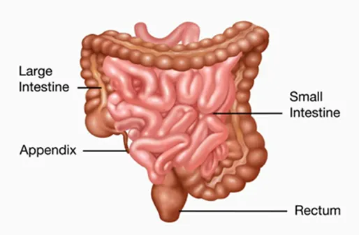
- The contents of the small intestine empty into the large intestine, which is also called the colon. While food travels through the colon, the water and salts are absorbed. Once the nutrients are removed from the food, the remaining materials become stool – which moves into the rectum to be expelled from the body as waste.
- The lower GI consists of the following organs: Small Intestine, Large Intestine.
Description:
Q1) Detailed description of the clinical condition selected
SELECTED CLINICAL CONDITION:- GERD (Gastroesophageal Reflux Disease)
a-Normal function of the organ
- The gastrointestinal system has the basic function of digesting food into nutrients and then eliminating the waste.
- The organs involved in this disorder are stomach and esophagus.
Esophagus
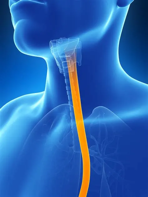
- food moves along the esophagus by peristalsis. This is the rhythmic, involuntary contraction of the gut wall in a curvy manner, propelling its content along it.
- The gut wall comprises of smooth (involuntary) muscles, some of which are arranged longitudinally while others are circular.
Stomach
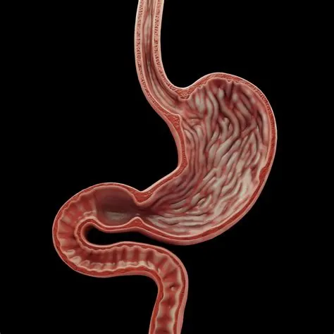
- It is found in the left upper part of the abdominal cavity, just below the diaphragm. Its wall, unlike that of the other parts of the gut, comprises of three layers of smooth muscles, which all help to churn(mix) the food in it. The muscles are: the outer longitudinal, the middle circular and the inner oblique(angular) muscles.
- The functions of the stomach are to store and digest it. It lining possess three types of gastric glands, producing three types of secretions:
- The zymogrn or chief cells secrete pepsinogen.
- The oxyntic or parietal cells secrete hydrochloric acid
- The mucus-secreting cells secrete mucus
b-Current pathological state in detail
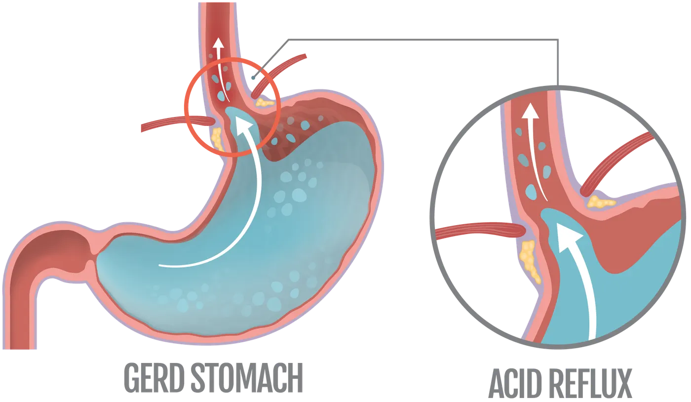
- Gastroesophageal reflux disease (GERD) occurs when stomach acid repeatedly flows back into the tube connecting your mouth and stomach (esophagus). This backwash (acid reflux) can irritate the lining of your esophagus.
- Many people experience acid reflux from time to time. However, when acid reflux happens repeatedly over time, it can cause GERD.
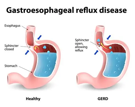
- occurs when the lower esophageal sphincter, a ring-shaped muscle at the lower end of the esophagus, malfunctions. When it is working normally, this muscle relaxes and opens to allow food to pass into the stomach and contracts, or closes, to prevent stomach contents from flowing back into the esophagus.
- When the lower esophageal sphincter is too relaxed, stomach acid can splash up into the esophagus. This backward flowing of stomach contents is called gastroesophageal reflux, or sometimes acid reflux.
- The stomach has a special lining that protects it from being damaged by gastric acid, but the esophagus does not. As a result, when the esophagus is exposed to stomach acid, it becomes irritated and inflamed. This results in heartburn, a painful burning sensation in the chest or in the throat.
- Acid reflux happens to just about everyone from time to time and usually causes only mild symptoms. But GERD occurs when acid reflux is chronic and repeatedly causes heartburn or other symptoms or complications.
Symtoms
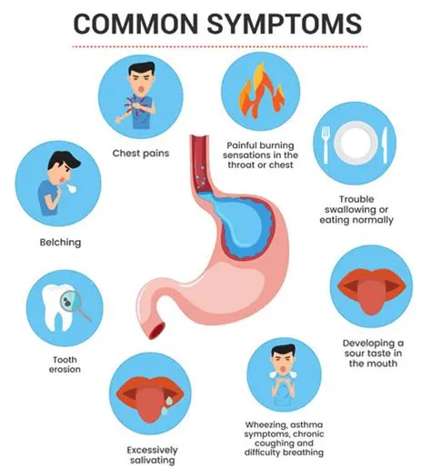
Common signs and symptoms of GERD include:
- A burning sensation in your chest (heartburn), usually after eating, which might be worse at night or while lying down
- Backwash (regurgitation) of food or sour liquid
- Upper abdominal or chest pain
- Trouble swallowing (dysphagia)
- Sensation of a lump in your throat
If you have nighttime acid reflux, you might also experience:
- An ongoing cough
- Inflammation of the vocal cords (laryngitis)
- New or worsening asthma
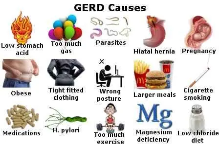
Several factors can increase the risk of developing GERD, including:
- Weight gain, being overweight or obese
- Family history of GERD
- Hiatal hernia (condition in which the top of the stomach at the esophageal junction bulges into the chest)
- Smoking or inhaling secondhand smoke
- Eating fatty foods
- Drinking alcohol, coffee, or carbonated beverages
d-Effect of homoeostasis imbalance on human body
- GERD greatly influences a disruption in homeostasis due to one major effect-heartburn. Heartburn, one of the effects brought upon by GERD, which is characterized by a burning pain in the upper chest, stands as the main disrupter of homeostasis.
- The heart plays a major role in the human body by continuously pumping blood to every corner of the human body; when a disruption (such as heartburn) rouses, the human body’s ability to maintain a stable temperature that corresponds to that of the outside environment becomes inefficient.
- The ceaselessly potent burning that inflames and severely damages the esophagus causes major disruptions in one’s daily life as medications and precautions must be taken to ensure no uncomfortable feelings will prevent one from going to his daily life as well as any unfortunate event like a heart attack take place.
- Homeostasis and the digestive system are greatly disrupted as the esophagus inflammation and prevent the body from carrying out its tasks due to an abnormal burning of the chest, esophagus, and even mouth.
- Aside from heartburn, the internal attrition of the esophagus may cause several other long-term issues, such as esophagitis. Esophagitis may bring upon difficulty whilst swallow and even ulcers.
- Persistent acid reflux, or gastroesophageal reflux disease (GERD), can make mealtimes unbearable. It may be difficult to eat anything without experiencing burning chest pain, nausea, and difficulty swallowing.
- Sleeping flat with acid reflux can be nearly impossible. Acid and stomach contents can creep up the esophagus, causing burning pain. People with severe GERD may sleep very poorly and wake up often during the night.
Q2)Description of the diagnostic method selected by the surgeon for assessing the condition.
Common diagnostic methods:-
- Upper GI series(Barium swallow)
- Gastroscopy
- Endoscopic Retrograde Cholangiopancreatography(ERCP)
- Endoscopic ultrasound
- pH Monitoring
a-Selected Diagnostic method:- pH Monitoring
Why:
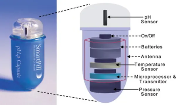
- Health care professionals use pH monitoring to test for signs of stomach acid in the oesophagus. It can help to determine severity of symptoms related to the pH balance of GI system, such as Gastroesophageal Reflux Disease (GERD).
- This diagnostic test can help establish whether our current acid suppression or antacid therapy is working effectively or needs updating or modification.
- If we are experiencing chest pain, sore throat, cough, or hoarseness, this can help physician figure out if it is due to heartburn or other causes.
Preparing:
- we don't need to prepare much for this test.
- Follow the instructions that health care team provides but, generally, we will go about our day normally.
How:
- physician will guide a thin, plastic catheter, with a sensor at the end, down the nose and into the throat, and leave it there for the next 24 hours.
- They will also provide us with an accompanying monitor. we will need to record any symptoms we experience for the next 24 hours as the probe tests internal pH balances.
- Wireless pH monitoring is also sometimes available, which involves placing a small capsule with a transmitter in the oesophagus.
- we will then record any symptoms, while the capsule transmits results to an externally attached receiver that we carry, and the capsule will eventually pass through the digestive system.
- At the end of the testing period for a wireless pH test, the physician will retrieve the equipment and accompanying notes and review them to form a diagnosis.
Complications:
- we might experience some discomfort from the catheter, but pH monitoring is an otherwise safe procedure.
- Some individuals are uncomfortable with their appearance during this time, because part of the catheter is visible.
b-Data acquisition method
- By referring the images from above method
- Most commercially available systems sample data points every 4 to 5 seconds.
- transmits information for up to 48 hours to a receiver worn around the waist.
- With the touch of a button on your monitor, it will record the following information:
- The occurrence of symptoms
- The times when you eat and lie down
- Record your primary symptoms, as discussed with your doctor, by pressing the appropriate buttons on the monitor when the symptoms occur.
- Record the times when you are eating, drinking anything except water, or lying down (you do not have to be sleeping or intending to sleep when lying down). Record the following:
- The time when you start eating
- The time when you finish eating
- The time when you start lying down
- The time when you finish lying down
c-How the doctor identifies the problem from data , parameters he considered
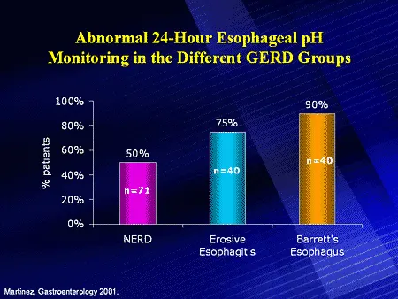
- The doctor identifies the problem based on the readings obtain from the monitor.
- the pH should be in the range of 1.5 to 3.5. If your test results fall more than this normal range, then you have diagnosed as high stomach acid.
- Your pH monitoring may reveal that your pH is normal, in which case another type of testing may be recommended.
- If there is increased acid in your esophagus it could present as a few different conditions, ranging from esophagitis, to scarring (fibrosis), to Barett's esophagus.
- These issues are a consequence of increased acid exposure to the esophagus. At your visit, your healthcare provider will discuss what this means and whether further testing or treatment is needed.
d-Result to the patient
- Most of the time people go home after the test and will have an appointment scheduled at which time they will hear about the results of the pH test.
- Your pH monitoring may reveal that your pH is normal, in which case another type of testing may be recommended.
- If there is increased acid in your esophagus it could present as a few different conditions, ranging from esophagitis, to scarring (fibrosis), to Barett's esophagus.
- These issues are a consequence of increased acid exposure to the esophagus. At your visit, your healthcare provider will discuss what this means and whether further testing or treatment is needed.
- Depending on your results, your healthcare provider may recommend lifestyle changes as well as medications to ease your symptoms and prevent complications or in very severe cases surgery might be required.
Q3) What are all the available clinical solutions to the pathology condition. Consider worst case also
A)Minor severity of the condition - Medication or lifestyle change
Lifestyle Modification
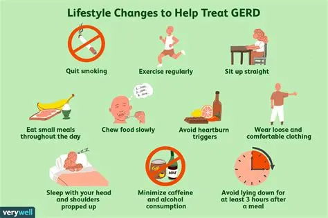
A number of lifestyle and dietary changes may help relieve symptoms. These include:
- Losing weight
- Elevating the head of your bed
- Avoiding overeating
- Avoiding lying down within 3 hours of eating
- Quitting smoking and avoiding exposure to secondhand smoke
- Avoiding foods and beverages that can trigger reflux, including chocolate, peppermint, fatty or spicy foods, citrus fruits, tomatoes, coffee, tea, alcohol, and carbonated drinks
Medications
Three classes of medications, all of which work by reducing acid levels in the stomach, may alleviate symptoms caused by GERD.
- Antacids. These drugs neutralize stomach acid so, even if it regurgitates into the esophagus, its harmful effects will be lessened. Antacids are available over-the-counter.
- H2 Blockers. These medications work by reducing the stomach’s production of acid. They are available by prescription or over-the-counter.
- Proton pump inhibitors (PPIs). These medications also reduce the stomach’s production of acid. They are available by prescription or over-the-counter.
B)Major severity condition - Surgery
- If symptoms persist in spite of other treatments, or if patients cannot tolerate medications or do not wish to use medications over the long-term, a doctor may recommend surgery. The goals of surgery may be to strengthen the lower esophageal sphincter or correct hiatal hernia.
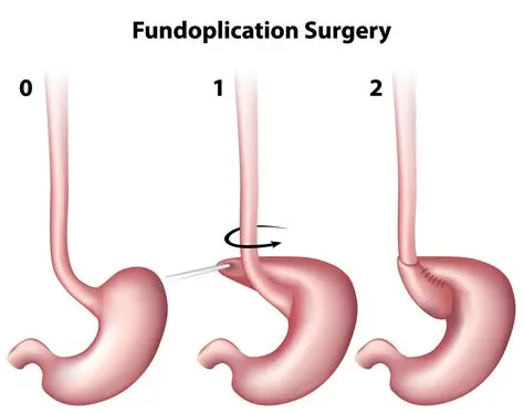
- There are multiple types of surgery available to treat GERD, the most common include fundoplication (during which the top of your stomach is sewn around your esophagus), and bariatric surgery (usually recommended when a doctor has concluded that your GERD may be exacerbated by too much excess weight).
Fundoplication surgery
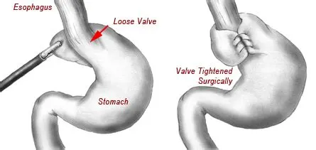
- Fundoplication is one of the most common surgeries used to treat heartburn caused by GERD (Gastroesophageal Reflux Disease). GERD is a chronic backup of stomach acid or contents into your esophagus, the tube that food goes down when you eat.
- GERD can weaken the muscles that help move food down into your stomach, including the sphincter that closes the opening between the esophagus and stomach. Fundoplication helps strengthen this opening to prevent food and acid from going back up.
- This procedure is usually successful and has a good long-term outlook.
Bariatric surgery
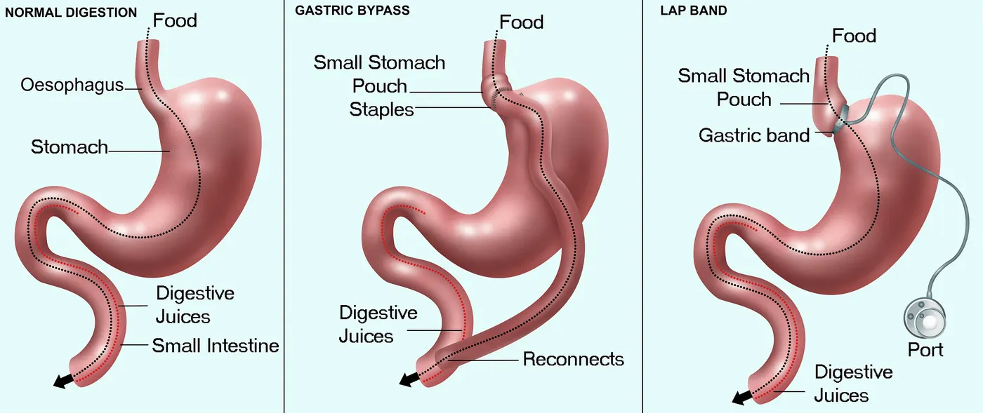
- Bariatric surgery is a term used to describe weight loss surgeries like gastric bypass and gastric sleeve. These surgeries work to treat obesity by altering the structure of your digestive tract. These changes help limit the amount of food you can comfortably eat. They can also influence hunger hormones and appetite.
- Bariatric surgery can be a good option for people with obesity who’ve struggled to lose weight with diet and exercise alone, including people who repeatedly regain the weight they do lose. Bariatric surgery can also a good option for people experiencing obesity-related health conditions, such as type 2 diabetes.
- But this approach isn’t right for everyone, and there are risks to consider in the short and long term.
Q4) Surgical solution in detail
a-Name of surgery
Fundoplication surgery
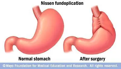
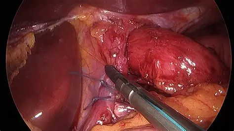
- Fundoplication is one of the most common surgeries used to treat heartburn caused by GERD (Gastroesophageal Reflux Disease). GERD is a chronic backup of stomach acid or contents into your esophagus, the tube that food goes down when you eat.
- GERD can weaken the muscles that help move food down into your stomach, including the sphincter that closes the opening between the esophagus and stomach. Fundoplication helps strengthen this opening to prevent food and acid from going back up.
b-Surgical position
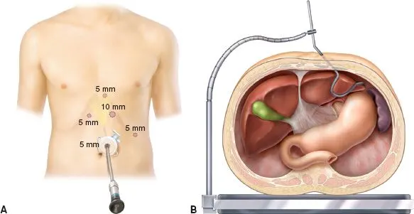
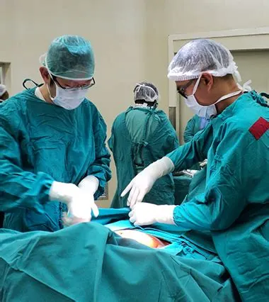
- Surgical technique The patient is placed on the operating table in the modified lithotomy position, in 20 to 45° of reverse Trendelenberg which allows the abdominal viscera, specifically the transverse colon and small bowel, to fall down toward the pelvis out of the operative field.
c-Surgical procedure in detail
Preparation:-
- If a doctor recommends fundoplication, they will explain the necessary preparation for the surgery. For example, they may ask the person to consume only clear liquids prior to surgery. The person may also need to avoid certain medications.
Procedure:-
- When you get to the hospital for the surgery, you’ll check in and be led into a room where you can change into a hospital gown.
- Then, your doctor will put Intravenous (IV) tubes into your veins for both fluid regulation and anesthesia during the surgery. You’ll be asleep during the entire procedure.
- Each type of fundoplication has slightly different steps. But each takes about two to four hours and follow a similar overall procedure. Here’s a general overview of a fundoplication surgery:
- Several small cuts are made through the skin and perotoneum, a layer of tissue around your gut.
- A thin, lighted tube with a camera and tiny surgical tools are inserted into the cuts.
- Your fundus is wrapped around tissue from your lower esophagus.
- Dissolvable stitches are used to attach the fundus to your esophagus.
- Any gas in the abdomen is vented out and all tools are removed from the surgical site.
- The cuts are closed with dissolvable stitches.
d-Implants or instruments required during the surgery with relevant images
Allis
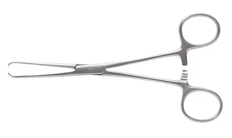
Allis forceps are toothed surgical instruments, used to grasp firm tissues (such as fascia).
Babcock
Babcock forceps are jawed instruments with a smooth end, allowing for more delicate structures to be held in an atraumatic manner.
Dunhill
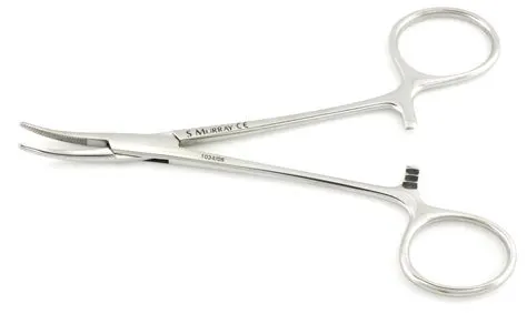
Dunhill forceps are small forceps with curved serrated ends, often used to grasp vessels prior to ligation.
Lane tissue forceps
Lane tissue forceps have interlocking teeth, allowing the grasping of tough tissues, such as fascia.
Littlewood forceps
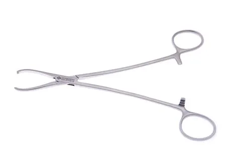
Littlewood forceps have blunt ended teeth, used to grasp tough tissues, such as fascia. They are often used to gain entry via the umbilicus for laparoscopic surgery.
Sawtell
Sawtell forceps are curved forceps with a serrated end, often used to grasp vessels prior to ligation.
Spencer Wells
Available as curved or straight, Spencer Wells forceps can be used to clamp medium to large sized vessels prior to ligation.
Q5-Brief description about - Post surgical care required for the patients
- After fundoplication surgery, a person can typically expect to go home from the hospital . They will receive special instructions on how to care for their surgical wounds.
- These instructions often include not bathing for the first days after being discharged, regularly changing the dressings on wounds, and washing the wounds with gentle soap.
Diet
- Start with a clear liquid diet for the first 24 hours. Drink at least 4 – 6 cups of clear, non-carbonated liquids to meet fluid needs.
- Start with a clear liquid diet for the first 24 hours. Drink at least 4 – 6 cups of clear, non-carbonated liquids to meet fluid needs.
Medicines
- You will receive a prescription for a pain reliever. Take your pain medicine as directed and as needed.
Wound care
-
Tape strips (steri-strips) may be used to close your skin. Do not try to remove these.
-
When you take a shower, cover the tape strips, sutures, staples or glue with a plastic wrap the first week. Avoid taking a bath, going swimming or going into a hot tub until your health care provider tells you.
Activity
-
Walk several times a day.
-
Plan on taking 1 – 2 weeks off from work to recover from surgery. The time may need to be longer if you have a strenuous job.
-
Avoid exercise, and limit any exertion for at least 2 week following surgery. This includes no bending, lifting (more than 10 pounds) or straining. Your surgeon will be able to advise you when it is safe to begin exercising again.
Q6) List all the medical devices used in this case study or project with a single line description of its intended use (with images)
1.pH Monitoring
- Health care professionals use pH monitoring to test for signs of stomach acid in the oesophagus. It can help to determine severity of symptoms related to the pH balance of GI system, such as Gastroesophageal Reflux Disease (GERD).
2.Cutting and Dissecting Instruments
- These surgical instruments as the name implies are used for cutting and dissecting the skin, tissue and suture material.
Types-
a-Scalpels:-A scalpel is a fine-edge cutting instrument with a blade that allows the surgeon to exert maximum control while cutting tissues and is used to make incisions in the skin or other tissues.
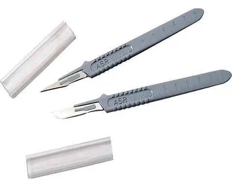
b-Scissors:-Surgical scissors are surgical instruments usually used for cutting.
3.Grasping or Holding Instruments
- Grasping and holding instruments are used to hold tissue, drapes or sponges. Examples include Forceps, Towel Clamps, vascular clamps, and organ holders.
Types:-
Forceps:- Surgeons use forceps during surgical procedures to hold onto or manipulate tissues and to clamp blood vessels.
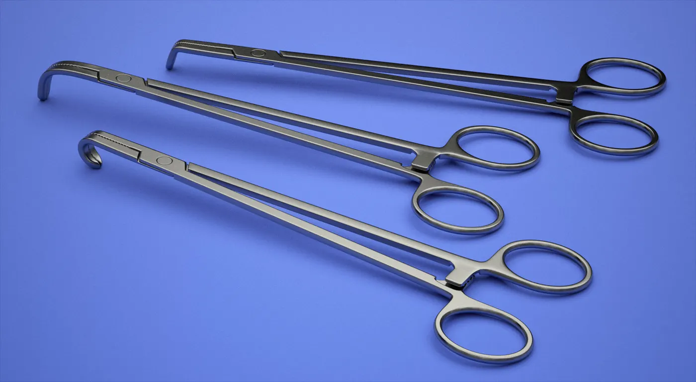
Towel Clamps:-A towel Clamp is a perforating clamp used for grasping tissue, securing towels or drapes and holding or reducing small bone fractures.
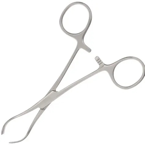
Needle Holder:-A needle holder, also called a needle driver, is a surgical instrument, similar to a hemostat, used by doctors and surgeons to hold a suturing needle for closing wounds during suturing and surgical procedures.
4-Tissue Unifying Instruments
- This is another class of surgical instruments that includes tissue unifying instruments and materials such as needle holders, surgical needles, staplers, clips, adhesive tapes.
Types:-
Needle Holders:-Needle holders are hinged instruments that have a carbide insert in the tip to prevent slipping.
Surgical Needles:-Surgery requires the use of several different types of needles. Needles need to be strong enough to pass through tough tissue while causing minimal trauma to delicate tissues and reducing tissue reactions.
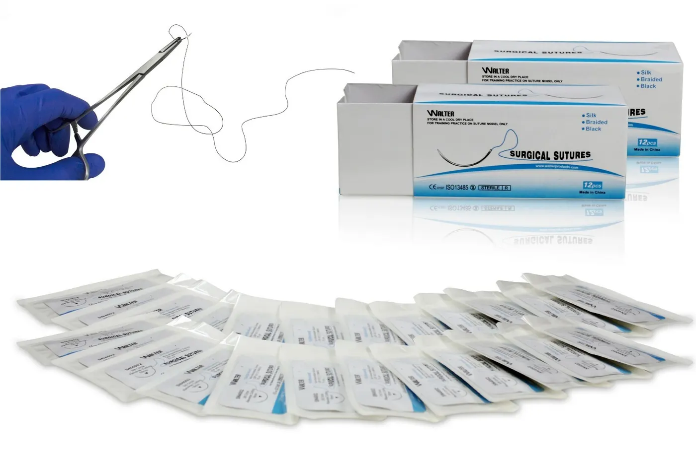
Surgical suture:- to tie the cut parts
PROJECT(B)
SELECTED SYSTEM:- CARDIOVASCULAR SYSTEM
CARDIOVASCULAR SYSTEM
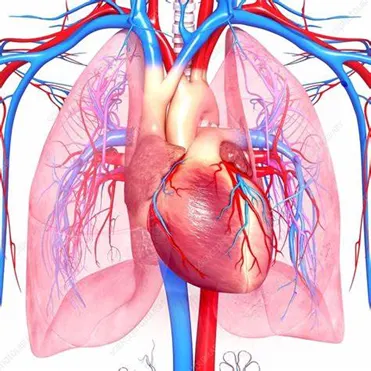
- The cardiovascular system consists of the heart, blood vessels, and blood. Its primary function is to transport nutrients and oxygen-rich blood to all parts of the body and to carry deoxygenated blood back to the lungs.
- The heart consists of four distinct chambers: two upper chambers called “atria” and two lower chambers called “ventricles.” A wall or “septum” separates the atria and ventricles. Valves control the flow of blood within the different chambers.
- It consists of the following organs and tissues:
- The heart: A muscular pump that forces blood around the body.
- A closed system of blood vessels: These vessels include:
- Arteries: Vessels that carry blood away from the heart.
- Veins: Vessels that bring blood back to the heart.
- Capillaries: Tiny vessels that branch off from arteries to deliver blood to all body tissues.
- There are two blood circulatory systems in the body. The first is the systemic circulatory system. This is the main blood circulatory system that transports blood to the organs, tissues, and cells throughout the body.
- The second is the pulmonary circulatory system. This circulatory system moves blood between the heart and lungs. It is where oxygen enters the blood and carbon dioxide leaves the blood.
Description:
Q1) Detailed description of the clinical condition selected
SELECTED CLINICAL CONDITION:- HEART ATTACK (Myocardial infarction )
a-Normal function of the organ
- As a central part of the circulatory system, the heart is primarily responsible for pumping blood and distributing oxygen and nutrients throughout the body. Because of this task, the heart may be considered one of the most important organs of the body, such that even small dysfunctions or abnormalities may cause drastic changes or effects in the human organism.
- he organ is divided into several chambers that take in and distribute oxygen-poor or oxygen-rich blood. These chambers are accompanied by veins and arteries that facilitate the same function. With all of its parts working together towards the same goal, the heart successfully pumps blood with ease.
- Normally, a good-functioning adult heart could go on three cardiac cycles or 72 beats per minute—this rate changes for children whose heart rates are normally and relatively faster.
b-Current pathological state in detail
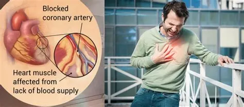
- Myocardial infarction (MI) is the condition in which insufficient blood flows to the heart (or a part of the heart) and causes damage to the cardiac muscles.
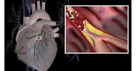
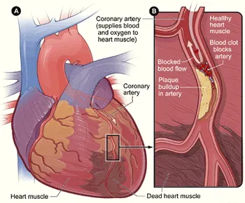
- The blockage is usually caused when a plaque ruptures. If blood flow isn’t restored quickly, either by a medicine that dissolves the blockage or a catheter placed within the artery that physically opens the blockage, the section of heart muscle begins to die. the following are the stages:-
Occlusion
The first stage in a heart attack is occlusion. This is the blocking of an artery that supplies the heart muscle with blood and oxygen. a person doesn't even realize anything is happening at this stage.
 |
Infarction
If the occlusion is not removed, then heart muscle cells begin to die. This is called infarction. person is now experiencing growing chest pain that radiates down his left arm. In his heart, more and more cells are dying due to lack of oxygen. This causes his heart to pump less efficiently and causes him to experience shortness of breath, dizziness, and hypotension (low blood pressure).
Acute phase
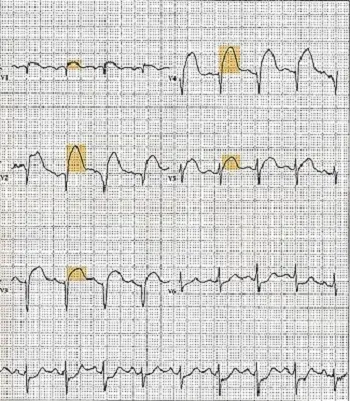
The acute phase of a heart attack begins as soon as the event occurs and lasts about seven days. The first clinical sign of a heart attack is a change in the EKG. Because the dead heart muscle does not conduct electrical signals like the healthy cells, EKG changes occur, usually in the form of elevation or depression. The most common is ST segment elevation. The ST segment is the space between the S wave and the T wave and represents the period of time right after both ventricles have contracted.
This is helpful to determine if an infarcted area or EKG change is new or an old myocardial infarction. As the muscle dies, a protein called CK-MB is released. This helps to differentiate between muscle injury or a cardiac event. CK-MB becomes elevated about four hours after the event and peaks at 24 hours.
 |
Healing phase
The healing phase begins around day seven and lasts around a month. During this time the infarcted area becomes a scar on the heart. These scars can cause heart failure and dangerous heart rhythm, the patient must be monitored for any complications and started on treatment to prevent another heart attack from occurring.
Healed phase
Typically, the healed phase begins about a month after the heart attack. By now the cardiac enzymes have gone back to normal levels.
Symtoms
- An abnormal heart rate (palpitation)
- Sudden breathlessness (dyspnea)
- Difficulty breathing while lying down (orthopnea)
- Sleep apnea (breathlessness during sleep)
- A cough with blood in sputum due to the accumulation of fluid in lungs (pulmonary edema)
- Swelling of feet, ankles, legs, abdomen (ascites) due to fliud accumulation.
- Extreme fatigue and dizziness
- Bluish discoloration of the skin (cyanosis)
- A wheezing cough after slight physical exertions.
c-Reason for the pathological condition
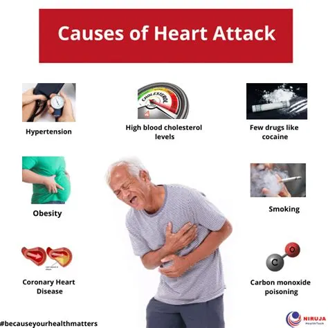
A heart attack is precipitated by a wide range of physical, mental, environmental and dietary factors. The most important ones are listed below-
- Coronary Artery Disease (CAD): Excess deposition of fats on the inner wall of the artery (atherosclerosis) supplying blood to heart muscles causes narrowing and blockage of the arterial passage. Plaques are formed, which can rupture any time, leading to obstructed blood circulation and heart attack.
- High blood pressure: Under high blood pressure conditions, the heart has to struggle more to pump sufficient blood to the organs. This leads to gradual thickening of the heart muscles, which become stiff and weak and fail to pump blood efficiently.
- Defective heart valves: Damaged auricular or ventricular valves fail to maintain blood flow in the proper direction. This can cause backflow of blood to the veins and cause a heart attack.
- Cardiomyopathy: Dilatation, thickening or stiffening of the heart muscles predispose the patient to myocardial infarction.
- Myocarditis: Inflammation of heart muscles occurs in few cases that might lead to myocardial infarctions (heart attack).
- Cardiac arrhythmia: Abnormal heart rates interfere with the normal mechanism of blood circulation and can cause heart failure.
- Diet: Diet rich in fat, cholesterol, and sodium can lead to heart attack.
d-Effect of homoeostasis imbalance on human body
- Stasis of waste products of cellular metabolism in addition to a lack of oxygen delivery, leading to cellular damage.
- During a heart attcak, your heart won’t be able to function normally. That’s bad news for your entire body. Your vital organs—meaning your brain, lungs, kidneys, and liver—depend on a steady diet of blood, oxygen, and nutrients, which the heart pumps to them. If your organs go without these essential supplies for too long, they too will become permanently damaged and begin to shut down.
- The more severe the heart attack—meaning the bigger the blockage and the more time it takes to treat it—the faster this occurs.
- blood pressure levels drop significantly.
- The amount of blood and oxygen that reaches the brain drops significantly, too. Without oxygen, the brain cells begin to die, a process called cerebral infarction. The signs that this has begun to happen include vision loss, trouble moving, and impaired speech. It also may lead to unconsciousness, and it could cause the heart to stop beating altogether, a quickly fatal condition known as cardiac arrest.
Q2)Description of the diagnostic method selected by the surgeon for assessing the condition.
Common diagnostic methods:-
- Chest Radiograph (to detect cardiac enlargement or pulmonary edema)
- Echocardiogram (measures systolic and diastolic ventricular contractile functions)
- Electrocardiogram ( can detect the presence of coronary artery disease or ventricular hypertrophy)
- Endomyocardial biopsy (to detect myocarditis or cardiotropic viruses)
- Chest X-ray
- Lipid profile test
a-Selected Diagnostic method:-Echocardiogram
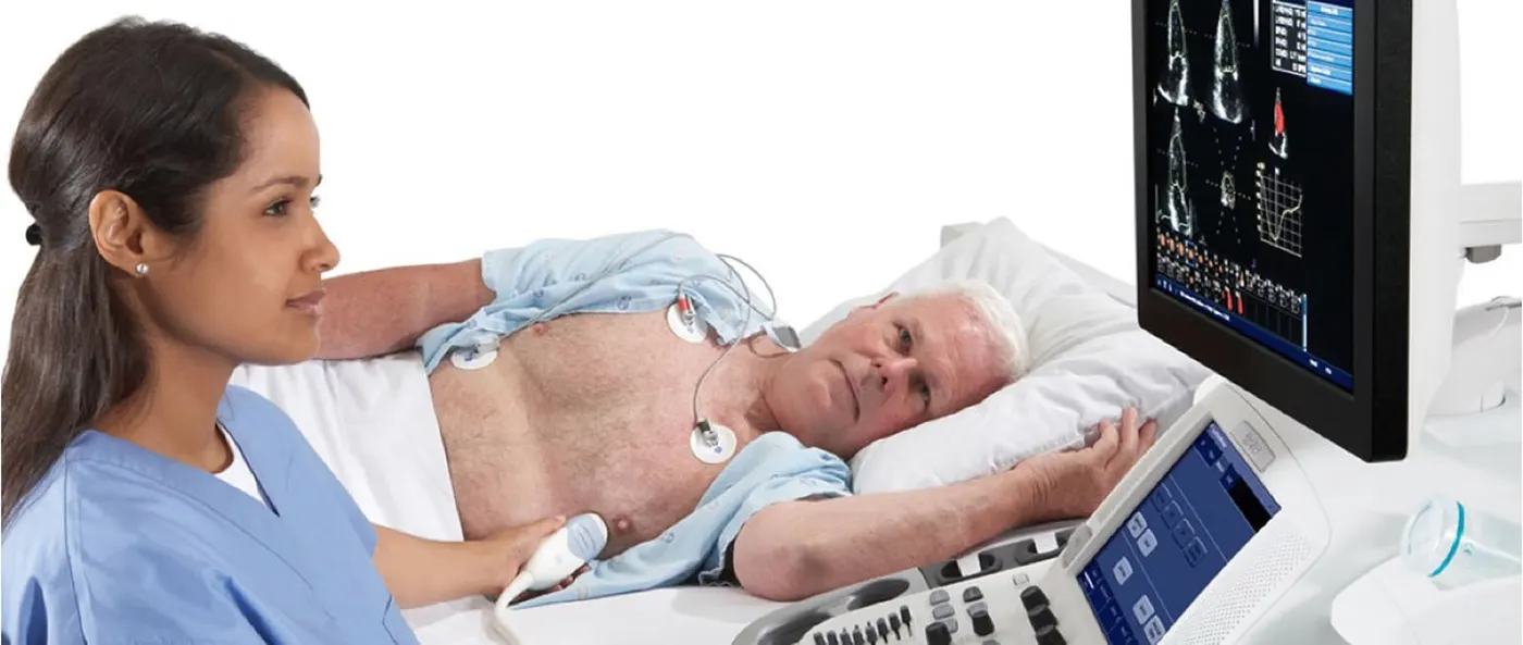
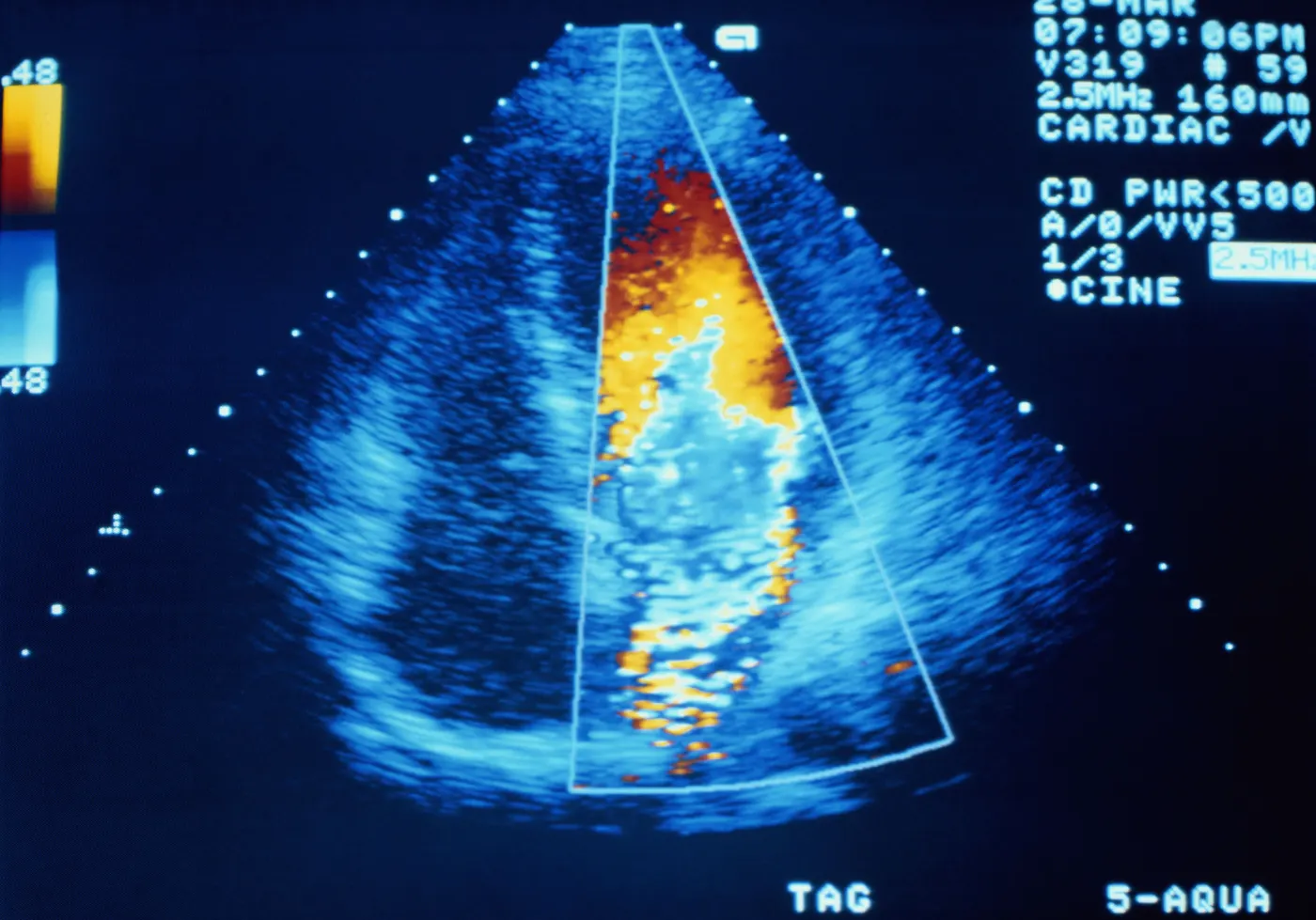
- Echocardiography uses ultrasound waves to create a picture of the heart, called an echocardiogram (echo).
- It is a noninvasive medical procedure that produces no radiation and does not typically cause side effects.
Why
- Determine how well the heart is pumping blood
- diagnose heart disease-including weak pumping or stiffening of the heart muscle, leaky or blocked heart valves, and chamber enlargement-in adults
- locate blood clots or tumors
- assess the pressure in the heart to diagnose a condition called pulmonary hypertension.
Time
- 10 to 15 mins.
About instrument-how it works
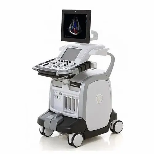
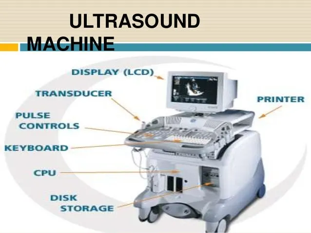
An ultrasound machine uses high frequency sound waves and their echoes to help determine the size, shape and depth of an abnormality. In a typical ultrasound, millions of pulses and echoes are sent and received from the probe each second.
A ultrasound machine has the following parts:
- Transducer probe - The probe is the mouth and ears of the ultrasound machine. In the probe, there are one or more quartz crystals called piezoelectric crystals. When an electric current is applied to these crystals, they change shape rapidly. The rapid shape changes, or vibrations, of the crystals produce sound waves that travel outward. The sound waves travel into the patient being scanned. The sound waves bounce back at various intervals depending on the type of material they pass through.
- Central Processing Unit (CPU) - The CPU is the brain of an ultrasound machine. The CPU is a computer that contains the microprocessor, memory, amplifiers and power supplies for the microprocessor and transducer probe. The transducer receives electrical currents from the CPU and sends electrical pulses that are created by returning echoes.
- Transducer pulse controls - The operator, called the ultrasonographer, changes the amplitude, frequency and duration of the pulses emitted from the transducer probe
- Display - Displays the image from the ultrasound data processed by the CPU. This image can be either in black-and-white or color, depending upon the model of the ultrasound machine
- Keyboard/cursor - Ultrasound machines have a keyboard and a cursor. The keyboard allows the operator to add notes and to take measurements of the image.
- Disk storage device - The processed data and/or images can be stored on disks (hard disks, floppy disks, CD, or DVD). They can also be stored digitally so they can be accessed from the office or anywhere you have an Internet connection.
- Printer - Most ultrasound machines have thermal printers. These can be used to capture a printed picture of the image from the monitor.
A computer compiles these returning sound waves, or echoes, and turns them into a picture of the heart.
Complications
- safe without any risks.
b-Data acquisition method
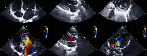
- computer compiles the returning sound waves, or echoes, and turns them into a picture of the heart.A technician or a doctor will perform your echo.
- Often, a technician does some or all of the test, but a doctor, usually a cardiologist, will look at your heart images while you are having your echo.
- They may want to adjust the transducer—the handheld device used—to visualize additional views, if necessary.
c-How the doctor identifies the problem from data , parameters he considered
Parameters considered to identify the problem:-
- Problems with the heart valves: how well your hearts valves are functioning.
- The velocity, or speed, of blood flow within the heart: This is helpful in measuring problems with blood flow in conditions such as aortic stenosis.
- Anatomical defects: Congenital heart conditions such as tetralogy of fallot and atrial septal defect are conditions that are present from birth in which the heart does not develop properly.
- Left ventricular ejection fraction: "Ejection fraction" is a term used to describe how strong the heart is and how well it pumps blood.
- Cardiac arrythemia: An echo can assess your heartbeat. This may help in determining the exact cause and best treatment if you do have an arrhythmia, or irregular heart rhythm.
d-Result to the patient
- The results of your echo will be prepared in a written report by your doctor.
- The report will describe the heart anatomy, heart movements, and any defects observed during the test.
- Often, because the results are so detailed, your doctor may schedule an appointment with you to discuss the results and next steps.
- In general, follow-up after an echo is focused on the treatment of your heart condition. For example:
- If the test was used to diagnose a congenital heart condition, the next steps may include surgical repair.
- If it detected heart failure, medication adjustments may be needed.
- If it is used for evaluation of an arrhythmia, medication, surgery, or a pacemaker may be needed.
Q3) What are all the available clinical solutions to the pathology condition. Consider worst case also
A)Minor severity of the condition - Medication or lifestyle change
Lifestyle Modification

- Stop smoking
- Choose good nutrition
- dont consume alcohol
- walk dailyly for atleast 30 mins.
- reduce fat and oily foods.
- Find ways to manage stress.
- take medications on time
Medications
- Beta-blockers:- are a class of medications commonly used to treat heart problems such as high blood pressure, chest pain, abnormal heart rhythm, congestive heart failure, and heart attacks.These medications block the effects of adrenaline, which makes it easier for your heart to do its job.
- Angiotensin-converting enzyme (ACE) inhibitors:- treat high blood pressure and other conditions such as heart failure and heart attack. They block, or inhibit, the production of an enzyme that causes your vessels to narrow. This can help improve your blood flow by relaxing and widening your blood vessels. e.g.enalapril (Vasotec), lisinopril(Prinivil, Zestril),quinapril (Accupril).
- Antiplatelet agents:- prevent clotting in your arteries by keeping blood platelets from sticking together, which is usually the first step in blood clot formation. Aspirin is the most well-known type of antiplatelet medication.
- Antocoagulant drugs:- are blood thinners, which means they reduce the risk of clotting in people who have had a heart attack. Unlike antiplatelets, they work by affecting the coagulation factors that are also involved in the blood clotting process.e.g. heparin, warfarin.
B)Major severity condition - Surgery
1- Angioplasty
- Special tubing with an attached deflated balloon is threaded up to the coronary arteries.
- The balloon is inflated to widen blocked areas where blood flow to the heart muscle has been reduced or cutoff. Often combined with implantation of a stent (see below) to help prop the artery open and decrease the chance of another blockage.
- Considered less invasive because the body is not cut open. Lasts from 30 minutes to several hours. May require an overnight hospital stay.
2-Angioplasty, laser
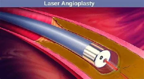
- Laser angioplasty is one method to remove atherosclerotic plaques.
- Similar to angioplasty except that the catheter has a laser tip that opens the blocked artery. Pulsating beams of light vaporize the plaque buildup.
3-Bypass surgery
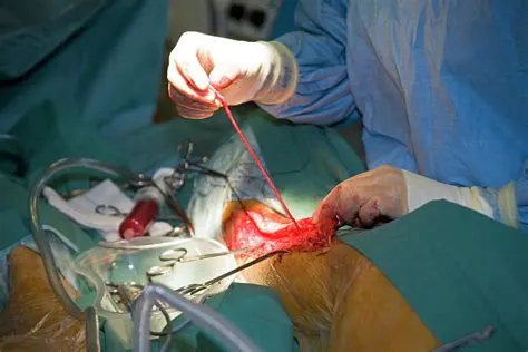
- Treats blocked heart arteries by taking arteries or veins from other parts of your body — called grafts — and using them to reroute the blood around the clogged artery to supply blood flow to your heart muscle.
- A patient may undergo one, two, three or more bypass grafts, depending on how many coronary arteries are narrowed. Requires several days in the hospital.
Q4) Surgical solution in detail
a-Name of surgery
Angioplasty
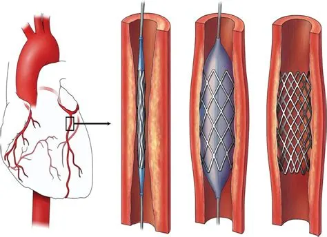
- Angioplasty, is also known as balloon angioplasty and percutaneous transluminal angioplasty (PTA), is a minimally invasive endovascular procedure used to widen narrowed or obstructed arteries or veins, typically to treat arterial atherosclerosis.
- During angioplasty, a surgeon inserts a tube into an artery in the groin or wrist. They then thread the tube towards the affected artery around the heart. Finally, they insert a balloon or stent(metal tube) to open the artery.
b-Surgical position
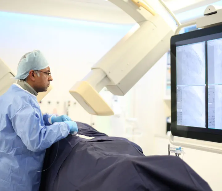
- You will be placed in a supine (on your back) position on the procedure table.
- You will be connected to an ECG monitor that records the electrical activity of the heart and monitors the heart during the procedure using small, adhesive electrodes.
c-Surgical procedure in detail
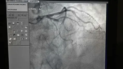
Preparation
- Angioplasty is a minimally invasive procedure, but it is still surgery, and people must follow their doctor’s instructions carefully beforehand.
- People need to inform their doctor about any medications and supplements they are taking. In some cases, they may need to stop taking these drugs, especially blood thinners, before the procedure.
- Also, an individual may need to avoid food or drinks for several hours before the angioplasty procedure as doctors will need to sedate them.
Procedure
- Before beginning angioplasty, a healthcare professional will clean and numb the area where the catheter enters the body, usually the groin but sometimes the wrist.
- Next, a doctor inserts the catheter into the artery and directs it towards the coronary artery, watching its progress on an X-ray feed.
- Once the catheter is in position, the doctor injects a contrast dye through the artery, which helps identify blockages around the heart. Once they locate the blockages, the doctor inserts a second catheter and a guidewire, usually with a balloon at the tip.
- When the second catheter is in position, the doctor inflates the balloon, which pushes the plaque buildup away and opens up the artery. The surgeon may insert a stent to keep the artery propped open.
d-Implants or instruments required during the surgery with relevant images
STENTS
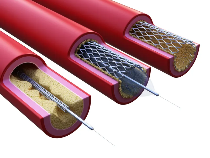
- Stents can be defined as small mesh tubes inserted into an artery to prevent it from collapsing and keep it open after an angioplasty surgery.
- The total coronary stent market comprises bare-metal stents, drug-eluting stents, covered stents, and bifurcated stents.
CATHETER
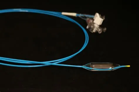
- small catheter-a thin hollow plastic tube with a diameter of 2-3 mm.
- Angioplasty uses a balloon-tipped catheter to open a blocked blood vessel and improve blood flow.
- The doctor uses medical imaging, typically live x-rays, to guide the catheter across the blockage. Once the balloon spans the blockage, it is inflated to open the blocked vessel and improve blood flow.
Q5-Brief description about - Post surgical care required for the patients

- Once you leave the hospital, your post-procedure care will be two-fold. You will need to take care of yourself by taking medications exactly as prescribed, as well as making lifestyle changes such as exercising, improving your diet and quitting smoking (if you smoke).
- You should also have at least one follow-up appointment with your treating interventional cardiologist or another qualified specialist if you are not able to return to your treating physician. At this appointment, your interventional cardiologist will examine the catheter insertion site to be sure it is healing properly.
- Irrespective of the type of angioplasty, there are certain precautions after angioplasty required to stay healthy after the procedure. You need great care after angioplasty to avoid any serious consequences.
- These lifestyle changes include dietary changes as well as a change in exercise routine for better heart health.
- Start Slow
- Follow a well-rounded program
- Stop smoking
- Choose good nutrition
- dont consume alcohol
- walk dailyly for atleast 30 mins.
- reduce fat and oily foods.
- Find ways to manage stress.
- take medications on time
- Track your progress
Q6) List all the medical devices used in this case study or project with a single line description of its intended use (with images)
1. ECHOCARDIOGRAM
An ultrasound machine uses high frequency sound waves and their echoes to help determine the size, shape and depth of an abnormality. In a typical ultrasound, millions of pulses and echoes are sent and received from the probe each second.
2.STENTS
- Stents can be defined as small mesh tubes inserted into an artery to prevent it from collapsing and keep it open after an angioplasty surgery.
3.CATHETER
- small catheter-a thin hollow plastic tube with a diameter of 2-3 mm.
- Angioplasty uses a balloon-tipped catheter to open a blocked blood vessel and improve blood flow.