Project 1
Part A - Detailed study of anatomical basis of surgical procedures or Clinical management in association with any of the organ system .
SELECTED 3 SYSTEMS:-
A) CARDIOVASCULAR SYSTEM
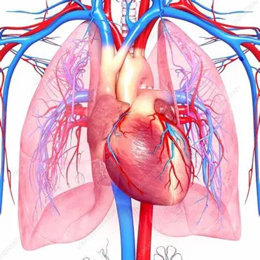
- The cardiovascular system consists of the heart, blood vessels, and blood. Its primary function is to transport nutrients and oxygen-rich blood to all parts of the body and to carry deoxygenated blood back to the lungs.
- The heart consists of four distinct chambers: two upper chambers called “atria” and two lower chambers called “ventricles.” A wall or “septum” separates the atria and ventricles. Valves control the flow of blood within the different chambers.
- It consists of the following organs and tissues:
- The heart: A muscular pump that forces blood around the body.
- A closed system of blood vessels: These vessels include:
- Arteries: Vessels that carry blood away from the heart.
- Veins: Vessels that bring blood back to the heart.
- Capillaries: Tiny vessels that branch off from arteries to deliver blood to all body tissues.
- There are two blood circulatory systems in the body. The first is the systemic circulatory system. This is the main blood circulatory system that transports blood to the organs, tissues, and cells throughout the body.
- The second is the pulmonary circulatory system. This circulatory system moves blood between the heart and lungs. It is where oxygen enters the blood and carbon dioxide leaves the blood.
B) EXCRETORY SYSTEM
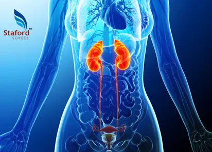
- The Excretory System is defined as a biological system that helps in the elimination of the nitrogenous waste products of metabolism and also helps in maintaining homeostasis in our body.
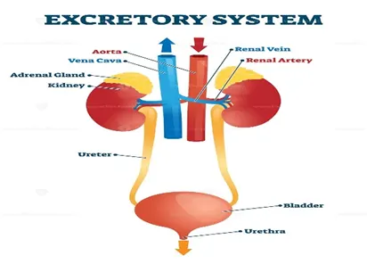
- The human excretory system organs include: A pair of kidneys, A pair of ureter, A urinary bladder, A urethra.
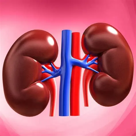
- Kidneys are bean-shaped structures located on either side of the backbone and are protected by the ribs and muscles of the back. Each human adult kidney has a length of 10-12 cm, a width of 5-7 cm and weighs around 120-170g.
- Ureter are a pair of thin muscular tubes called the ureter comes out of each kidney extending from the renal pelvis. It carries urine from the kidney to the urinary bladder.
- Urinary bladder is a muscular sac-like structure, which stores urine. The urinary bladder is emptied by the process of micturition, i.e. the act of urination.
- Urethra tube arises from the urinary bladder and helps to expel urine out of the body. In males, it acts as the common route for sperms and urine. Its opening is guarded by sphincter muscles.
- It helps in the elimination of toxic nitrogenous wastes from our body.
- It also helps in the filtration of blood by ultrafiltration.
- It helps in osmoregulation (i.e., maintaining salt and water balance in our body).
3) GASTROINTESTINAL SYSTEM
- The gastrointestinal system is also known as the digestive system. It is a collective name of the alimentary canal and accessory organs.
- The alimentary canal forms a continuous tube that is open to the outside environment at both ends.
- The organs of the alimentary canal are the mouth, pharynx, esophagus, stomach, small intestine, and large intestine.
- The overall process of digestion and absorption occurs in the alimentary canal.
- Accessory structures include the teeth, tongue (in the oral cavity), salivary glands, liver, gallbladder, and pancreas.
UPPER GI TRACT
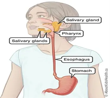
- The upper GI tract is responsible for starting digestion and preparing it for the lower GI tract to process and absorb nutrients.
- The upper GI consists of the following organs: Mouth, Pharynx, Oesophagus, Stomach, Duodenum.
LOWER GI TRACT
- The contents of the small intestine empty into the large intestine, which is also called the colon. While food travels through the colon, the water and salts are absorbed. Once the nutrients are removed from the food, the remaining materials become stool – which moves into the rectum to be expelled from the body as waste.
- The lower GI consists of the following organs: Small Intestine, Large Intestine.
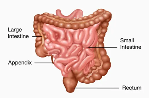
Description:
Q1. Describe one common clinical condition or disorder associated with the system selected.
SYSTEM SELECTED:- EXCRETORY SYSTEM
Common disorders:-
Uremia
- Under this condition, the urea accumulation is comparatively high. In such patients, excess urea is removed by hemodialysis.
- In hemodialysis, the blood is drained carefully from whichever artery is convenient and is sent to the dialyzing unit. The unit contains a porous membrane which allows passage of molecules on the basis of the concentration gradient.
Renal failure
- Renal failure is the decrease in the glomerular filtration in humans. Both the kidneys are damaged and stop working in case of acute renal failure. The main feature of acute renal failure is scanty urine production.
- The kidney transplant is the ultimate treatment for the correction of acute renal or kidney failure. The process is elaborate and relies heavily on the availability of a functioning kidney from donors. There are numerous complications that could occur. To avoid these, the kidney from a close or distant relative is preferred. But due to modern technology, the complications related to kidney transplant have been reduced to a greater deal.
Renal calculi or kidney stones
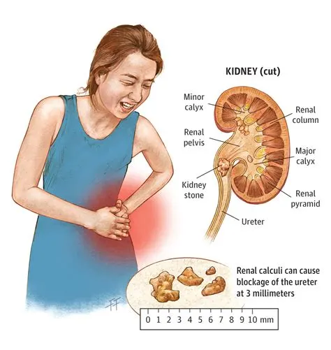
- Kidney stones are the result of the deposition of dissolved minerals or insoluble salts on the inner lining of the kidney. These increase in size and cause immense pain in the patients when passed.
Nephritis or Brights disease
- Nephritis is generally the inflammation of the kidney. Glomerulonephritis is characterised by inflammation in the glomeruli of the kidney. In this case, the glomeruli are completely filled in blood. If many glomeruli become non-functional, the patient needs an artificial kidney.
Renal Tabular Acidosis
- In this condition, the person is unable to secrete optimum quantities of hydrogen ions due to which a large amount of sodium bicarbonate ions are lost during urination.
RENAL CALCULI OR KIDNEY STONES
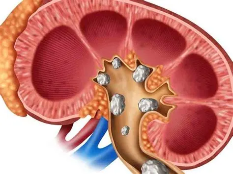
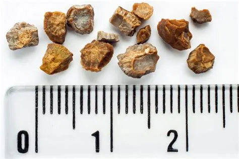
- Kidney stones, or renal calculi, are solid masses made of crystals. Kidney stones usually originate in your kidneys. However, they can develop anywhere along your urinary tract, which consists of these parts: kidneys, ureters, bladder, urethra
- Kidney stones can be a painful medical issue. The causes of kidney stones vary according to the type of stones.
- There are four types of kidney stones: calcium oxalate, uric acid, struvite, and cystine.
- A kidney stone may be treated with shockwave lithotripsy, uteroscopy, percutaneous nephrolithomy or nephrolithotripsy.
- Common symptoms include severe pain in lower back, blood in your urine, nausea, vomiting, fever and chills, or urine that smells bad or looks cloudy.
- Each year, more than half a million people go to emergency rooms for kidney stone problems. It is estimated that one in ten people will have a kidney stone at some time in their lives.
Types of stones
- Calcium oxalate: The most common type of kidney stone which is created when calcium combines with oxalate in the urine. Inadequate calcium and fluid intake, as well other conditions, may contribute to their formation.
- Uric acid: This is another common type of kidney stone. Foods such as organ meats and shellfish have high concentrations of a natural chemical compound known as purines. High purine intake leads to a higher production of monosodium urate, which, under the right conditions, may form stones in the kidneys. The formation of these types of stones tends to run in families.
- Struvite: These stones are less common and are caused by infections in the upper urinary tract.
- Cystine: These stones are rare and tend to run in families.
What happens
- Urine has various wastes dissolved in it. When there is too much waste in too little liquid, crystals begin to form.
- The crystals attract other elements and join together to form a solid that will get larger unless it is passed out of the body with the urine.
- Usually, these chemicals are eliminated in the urine by the body's master chemist: the kidney.
- In most people, having enough liquid washes them out or other chemicals in urine stop a stone from forming. The stone-forming chemicals are calcium, oxalate, urate, cystine, xanthine, and phosphate.
- After it is formed, the stone may stay in the kidney or travel down the urinary tract into the ureter.
- Sometimes, tiny stones move out of the body in the urine without causing too much pain.
- But stones that don't move may cause a back-up of urine in the kidney, ureter, the bladder, or the urethra. This is what causes the pain.
Causes
- Possible causes include drinking too little water, exercise (too much or too little), obesity, weight loss surgery, or eating food with too much salt or sugar.
- Infections and family history might be important in some people.
- Eating too much fructose correlates with increasing risk of developing a kidney stone. Fructose can be found in table sugar and high fructose corn syrup.
Symtoms
- severe pain on either side of your lower back
- more vague pain or stomach ache that doesn't go away
- blood in the urine
- nausea or vomiting
- fever and chills
- urine that smells bad or looks cloudy
ORGAN INVOLVED:- KIDNEY
Gross anatomy
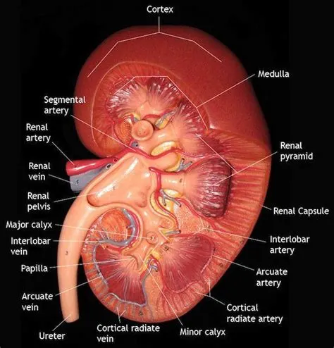 |
|
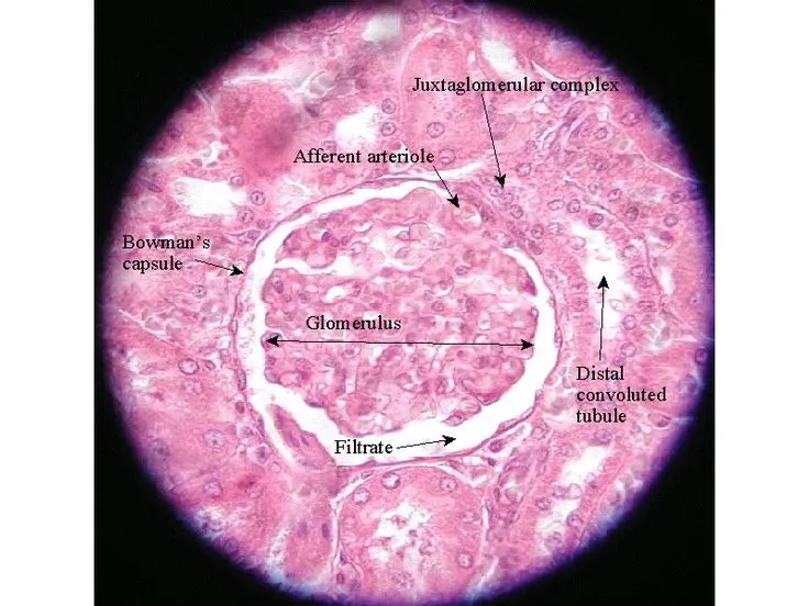
Microscopoc veiw
 |
Kidney Structures
Glomerulus
Nephron
|
Macroscopic view
|
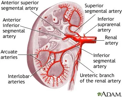
Blood and Nerve suppy
 |
|
Q2. Describe the common disorders in association with the organ
ORGAN:- KIDNEY( EXCRETORY SYSTEM)
Common Disorders
Chronic kidney disease(CKD)
- CKD slowly damages the kidneys over time. CKD affects about 37 million adults in the United States. The disease is divided into different stages depending on the severity of kidney damage.
- Diabetes is the most common cause of CKD. Other causes include High BP, heart diseases, and certain genetic conditions such as polycystic kidney disease.
Acute kidney injury
- Also called acute renal failure (ARF). AKI causes kidney damage or failure within a short period of time. AKI may damage other organs as well, such as the heart, lungs, and brain.
- Causes may include major burns, shock (a sudden drop in blood pressure), excessive bleeding, severe dehydration, or medication use.
Kidney stones
- Also called reanl stones are hard deposits of minerals or salts formed inside the kidneys. They can form inside the ureter (tubes that carry urine from the kidneys) or urinary bladder (structure that receives urine from the kidneys via the ureters).
- Kidney stones generally occur when there is an excess of mineral salts, such as calcium oxalate or calcium phosphate, or uric acid in the body.
Kidney infection
- Kidney infections or pyelonephritis is a special type of UTI, which typically occurs in any part of the urinary tract (kidneys, ureters, urinary bladder or urethra).
- Kidney infections occur when infection from the urethra or the urinary bladder travels upward to the kidneys.
Kidney cysts
- A kidney cyst is a fluid-filled pouch or sac-like structure. Also called simple kidney cysts, these are different from a genetic condition called polycystic kidney disease that causes the formation of numerous cysts that damage the kidneys.
Kidney cancer
- Kidney cancer occurs when the cells in the kidney grow out of control. The most common type of kidney cancer is called renal cell carcinoma , accounting for about 90% of all kidney cancers.
KIDNEY STONES
- Kidney stones, or renal calculi, are solid masses made of crystals. Kidney stones usually originate in your kidneys. However, they can develop anywhere along your urinary tract, which consists of these parts: kidneys, ureters, bladder, urethra
- Kidney stones can be a painful medical issue. The causes of kidney stones vary according to the type of stones.
- There are four types of kidney stones: calcium oxalate, uric acid, struvite, and cystine.
- A kidney stone may be treated with shockwave lithotripsy, uteroscopy, percutaneous nephrolithomy or nephrolithotripsy.
- Common symptoms include severe pain in lower back, blood in your urine, nausea, vomiting, fever and chills, or urine that smells bad or looks cloudy.
- Each year, more than half a million people go to emergency rooms for kidney stone problems. It is estimated that one in ten people will have a kidney stone at some time in their lives.
What happens
- Urine has various wastes dissolved in it. When there is too much waste in too little liquid, crystals begin to form.
- The crystals attract other elements and join together to form a solid that will get larger unless it is passed out of the body with the urine.
- Usually, these chemicals are eliminated in the urine by the body's master chemist: the kidney.
- In most people, having enough liquid washes them out or other chemicals in urine stop a stone from forming. The stone-forming chemicals are calcium, oxalate, urate, cystine, xanthine, and phosphate.
- After it is formed, the stone may stay in the kidney or travel down the urinary tract into the ureter.
- Sometimes, tiny stones move out of the body in the urine without causing too much pain.
- But stones that don't move may cause a back-up of urine in the kidney, ureter, the bladder, or the urethra. This is what causes the pain.
Causes
- Possible causes include drinking too little water, exercise (too much or too little), obesity, weight loss surgery, or eating food with too much salt or sugar.
- Infections and family history might be important in some people.
- Eating too much fructose correlates with increasing risk of developing a kidney stone. Fructose can be found in table sugar and high fructose corn syrup.
Symtoms
- severe pain on either side of your lower back
- more vague pain or stomach ache that doesn't go away
- blood in the urine
- nausea or vomiting
- fever and chills
- urine that smells bad or looks cloudy
Common diagnostic methods
KIDNEY STONE
- SONOGRAPHY
- X-RAYS
- CT SCANS
KIDNEY DISEASES
- CREATININE TEST
- BLOOD AND URINE ANALYSIS
- GLOMERULAR FILTERATION TEST
1) SONOGRAPHY (RENAL ULTRASOUND)
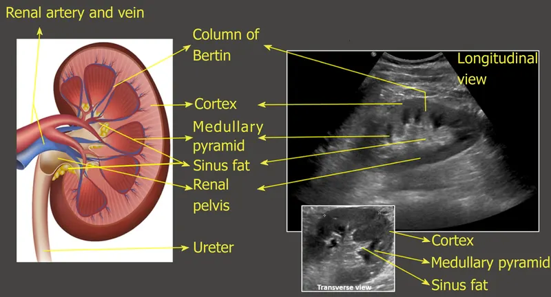
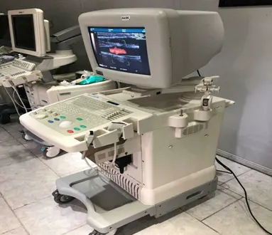
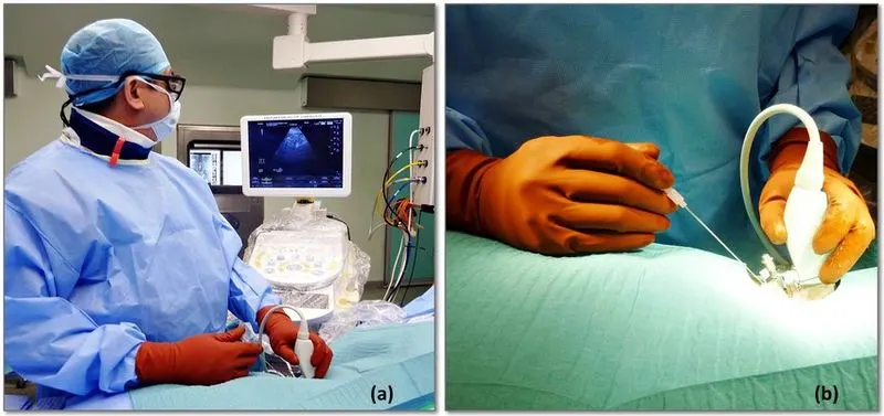
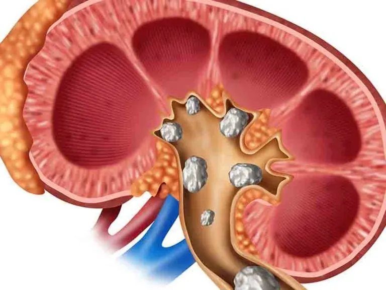
- A kidney ultrasound is a non-invasive exam that uses sound waves to produce images of kidneys.
- Ultrasound or sonography, uses high frequency sound waves sent out by a transducer (wand) pressed against skin. The sound waves move through the body, bouncing off organs back to the transducer.
- These echoes are recorded and digitally turned into video or images of the tissues and organs selected for examination.
- Ultrasound images can help doctor evaluate: the location of your kidneys, the size of your kidneys, the shape of your kidneys, blood flow to your kidneys, injury, kidney stones, tumours.
Why
- Your doctor may recommend a kidney ultrasound if they think you have a kidney problem like kidney abscess, a blockage in your urinary tract, kidney stones, kidney infection.
Preparation
- If doctor orders a kidney ultrasound, they’ll give specific instructions about how to prepare and what to expect.
- It’s possible that we may be asked to drink several glasses of water in the hours before exam. This can help to ensure that good-quality images are collected during the ultrasound.
- we need not have to fast before a kidney ultrasound.
Time
- A kidney ultrasound is a short, non-invasive procedure. It typically takes about 20 to 30 mins.
Steps
- When you arrive for your ultrasound, you’ll be asked to remove your clothing and jewellery and change into a medical gown.
- You’ll then be taken to the room where you will get the ultrasound. This room is typically kept dark.
- You’ll be instructed to lie down on an exam table that’s located next to the ultrasound equipment.
- The technician doing the ultrasound will spread a gel onto your skin in the area of the kidneys. This gel helps to better conduct the sound waves produced by the transducer.
- The technician will then rub the transducer against your abdomen. At certain points during the ultrasound, they may ask you to hold your breath or roll onto your side.
- The sound waves generated by the transducer will bounce back from your organs. A computer will capture these echoes and create images of your kidneys and bladder.
Complications
- Ultrasound isn’t hazardous and there are no known harmful side effects.
- Overall, the procedure is non-invasive and painless. You might feel uncomfortable lying on the table, and the gel and transducer might feel cold.
Examination technique/instrument
- The transducer used for renal ultrasonography is a sector or curved array probe with 3.5–5 MHz.
- The examination of the kidneys is done in supine position and with inspiration of the patient.
- In case of unfavourable conditions, a lateral decubitus position is helpful, whereby the side to be examined is raised by 30 degrees. Each kidney is screened in sagittal and transverse plane. The upper pole is further dorsal than the lower pole, the transducer must be tilted accordingly.
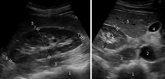
FIG:-Normal findings in renal ultrasonography (right side): sagittal plane (left) and transverse plane (right). Length and width are measured in the sagittal plane and the thickness or parenchymal pyelone index in the transverse plane. Spine (1), vena cava (2), psoas muscle (3), gall bladder (4), liver (5), upper pole (6), lower pole (7).
Normal findings
- size/shape of kidney
- doppler studies
- reanl parenchyma pyelon index etc.
Q3. Describe the management and treatment plan utilized in the disorder
DISORDER:- KIDNEY STONES OR RENAL CALCULI
Medical management
Evaluation
- A clinician should perform a screening evaluation consisting of a detailed medical and dietary history, serum chemistries and urinalysis on a patient newly diagnosed with kidney or ureteral stones.
- Clinicians should obtain serum intact parathyroid hormone (PTH) level as part of the screening evaluation if primary hyperparathyroidism is suspected.
- When a stone is available, clinicians should obtain a stone analysis at least once.
- Clinicians should obtain or review available imaging studies to quantify stone burden.
Diet Therapies
- Clinicians should recommend to all stone formers a fluid intake that will achieve a urine volume of at least 2.5 liters daily.
- Clinicians should counsel patients with calcium stones and relatively high urinary calcium to limit sodium intake and consume 1,000-1,200 mg per day of dietary calcium.
- Clinicians should counsel patients with calcium oxalate stones and relatively high urinary oxalate to limit intake of oxalate-rich foods and maintain normal calcium consumption.
- Clinicians should encourage patients with calcium stones and relatively low urinary citrate to increase their intake of fruits and vegetables and limit non-dairy animal protein.
Pharmacologic Therapies
- Clinicians should offer thiazide diuretics and/or potassium citrate to patients with recurrent calcium stones in whom other metabolic abnormalities are absent or have been appropriately addressed and stone formation persists.
- Clinicians should offer potassium citrate to patients with uric acid and cystine stones to raise urinary pH to an optimal level.
- Clinicians should not routinely offer allopurinol as first-line therapy to patients with uric acid stones.
- Clinicians should offer cystine-binding thiol drugs, such as alpha-mercaptopropionylglycine (tiopronin), to patients with cystine stones who are unresponsive to dietary modifications and urinary alkalinization, or have large recurrent stone burdens.
- Clinicians may offer acetohydroxamic acid (AHA) to patients with residual or recurrent struvite stones only after surgical options have been exhausted.
Follow-up
- Clinicians should obtain a single 24-hour urine specimen for stone risk factors within six months of the initiation of treatment to assess response to dietary and/or medical therapy.
- After the initial follow-up, clinicians should obtain a single 24-hour urine specimen annually or with greater frequency, depending on stone activity, to assess patient adherence and metabolic response.
- Clinicians should obtain periodic blood testing to assess for adverse effects in patients on pharmacological therapy.
- Clinicians should obtain a repeat stone analysis, when available, especially in patients not responding to treatment.
- Clinicians should monitor patients with struvite stones for reinfection with urease-producing organisms and utilize strategies to prevent such occurrences.
Surgical procedures applicable for the disorder
Comman surgical methods are:-
- Ureteroscopy(URS)
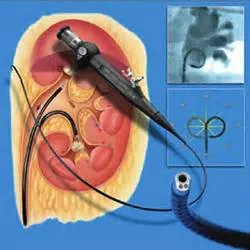
- Retrograde Intrarenal Surgery(RIRS)
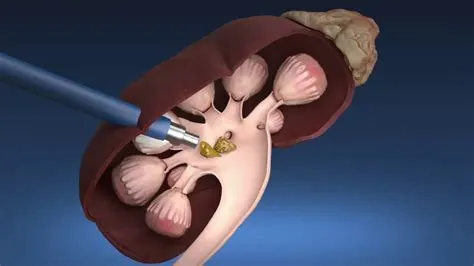
- Percutaneous Nephrolithotripsy (PCNL)
- Laser method
- Kidney transplant
1) Ureteroscopy(URS)
 |
|
2) Retrograde Intrarenal Surgery(RIRS)
 |
|
3) Percutaneous Nephrolithotripsy (PCNL)
 |
|
Q4. Describe the surgical management of the disorder.
DISORDER:- KIDNEY STONES OR RENAL CALCULI
SURGICAL METHOD:- URETEROSCOPY(URS)
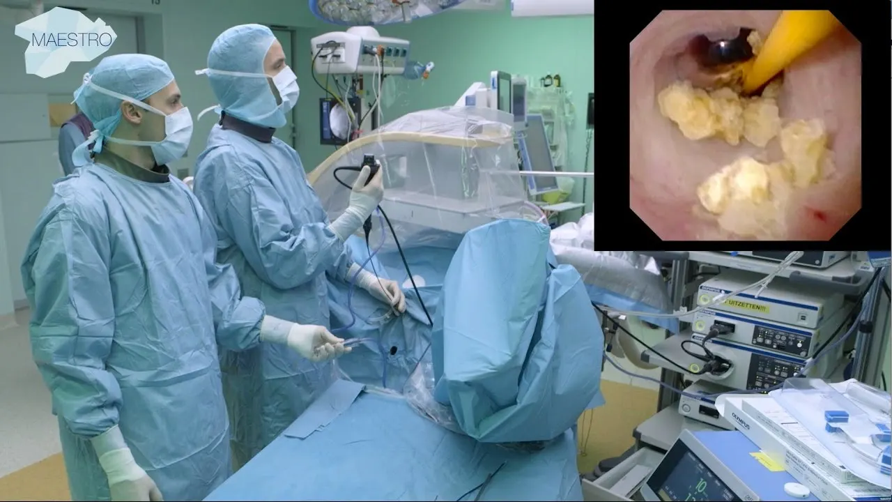
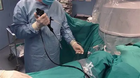
- Is a procedure which involves examination of the unrinary tract with help of uteroscope.
- it is inserted into the bladder through urethra and which helps the surgeaon to veiw the insides in a computer screen.
Why
- doctor may recommend URS surgery if you have a large stone in your kidney or ureter.
- any kidney cysts formation or infections.
Preparation
- doctors may check vital signs, such as bp, pulse rate, temperature etc.
- urine test to check levels of urea and electrolytes.
- xray,MRI, CT scan to get a clear picture of stones regarding shape, size,lacation etc.
- about food.
Time
- The surgery typically takes 30 minutes to two hours and can be done as an outpatient procedure. You should be able to resume normal activities in two to three days.
Steps and anatomical postion involved
-
After you are under anesthesia, your doctor inserts a telescope-like instrument, called a ureteroscope, through the opening of your urinary tract and into the bladder, which means there are no surgical cuts or incisions made. -
Your doctor uses the scope to examine your urinary tract – including your kidneys, ureters and bladder – then locates the kidney stone and breaks it apart using laser energy or removes the stone with a basket-like device. -
To help control swelling and allow the kidney to drain urine, your doctor may insert a small plastic tube, called a ureteral stent, in your urinary tract at the end of the procedure. The stent will be removed a follow-up appointment. - Two positions have been reported for ureteroscopy (URS): dorsal lithotomy (DL) position and dorsal lithotomy position with same side leg slightly extended (DLEL).
Complications
- Chills, abnormal bleeding while urinating, nausea, fatigue,swelling.
Instruments
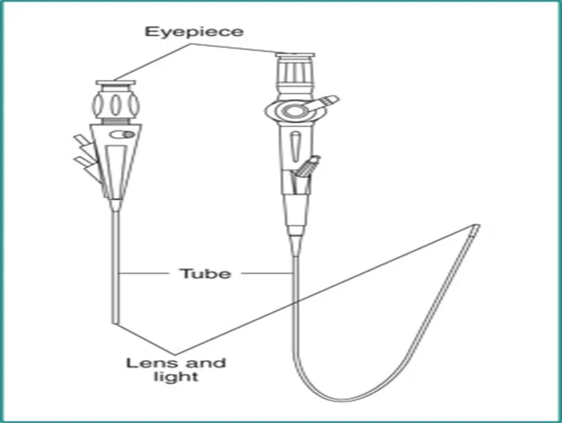
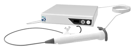
- The ureteroscope generally consist of eyepiece, tube and a lens and light.
- There are two types of ureteroscopes, rigid and flexible.
- Rigid ureteroscopes, as their name implies, are firm and preferred for treatment of stones lodged in the lower ureter which can usually be accessed in a straight path. semirigid ureteroscopy, 365-µm fibers are more suitable, although they can be used with flexible nephroscopy if minimal deflection is required. Smaller semirigid ureteroscopes have smaller working channels, and smaller laser fibers (<300 µm fibers) are preferred to allow for adequate irrigant flow.
- When stones are located in the upper ureter and/or kidney, they often require flexible ureteroscopes that accommodate to the shape of the ureter and renal collecting system.
- Though more difficult to maneuver, flexible scopes allow the urologist to inspect nearly the entirety of the inner kidney to find stones, treat them, and remove them using a variety of techniques. Typically uses 200-µm laser fibers, which have a minimal impact on scope deflection.Innovation this area is brisk and modern digital ureteroscopes offer increasingly superior image quality, lighting and wide fields of view.
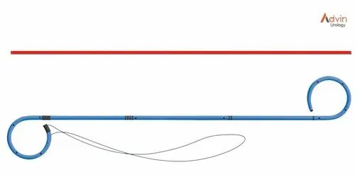
- A double j stent is used to ensure swelling does not block urine.
Post operative care
- The postoperative care of the patient submitted to ureteroscopy depends on the type of instrumentation (rigid, flexible), on the size of the ureteroscope, on the type of the procedure (diagnostic, therapeutic for stone or tumor), on the anatomic location the procedure is focused (ureter or kidney), the duration of the operation, the use or not of upper urinary tract drainage, and finally on the occurrence of intraoperative complications.
- Despite the fact that no skin incisions were used, the area around the ureter and bladder is raw and irritated. If you have a stent, it is foreign to the body and will further irritate the bladder wall. This will cause you to have increased frequency of urination, both day and night, and an increase in the need to urinate. In some people, the urge to urinate is present almost always. Sometimes the urge is strong enough that you may not be able to stop yourself from urinating. These problems will resolve after the stent is removed.
- Activity:
Pay close attention to the color of your urine as you increase your activity. If the urine stays clear to light pink, you are doing the right amount of activity. Overexertion may cause urinary bleeding. If you start to pass clots or don’t improve, call us. - Diet:
You may return to your normal diet immediately. Because of the raw urinary tract surfaces, alcohol, spicy foods, and drinks with caffeine may cause some irritation or frequency of urination and should be used in moderation. To keep your urine flowing freely and to avoid constipation, drink plenty of fluids during the day (8-10 glasses.) Water is the best. - Medication:
You should resume your medications unless we tell you otherwise
Use your prescribed pain medication as directed, but do not take additional Tylenol with it.
Medications such as Pyridium may be given to help with burning and an intense desire to urinate. (Pyridium will stain urine and clothing orange.) - Problems you should report to the Urology Dept (contact info below):
Fever greater than 100.5 degrees F
Heavy bleeding or clots
Inability to urinate
Reactions to medication (hives, rash, nausea, vomiting, diarrhea) - Follow up:- Ultrasound, discussing laboratory results, removal JJ Stent.
-
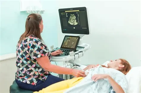
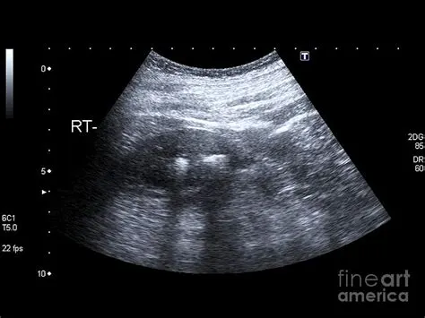
- Ultrasound can be efficient in visualizing clear uric acid stones and obstructions caused by any type of kidney stones. Most scientist agree on the fact that ultrasound simply is not sufficient enough to detect very small kidney stones. However, ultrasound scan remains a very useful first diagnostic step in emergency rooms.
Q5. Self study on recent advances in the field.
A High-powered Laser Facilities Stone removal
- A new high-powered laser uses a patented technology called "Moses" to make it easier to remove kidney stones. Procedures are faster and potentially safer. With Moses, the stone doesn't move as much after the laser hits it, and it is easier to accurately hit stones that are moving as the patient breathes. This makes it easier for surgeons to break down stones without damaging surrounding tissues and causing bleeding.
- Newer high-powered lasers, including those with Moses, also let surgeons remove kidney stones by "dusting" them (breaking stones into a fine powder). After surgery, people who have "dusting" may not need a stent in the ureter, which can be uncomfortable. Using high-powered lasers, the procedure can be performed more efficiently by removing more stone volume per unit time, which helps reduce the procedure time.
A Miniaturized Procedure Could Reduce Bleeding
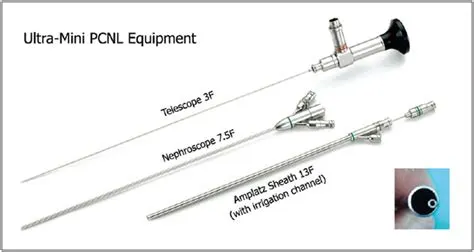
- In a procedure for larger kidney stones called percutaneous nephrolithotomy (PCNL), surgeons make small incisions in the lower back, and then use a small camera and instruments to break up and remove the stones. Now, with a technique called "mini PCNL" or "mini perc," incisions and instruments are smaller.
- This is part of a general trend to makes many types of surgery less traumatic, with less cutting. Theoretically, this means there is less bleeding and less pain during recovery. However, more evidence is needed before we can say if mini perc has these effects.
Single-use Scopes Bring Treatment Closer to Home
- New single-use, disposable ureteroscopes ("scopes") have the potential to give more patients access to ureteroscopy to treat their stones. Re-usable scopes are expensive, and smaller hospitals often have only one or two scopes. The scopes are also fragile, and often require repair after a certain number of cases. With single-use scopes, this is not a concern because the scope is only used once. Some hospitals keep some single-use scopes on hand in case their re-usable scope breaks. This prevents patients from having to have their surgeries canceled or delayed and improves access to care.
The super-pulsed thulium fiber laser is “a game-changer,”
- As technology has evolved in recent years, lasers have been used to break up the stones in a procedure called lithotripsy, often without any incision. The urologist passes a laser-bearing, flexible scope to view the stone and fragment it. These laser fibers are as thin as three human hairs.
- The thulium fiber laser can break kidney stones into pieces that are routinely 10 times smaller than those produced with a holmium laser, which is now used by most urologists. The smaller particles produced by the thulium laser are easier to flush or suction from the kidney.
- “In our studies, the holmium laser was able to clear 50% to 60% of stone fragments,” says Clayman, an internationally known urologist and pioneer of minimally invasive surgical techniques that have revolutionized treatment of kidney and other urinary tract diseases.
- “With thulium, we were able to clear more than 90% of fragments. If we can duplicate these results clinically, it opens the door for us to remove larger and larger stones with the ureteral approach, which is far less painful and costly than surgically removing them from the kidney,” he says.
Possible future technologies
1)Using Hydrgels to remove kidney stones
- Kidney stones are often removed via an endoscopic procedure. If a stone is too large, the urologists break it into pieces using a laser. The larger pieces can be removed with a grasping instrument, but this is not possible for the smaller fragments – instead, they remain in the kidney in the hope that they will leave the body naturally.
- In the future, doctors will be able to remove even the smallest of stone fragments from the kidney during endoscopy using a hydrogel. This biocompatible 2-component system was developed by Purenum GmbH, a spin-off of the Fraunhofer Institute for Manufacturing Technology and Advanced Materials IFAM.
- The product that the two researchers have developed to combat the tiny stone fragments is a hydrogel made from biocompatible materials. It consists of two liquid components that have been dyed blue and yellow to ensure a good color contrast. This allows the surgeon to control dispensing in an extremely precise manner.
2)Mobile and bio-artificial dialysis technologies.
- Advancing technology could render current dialysis technologies obsolete. Smaller, mobile technologies will provide temporary metabolic support. Devices will become wearable or take the form of artificial kidneys that automate metabolic functions. Patients will use mobile sensors that collect information on food intake and lifestyle habits, refining kidney function and treatment. Integrated use of 3D-printed tissue and smart materials will be able to replicate kidney design and function.
Part B – Case Study
1-Describe the type of bone the skull belongs to
Flat bones
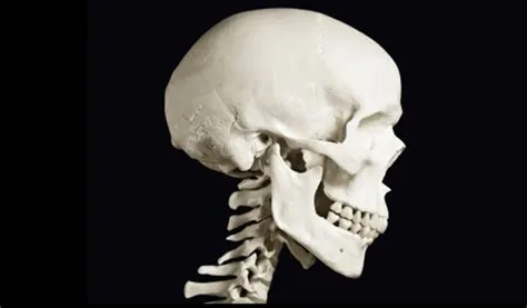
- expanded, elongated plates of bone found to outline the cavities of the body.
- Each bone consists of two plates of compact bone tissue, with intervening spongy bone and marrow.
- The intervening spongy tissue in the bone of vault of skull is known as diploe, which contains numerous veins.
- Frontal bone-This bone forms forehead and the upper portion of eye sockets.
- Parietal bones-two parietal bones on either side of the head. They form the top and sides of skull.
- Occipital bone-This bone forms the back of skull.
- Nasal bones-two nasal bones that form the bridge of the nose.
- Lacrimal bones- two small lacrimal bones that form part of eye socket.
- Vomer bone-This bone forms nasal septum, the space between nostrils.
2-Describe the microscopic anatomy of bone
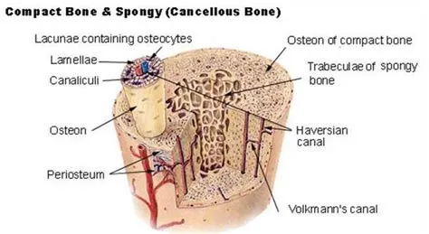
- Haversian system consists of a central canal called haversian canal, surrounded by osteocytes or bone cells are present in a circular fashion or in a concentric manner present in a lamella along with lacunae.
Osteocytes
- Mature bone cells
Perforating/Volkmann’s canal
- Canal perpendicular to the central canal
- Canals that communicate between the two haversian canal and carries blood and nerve vessels.
Central/haversian canal
- Haversian canal consists of blood, nerve and lymphatic vessels.
- Opening in the centre of an osteoma.
Canaculi
- Projections from osteocytes.
- Tiny canals.
- Radiate from the central canal to lacunae.
- Form a transport system.
Lacunae
- Cavities containing bone cells (osteocytes).
- Arranged in concentric rings called lacunae
- Rings around the central canal.
Q3-Explain the blood supply of bone
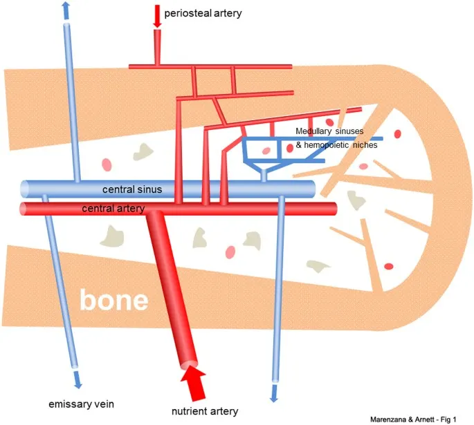
- Schematic diagram showing general arrangement of the vascular supply to healthy adult bone. The main blood supply is derived from one or more nutrient arteries, which penetrate to the medulla and connect to the smaller periosteal arterial supply to enable perfusion of cortical bone. The arterial branches drain into arterio-venous sinuses in the medulla that support hematopoietic and stromal cells. Blood exits the medullary cavity via multiple small veins that penetrate the cortex. Thus, perfusion is predominantly centrifugal, at least in young adult bone.
- Nutrient arteries breaks into smaller parallel arteries which run into metaphesis where they freely amnestomoses with the terminal branches of metaphyseal arteries.
- Radial branches enters the hervesin canal and the volkmann's canal and supply the cortex.
- Metaphyseal or Juksta epiphyseal artery are small arteries derived from the arterial annestomosis around the joint.
- Then they enter the bones by piercing its gen of attachment of capsular ligament.
- They penetrate into the epiphysis.
- Periosteal arteries these blood vessels lie within the periosteum and end by supplying superficial portion of the cortex.
- Short bones have only epiphysis and metaphyseal branches.
- Blood supply for flat bones is by nutrient artery and periosteal artery.
- Blood supply of irregular bones is by they pass through a basy vertibral parament and arteries pierce through antrolateral surface.
Q4-Elaborate on bone remodeling and new bone formation
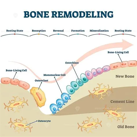
- Bone remodeling is a process of constantly resorbing bone by osteoclasts and replace it by osteoblast. Osteocytes act as mechanosensors and instruct the osteoclast where to resorb bone, and osteoblast when and where to form new bone.Bone remodeling consists of four phases.
- First the activation phase, this phase involves detection of a remodeling signal due to changes in physical forces recognized by the osteocytes or damage to the bone matrix due to osteocyte apoptosis, these are signals for the osteoclast.
- The second phase is the resorption phase, osteoclast breaks down the bone matrix.
- Third is the reversal phase, the mononuclear cell removes the undigested demineralized collagen matrix in the lacunae and prepare the bone surface for bone formation.
- The last phase is the bone formation by osteoblast. Mechanical stimulation stimulates bone formation and inhibits bone resorption. Resting conditions inhibit bone formation. The remodeling cycle concludes when an equal amount of resorbed bone has been replaced. Remodeling are most of the time develop randomly but also occur in areas that need repair.
Q5-Describe the anatomical basis of bone implant
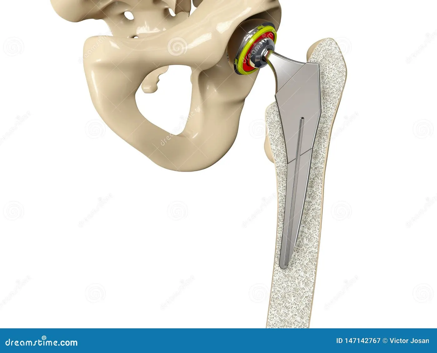
- Modern bone implant materials can be classified according to their biocompatibility into biotolerant, bioinert and bioactive materials.
- Dimensions between anatomical landmarks of maxillary and mandibular jaws
- Functional Implant zones
-
Implant size recommendation related to tooth anatomyPrinciples of implant positioningNumber of implants and key implant positions
-
Peri-implant bone modeling has been examined by a number of researchers. Davies has described two different forms of peri-implant bone modeling: appositional bone formation and intramembraneous ossification
Q6-Enumerate the indications and contraindications for the bone implant
Indications
- Bone graft materials are placed in different locations for various indications:1
- In alveolar sockets post extraction
- To refill a local bony defect due to trauma or infection
- To refill a peri-implant defect due to peri-implantitis.
Contradictions
- Implantable bone conduction devices are contraindicated if the SDS is lower than 60% and the bone PTA (in the better hearing ear) exceeds 55 dB. Implantable devices, moreover, are contraindicated in patients with previously irradiated bone and psychiatric disease.
- most frequent causes of the loss of bone substance are aggressive periodontitis and postextraction trauma related to wearing an unadapted removable prosthesis. The following clinical case shows the consequences of these bone pathologies.