Project 2
Project(a)
Gastrology system
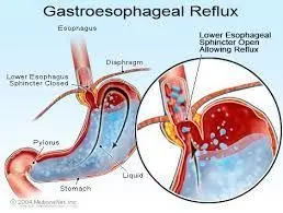
1. Pathology condition: Gastroesophageal reflux disease (GERD)
Main function of human Digestive system is to break down the food particle into smaller molecules that can be utilized for several prposes like energy, growth, repair and regulation.
It is a condition wherein the lower esophageal sphincter is not functioning properly and the gastric contents reflexal or enter back into the esophagus and the common complaint is heartburn.
GERD or Gastroesophageal Reflux Disease (a chronic disease that saps the digestive system) greatly influences a disruption in homeostasis due to one major effect--heartburn.
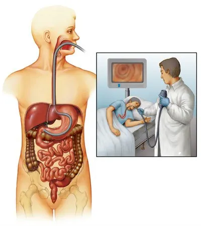
2. Diagnostic method: Upper gastrointestinal scopy
gastrointestinalscopy is a flexible tube with a camera at its lower base end and this is introduced via oral cavity and then it done under anaesthaetic condition and then the tube passes through the esophagus nd then finally into the stomach.
3. Medications: Prescription-strength treatments for GERD include: Prescription-strength proton pump inhibitors. These include esomeprazole (Nexium), lansoprazole (Prevacid), omeprazole (Prilosec), pantoprazole (Protonix), rabeprazole (Aciphex) and dexlansoprazole (Dexilant). Antacids. Over-the-counter antacids are best for intermittent and relatively infrequent symptoms of reflux. Histamine blockers.Proton pump inhibitors.
Prokinetic agents.
4. Surgical Solution:
A Nissen fundoplication is a surgery to treat gastroesophageal reflux disease (GERD).
During the procedure, a surgeon creates a sphincter (tightening muscle) at the bottom of the esophagus to prevent acid reflux.
5. Medical devices:
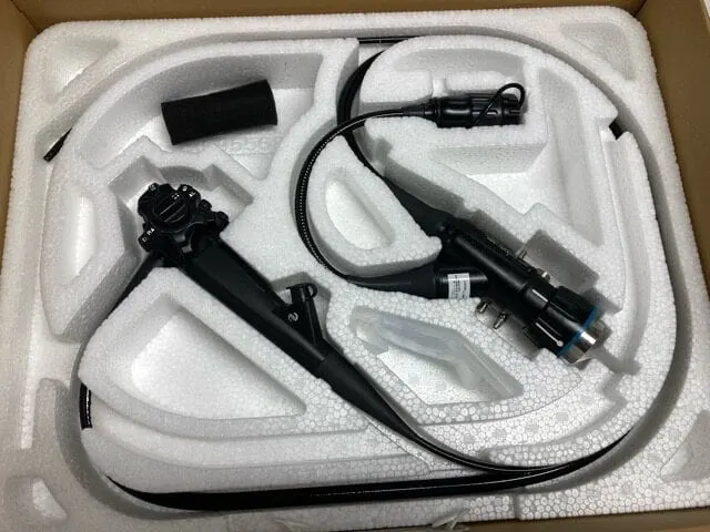
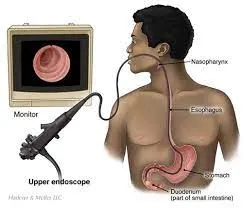
upper endoscope:visually examine upper digestive system
Project (b)
Cardiovascular system
1. Pathology conditions: Cerebrovascular accident(Stroke)
The primary function of the human cardiovascular system is to give oxygen, nutrients, and other essentials through the blood and also remove unwanted waste.
It is a stroke is when blood flow to a part of your brain is stopped either by a blockage or the rupture of a blood vessel.
During a stroke attack, interruption of circulation causes detrimental changes such as anoxia, oxygen and glucose deprivation (OGD), loss of adenosine triphosphate (ATP), and acidosis due to the anaerobic generation of lactate, and disrupted cell ionic homeostasis.
2. Diagnostic method: Magnetic Resonance Imaging(MRI)
An MRI can detect brain tissue damaged by an ischemic stroke and brain hemorrhages. Your doctor may inject a dye into a blood vessel to view the arteries and veins and highlight blood flow (magnetic resonance angiography or magnetic resonance venography).
3. Medications: One treatment for ischemic stroke is the FDA-approved drug, tissue plasminogen activator (tPA), which must be administered within a three-hour window from the onset of symptoms to work best.
4. Surgical Solution:
Ischemic stroke treatment clears the blockage, which can be done via endovascular surgeries.
In a hemorrhagic stroke, surgeons may need to relieve the pressure on the brain and repair the blood vessel.
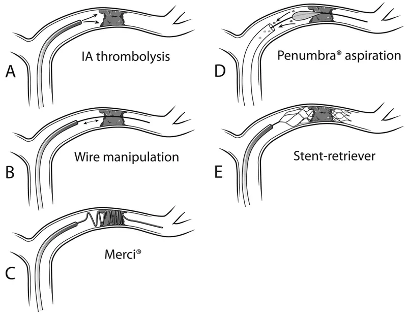
Illustration of different approaches to intraarterial (IA) treatment of patients with acute stroke due to large-vessel occlusion. An intracranial vessel with occlusive thrombus is shown.
A: Intraarterial pharmacological thrombolysis is done
by injecting a thrombolytic agent through a microcatheter directly into the clot
B: Microwire manipulation breaks down the
clot into smaller particles.
C: Mechanical thrombectomy with the Merci retriever works by wrapping around and capturing the
clot.
D: Aspiration thrombectomy using the Penumbra aspirator and separator breaks down the clot under constant negative
pressure.
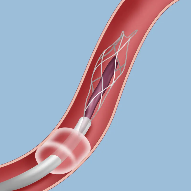
E: A stent retriever (Solitaire or Trevo) allows capture of the thrombus and instant restoration of blood flow. Both the
stent and clot are then retrieved together
5. Medical devices:
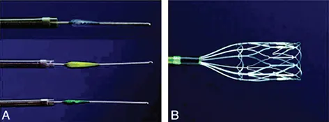
A. Aspiration catheters and separators. B. Thrombus removal ring.