Project 1
Part-A
Anatomical basis of the cardiovascular system, respiratory system, and digestive system:
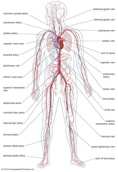
- Cardiovascular system: it is the transport system of the body and it comprises blood, heart, and blood vessels. This system supplies nutrients and removes waste products from various tissues or parts of the body. The conveying media is blood in form of liquid which flows in a closed tubular system. blood vessels include arteries, capillaries, and veins. The heart is a 4-chambered, hollow muscular organ located on the superior surface of the diaphragm left to the midline and anterior of the vertebral column posterior to the sternum. chambers of the heart include the right atrium and left atrium, right ventricle, and left ventricle. valves present are the atrioventricular valves and the aortic and pulmonary valves.
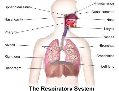
- Respiratory system: consists of the nose, pharynx, larynx, trachea, bronchi, and lungs. its primary function is to furnish oxygen for individual tissues, and cells and to take away the waste products and carbon dioxide produced by these cells. The nose is the 1st entry of the respiratory system, the pharynx is present between the base of the skull to the esophagus and the larynx is the voice box that extends from the root of the tongue to the upper end of the trachea. The bronchial tree is the subdivision of the tertiary bronchus that divides and gives rise to bronchioles. Lungs are pair of spongy air-filled respiratory organs located on either side of the chest.
- digestive system: gastrointestinal tract includes all those structures that are concerned with swallowing, digestion, and absorption of food. It consists of an alimentary canal that starts at the mouth and ends at anus. the oral cavity is meant for taste sensation and swallowing. The pharynx is the continuation of the posterior part of the oral cavity. this system mainly consists of the mouth, esophagus, stomach, small intestine, and large intestine.
clinical conditions/ the disorders associated with the cardiovascular system:
- Heart attack: A blockage of blood flow to the heart muscle. A heart attack is a medical emergency. A heart attack usually occurs when a blood clot blocks blood flow to the heart. Without blood, tissue loses oxygen and dies. Symptoms include tightness or pain in the chest, neck, back, or arms, as well as fatigue, lightheadedness, abnormal heartbeat, and anxiety. Women are more likely to have atypical symptoms than men.
a clinical condition associated with the respiratory system:
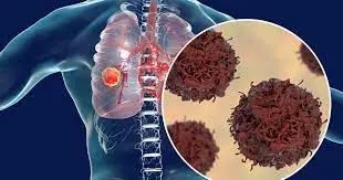
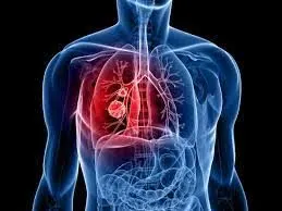
- lung cancer: Cancer that begins in the lungs and most often occurs in people who smoke.
Two major types of lung cancer are non-small cell lung cancer and small cell lung cancer. Causes of lung cancer include smoking, second-hand smoke, exposure to certain toxins, and family history. Symptoms include a cough (often with blood), chest pain, wheezing, and weight loss. These symptoms often don't appear until the cancer is advanced. Treatments vary but may include surgery, chemotherapy, radiation therapy, targeted drug therapy, and immunotherapy.
a clinical condition associated with digestive system:
- GERD(Gastroesophageal disease):
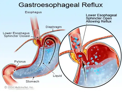
A digestive disease in which stomach acid or bile irritates the food pipe lining.This is a chronic disease that occurs when stomach acid or bile flows into the food pipe and irritates the lining. Acid reflux and heartburn more than twice a week may indicate GERD.Symptoms include burning pain in the chest that usually occurs after eating and worsens when lying down. Relief from lifestyle changes and over-the-counter medication is usually temporary. Stronger medication may be required. it is very common and more than 10 million cases per year (India) are seen.
2)
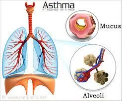
- Asthma
- The collapse of part or all of the lung (pneumothorax or atelectasis)
- Swelling and inflammation in the main passages (bronchial tubes) that carry air to the lungs (brnchitis)
- COPD
- Lung cancer
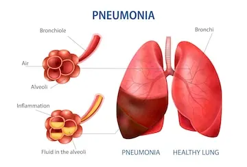
- Lung infection (pneumonia)
- Abnormal buildup of fluid in the lungs (pulmonary edema)
- Blocked lung artery (pulmonary embolus)
Various diagnostic techniques and investigations used in diagnosis of the disorder of lungs:
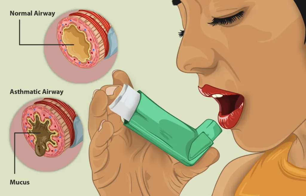
- Asthma: The most common treatment for asthma is rescue and controller inhalers, but other treatments and medications can be used. Doctors also recommend patients identify and reduce asthma triggers. Common triggers include allergies, viruses, exercise, cold weather and fumes. Patients are also often taught skills to monitor and manage their asthma.
- Chronic Cough: Some of the tests that may be used to diagnose the cause of a chronic cough may include a chest X-ray and other radiology tests, breathing tests, pH monitoring, swallow tests and upper GI endoscopy if reflux is associated with the cough. The treatment of chronic cough is usually directed at its cause.
- Chronic Obstructive Pulmonary Disease (COPD): The most common cause of COPD is smoking, although breathing in pollutants, dust or chemicals can also be the cause. For smokers, smoking cessation can help prevent the disease or keep it from getting worse. COPD can also be treated with inhalers, medications, oxygen therapy and pulmonary rehab. In severe cases, surgery may be an option.
- Lung Cancer: Lung cancer is diagnosed with a tissue sample or biopsy to determine the kind of cancer. The diagnosis is most commonly made by bronchoscopy or needle biopsy. Treatment of lung cancer depends on the type of cancer, the stage, the location and whether the cancer has spread. Treatment may include surgery, chemotherapy and/or radiation.
- Lung Nodules: Lung nodules are often found when tests are being done for another reason. Diagnostic tests include:
- Bronchoscopy
- Electromagnetic navigation bronchoscopy
- PET scan/CT scan
- Needle biopsy through the chest wall
- Surgical lung biopsy
- Pulmonary Hypertension: A series of tests may be needed to diagnose pulmonary hypertension such as:
Pulmonary function tests
Chest X-rays, lung perfusion scans, and other film studies
- Six-minute walk test
- Blood tests
- ECG (EKG)
- Echocardiogram
- There is no cure but there are treatment options to try to reduce the symptoms, slow the progression and improve the quality of life. If pulmonary hypertension is the side effect of another illness, treatment focuses on the primary cause. If pulmonary hypertension is the primary cause, medications can be used.
- Shortness of Breath: Diagnostic tests may include pulmonary function tests, chest X-ray, EKG, echocardiogram, bronchoscopy, blood tests or chest CT scan. Treatment for shortness of breath depends on the underlying cause and severity.
- Pulmonary Rehabilitation: Pulmonary rehab is for people with chronic breathing conditions that limit the quality of life. Gundersen exercise physiologists and respiratory therapists help you set goals, establish a safe exercise routine and learn how to exercise at home.
controller inhaler:
3)
Management and treatment plan utilized in the disorder of lungs:
- Bronchodilators: Bronchodilators are medications that usually come in inhalers — they relax the muscles around your airways. This can help relieve coughing and shortness of breath and make breathing easier. Depending on the severity of your disease, you may need a short-acting bronchodilator before activities, a long-acting bronchodilator that you use every day or both.
Examples of short-acting bronchodilators include:
- Albuterol (ProAir HFA, Ventolin HFA, others)
- Ipratropium (Atrovent HFA)
- Levalbuterol (Xopenex)
Examples of long-acting bronchodilators include:
- Aclidinium (Tudorza Pressair)
- Arformoterol (Brovana)
- Formoterol (Perforomist)
- Indacaterol (Arcapta Neoinhaler)
- Tiotropium (Spiriva)
- Salmeterol (Serevent)
- Umeclidinium (Incruse Ellipta)
- Inhaled steroids
- Inhaled corticosteroid medications can reduce airway inflammation and help prevent exacerbations. Side effects may include bruising, oral infections and hoarseness. These medications are useful for people with frequent exacerbations of COPD.
Examples of inhaled steroids include:
- Fluticasone (Flovent HFA)
- Budesonide (Pulmicort Flexhaler)
- Combination inhalers
Some medications combine bronchodilators and inhaled steroids. Examples of these combination inhalers include:
- Fluticasone and vilanterol (Breo Ellipta)
- Fluticasone, umeclidinium and vilanterol (Trelegy Ellipta)
- Formoterol and budesonide (Symbicort)
- Salmeterol and fluticasone (Advair HFA, AirDuo Digihaler, others)
Combination inhalers that include more than one type of bronchodilator also are available. Examples of these include:
- Aclidinium and formoterol (Duaklir Pressair)
- Albuterol and ipratropium (Combivent Respimat)
- Formoterol and glycopyrrolate (Bevespi Aerosphere)
- Glycopyrrolate and indacaterol (Utibron)
- Olodaterol and tiotropium (Stiolto Respimat)
- Umeclidinium and vilanterol (Anoro Ellipta)
- Oral steroids
For people who experience periods when their COPD becomes more severe, called moderate or severe acute exacerbation, short courses (for example, five days) of oral corticosteroids may prevent further worsening of COPD. However, long-term use of these medications can have serious side effects, such as weight gain, diabetes, osteoporosis, cataracts and an increased risk of infection.
Phosphodiesterase-4 inhibitors
A medication approved for people with severe COPD and symptoms of chronic bronchitis is roflumilast (Daliresp), a phosphodiesterase-4 inhibitor. This drug decreases airway inflammation and relaxes the airways. Common side effects include diarrhea and weight loss.
Theophylline
When other treatment has been ineffective or if cost is a factor, theophylline (Elixophyllin, Theo-24, Theochron), a less expensive medication, may help improve breathing and prevent episodes of worsening COPD. Side effects are dose related and may include nausea, headache, fast heartbeat and tremor, so tests are used to monitor blood levels of the medication.
Antibiotics
Respiratory infections, such as acute bronchitis, pneumonia and influenza, can aggravate COPD symptoms. Antibiotics help treat episodes of worseningCOPD, but they aren't generally recommended for prevention. Some studies show that certain antibiotics, such as azithromycin (Zithromax), prevent episodes of worsening COPD, but side effects and antibiotic resistance may limit their use.
Lung therapies
Doctors often use these additional therapies for people with moderate or severe COPD:
Oxygen therapy. If there isn't enough oxygen in your blood, you may need supplemental oxygen. There are several devices that deliver oxygen to your lungs, including lightweight, portable units that you can take with you to run errands and get around town.
Some people with COPD use oxygen only during activities or while sleeping. Others use oxygen all the time. Oxygen therapy can improve quality of life and is the only COPD therapy proved to extend life.
Pulmonary rehabilitation program. These programs generally combine education, exercise training, nutrition advice and counseling.
4)
Surgical management of the disorder of lungs: Surgery is an option for some people with some forms of severe emphysema who aren't helped sufficiently by medications alone. Surgical options include:
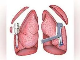
- Lung volume reduction surgery:In this surgery, your surgeon removes small wedges of damaged lung tissue from the upper lungs. This creates extra space in your chest cavity so that the remaining healthier lung tissue can expand and the diaphragm can work more efficiently. In some people, this surgery can improve quality of life and prolong survival.
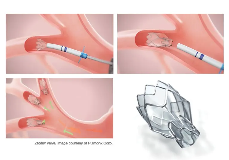
- Endoscopic lung volume reduction: a minimally invasive procedure ― has recently been approved by the U.S. Food and Drug Administration to treat people with COPD. A tiny one-way endobronchial valve is placed in the lung, allowing the most damaged lobe to shrink so that the healthier part of the lung has more space to expand and function.
- Lung transplant: A lung transplant may be an option for certain people who meet specific criteria. Transplantation can improve your ability to breathe and be active. However, it's a major operation that has significant risks, such as organ rejection, and you need to take lifelong immune-suppressing medications.
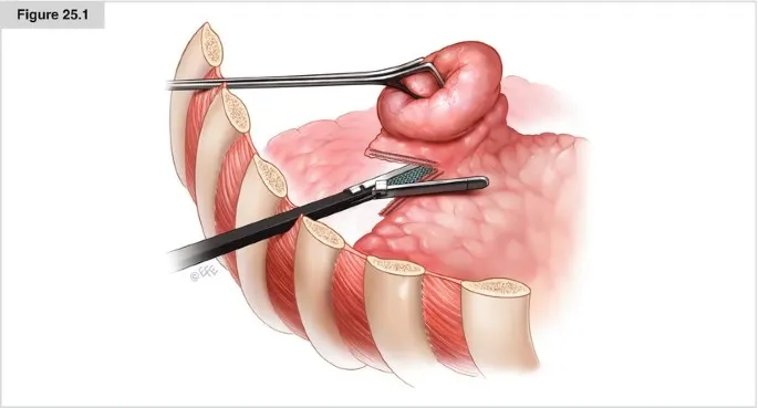
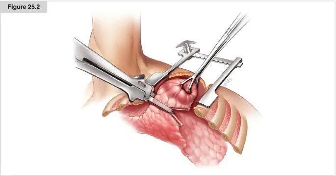
- Bullectomy: Large air spaces (bullae) form in the lungs when the walls of the air sacs (alveoli) are destroyed. These bullae can become very large and cause breathing problems. In a bullectomy, doctors remove bullae from the lungs to help improve airflow.
5)
Recent advancements in the field of treatment of lung diseases:
Breath measuring algorithms, smart inhalers, and wearable analytics are at the forefront of respiratory care technology. Lung cancers, including mesothelioma and asthma, account for 70% of the country’s breathing issues. Technological advancements are making it easier for specialists to diagnose and patients to administer self-care.
Smart Inhalers
Inhalers administer nearly 90% of the medications prescribed for respiratory illness. Smart inhalers are the newest tool that can help respiratory disease patients get their medications and relay vital information back to doctors.
Smart inhalers are embedded with AI technology that can collect data and monitor a patient’s condition. Technology is being tested that will alert users to potential environmental triggers and a reminder to take their medication on time. The data collected by these smart inhalers can be used by clinicians and doctors to better understand the needs of each patient.


Wearable Devices
One of the largest barriers to proper treatment is the patients’ own adherence to their treatment. Not all patients are great at taking their medications on the right schedule. With new technology like smart shirts or stickers that are worn on the body, patients can be monitored and reminded when it’s time for their treatment.

Part-B
The type of bone the skull belongs to Flat bone.
Flat bone includes skull, strenum(breast bone) and scapula(shoulder bone)
They are expanded,elongated plates of bones found at the outline cavities of the body.
Each bone consists of compact bone tissue with the intervaning spongy bone with a marrow.
Nutrient arteries breaks into smaller parallel arteries which run into metaphesis where they freely amnestomoses with the terminal branches of metaphyseal arteries.
Radial branches enters the hervesial canal and the vokman's canal and supply the cortex.
Metaphyseal or Juksta epiphyseal artery are small arteries derived from the arterial annestomosis around the joint.
Then they enter the bones by piercing its gen of attachment of capsular ligament.
Epiphyseal arteries are derived arterial annestomosis around the joint.
They penetrate into the epiphysis.
Periosteal arteries these blood vessels lye within the periosteum and end by supplying superficial portion of the cortex.
Short bones have only epiphysis and metaphyseal branches.
Blood supply for flat bones is by nutrient artery and periosteal artery.
Blood supply of irregular bones is by they pas through a basy vertibral parament and arteries pierce through antrolateral surface.
Titanium (Ti) and its alloys (mainly Ti-6Al-4V) have become the metals of choice for dental implants.
However, prosthetic components of the implants are still made from gold alloys, stainless steel, and cobalt-chromium and nickel-chromium alloys.
The most frequent causes of the loss of bone substance are aggressive periodontitis and postextraction trauma related to wearing an unadapted removable prosthesis. The following clinical case shows the consequences of these bone pathologies.
This radiographic example shows a 40-year-old woman in good general health but with severe chronic periodontitis that has been stabilized for 2 years with conventional periodontal treatment (ie, initial nonsurgical treatment, periodontal surgery, and replacement of tooth 16 with a conventional fixed bridge) .
For the past 15 years, she has received regular maintenance every 4 months. The periodontal situation after 15 years was acceptable. However, bone loss evolved on the distal of tooth 15, which also showed signs of occlusal trauma .
An occlusal adjustment was completed and maintenance care was reinforced. The patient returned for follow-up after 2 years (1997) with a marked mobility of her fixed prosthesis.
The radiograph showed terminal bone loss requiring the extraction of tooth 15 . A three-dimensional vertical and horizontal loss of substance is inevitable after healing.