Project 2
1.
A kidney transplant is surgery done to replace a diseased kidney with a healthy kidney from a donor. The kidney may come from a deceased organ donor or from a living donor.
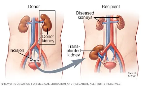
Robotic-assisted nephrectomy for living donation (RANLD) is a rapidly emerging surgical technique competing for supremacy with totally laparoscopic and laparoscopic hand-assisted techniques. Opinions about the safety of specific techniques of vascular closure in minimally invasive living kidney donation are heterogeneous and may be different for laparoscopic and robotic-assisted surgical techniques.
methods used for the Robotic-assisted surgery:
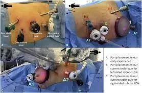
All robotic assisted donor nephrectomies were performed by two board certified urologic and transplant surgeon specialists using the da Vinci® Si Surgical System. As described earlier , the patient was placed in a left or right 45° lateral decubitus position, depending on the nephrectomy side. Following the team-timeout, under general anesthesia and sterile conditions, the 12mm camera trocar was placed on the hemiclavear line, next to the umbilicus. Capnoperitoneum was maintained at 12 mmHg. The 3 trocars (8 mm) for the robotic arms were subsequently placed in an imaginary semicircular line equidistant to the camera. Another 12 mm trocar used by the assistant for suction-irrigation, introduction and removal of suture materials and clip application was placed in the lower lateral abdomen. The “docking maneuver”, which virtually means connecting the robotic arms with all trocars, was performed after the robot was positioned lateral at the backside of the donor. Standard instruments used for all operations comprised hot shears (MSC), fenestrated bipolar forceps, Cadiere forceps and a large SutureCut needle driver (Endowrist®, Intuitive Surgical).
In short, for left donor nephrectomy (LDN) the descending colon was mobilized and displaced medially to allow opening of the Gerota’s fascia. In a first step the ureter was identified at the level of the iliac axis and dissected free up to the lower pole of the kidney preserving the periureteral tissue commonly known as “golden triangle”. After dissection of the lateral and posterior region of the kidney the renal vein was dissected free until its outlet into the inferior vena cava, and its branches (lumbar, gonadal and adrenal) are clipped and transected free. In a subsequent step the renal artery was identified and dissected free up to the level of its aortic origin. When the kidney was completely mobilized the ureter got clipped with one hemlock clip and was divided leaving the clip in situ. Subsequently the kidney was placed into an endobag for extraction. Before removal of the wrapped kidney through a Pfannenstiel or Rutherford-Morrison incision, the renal artery and renal vein was either double clipped with locking hemlock clips or addressed by one locking hemlock clip and one non-transfixing ligature (Vicryl 3-0).
For right donor nephrectomy (RDN), in a similar fashion the ascending colon was mobilized and displaced medially to allow access to the Gerota’s fascia and perirenal fat. In contrast to the left side the renal vascular stalk, especially the right renal vein is shorter. Ligation and clipping close to the inferior vena cava is even more crucial for a simple and successful subsequent transplant procedure. Again, the kidney was extracted manually in an endo-bag and afterwards flushed with 200 mL of heparinized ice-cold perfusion solution (Custodiol®). Before wound closure the renal bed and vascular stumps were inspected for bleeding. All trocars were removed under direct vision during evacuation of the pneumoperitoneum and occasionally a 12 Chr. Robinson drain was inserted into the wound bed depending on the surgeon’s decision.
[1]
Number of performed RANLDs per year beginning from 11/2013 to 08/2018, total n=40. (reference date: August 31st 2018)University Hospital of Leipzig, Leipzig, Germany;
From November 2013 till August 2018, forty consecutive RANLD were performed at the University Hospital of Leipzig, Leipzig, Germany; . Within this time period an exponential increase of donation surgeries could be detected (Figure 1). A number of 13 cases alone was performed within the last 8 months, which represents 32.5% of the total patient volume. Sex distribution among donors was n=21 (52.5%) women and n=19 (47.5%) men, with a donor’s average age of 53.075±11.68 years (range, 28–70) and an average body mass index of 25.99±3.58 kg/m2 (range, 19.9–35.3 kg/m2). All donors were Caucasian. Relation to the recipient was distributed in parent (n=18, 45%), sibling (n=5, 12.5%), spouse (n=9, 22.5%) and children (n=1, 2.5%) for direct relation as well as other (n=7, 17.5%) for a very close relationship. Preoperative laboratory parameters showed average creatinine values of 72.7±11.95 µmol/L and a mean glomerular filtration rate of 92.48±12.46 mL/min/1.73 m2
[1]
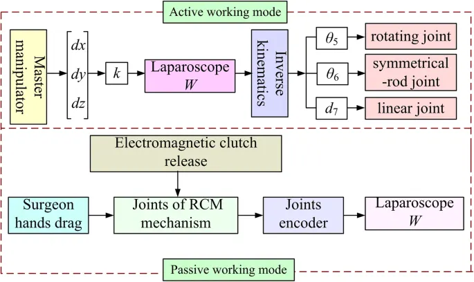
Control block diagram of the RCM mechanism
two assistants always help the surgeon accomplish a surgical operation: one holds the laparoscope to provide the surgeon with a visual display of the surgical site, while the other holds surgical instruments to perform some auxiliary work. For example, when the surgeon needs to cut a particular part of the body, the assistant uses an instrument to elevate the part to facilitate the cutting operation, because the tissues and the organs have a soft texture. The assistant may become tired, especially in some major surgeries, where instruments must be held for long durations. Consequently, the movements performed by hand may not provide sufficient stability, thereby affecting the surgical visual display or the tissue boundary dissociation.
to overcome this problem a robot that could perform the assistant’s work was designed . the robot had three arms: the middle arm and the left and right arms. The middle arm was used to hold the laparoscope and provide a visual display of the surgical site. This arm consisted of one linear joint, three positioning joints, and an RCM mechanism. The linear joint adjusted the height of the robotic arm relative to the operating bed, while the positioning joints enabled the laparoscope to reach the incision port. The RCM mechanism, which served as the end effector of the middle arm, held the laparoscope and facilitated its position change without causing pain at the incision port. The left and right arms were used to hold surgical instruments to accomplish auxiliary work. Unlike the laparoscope, the surgical instrument was designed to be flexible. As shown in Fig. 2, the instrument had a snake-like configuration with wire actuation, which allowed the instrument to achieve a position change in the patient’s body. Thus, the left and right arms did not need to have a RCM mechanism. Therefore, the key points of the robot development lay in the flexible design of the surgical instrument and the RCM mechanism. This study focused on the RCM mechanism design.
[2]
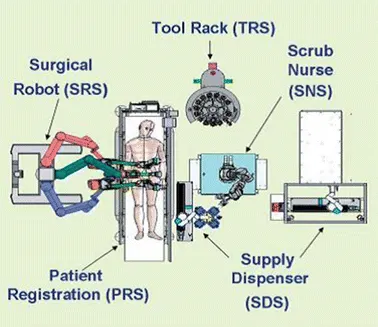
The learning curve to deliver laparoscopic radical prostatectomy (LRP) is estimated at 40–60 cases with skilled surgeons and 80–100 cases, with inexperienced surgeons. The robot shortens the prostatectomy learning curve for all surgeons, particularly experienced open prostatectomies. Interestingly, a surgeon skilled in open surgery was able to transfer his open skills to robotic surgery in 8–12 cases.
A fellowship-trained laparoscopic surgeon has a similar, short learning curve for robotic prostatectomy compared to an experienced open surgeon. The data showed a safe and reproducible surgery, interestingly even during the learning curve. Importantly, the outcomes were the same for early robotic surgery and a large cohort of open prostate surgery . There was an emphasis on having a good mentor, experienced in robotics being present during the initiation of the program.
Preoperative steps:
Correct selection of patients regarding their indications and comorbidities should be considered
before taking them for robotic surgery
It is advisable that the respective specialist should appropriately evaluate these patients before undergoing robotic surgery. Like any surgical procedure, a detailed discussion and counseling about the advantages
and disadvantages of robotic surgery should be done. Adequate information about the alternative
forms of treatment should also be made available to patients before robotic surgery. Informed consent should be obtained.
Appropriate preoperative blood investigations such as complete bloodcount, blood type, crossmatch, coagulation study, metabolic panel, chest X-ray, and electrocardiogram should be done.
Avoid blood thinners including aspirin for at least one week prior to surgery. The day before surgery, eat light meals until noon and then clear liquids. Stay well hydrated; the day of surgery, you should drink clear liquids until two hours before arrival at the hospital.
Perioperative steps:
One surgeon will remove the kidney from the donor, while another prepares you to receive the donated kidney. Your surgeon connects the transplanted kidney to your blood vessels and your bladder. Your blood flows through an artery into the transplanted kidney, and a vein takes filtered blood out.
Postoperative steps:
The postoperative nursing care of the renal transplant recipient is similar in many ways to the care of any patient who has undergone a major surgical procedure; the emphasis is on maintenance of fluid and electrolyte balance, wound care, pain management, good pulmonary toilet with incentive spirometry, early.
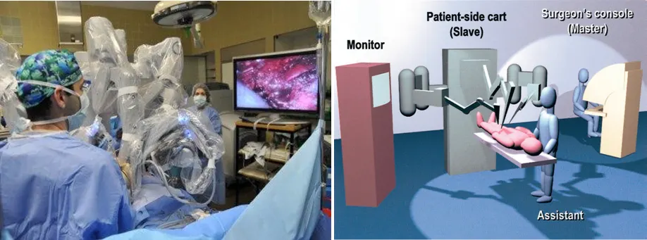
To operate using the Robotic system, your surgeon makes tiny incisions in your body and inserts miniaturized instruments and a high-definition three-dimensional camera, and sometimes skin incisions are not required at all. Then, from a nearby console, your surgeon manipulates those instruments to perform the operation.
[3]
transoral robotic surgery (TORS):
Advantages:
-
Allows the surgeon a true 3-dimensional endoscopic vision of the surgical field with accurate depth perception through the use of multiple endoscopes, cameras, and dual eye pieces.
-
Enhancement of the limited visualization of the 2-dimensional endoscope and line-of-sight microscope which improves the quality of microsurgery.
-
Allows multiple degrees of motion (flexion, extension, supination, pronation) and through robotic scaling converts large movements of the surgeon's hands into small movements of the robotic instruments.
-
Filters hand tremors through frequency filters and it is resistant to fatigue.
-
Minimally invasive surgery reduces blood loss and blood transfusion, reduces the risk of infection and complications.
-
Less postoperative pain and shortened hospital stay.
-
Permits surgery to be completed with smaller incision which improves the cosmetic outcome.
Drawbacks:
-
Reliance on visual perception rather than tactical stimulus for assessment of tumor resection.
-
High cost of acquisition and maintenance of the robotic system compared to open surgery.
-
Occupies large amount of space in the OR.
-
Any movement can prove disastrous after the robot is docked into the patient.
-
Needs to be performed widely to prove its safety and efficacy.
[4]
Minimally invasive surgery has well-established advantages: shorter length of hospital stay, markedly reduced postoperative pain, fast return to preoperative state of activity, lowered postoperative ileus, and a preservation of immune function . Importantly, laparoscopic urology has been superseded by the robot-assisted laparoscopic surgery The main reasons for this significant change from pure laparoscopic urology to robot-assisted laparoscopy are mainly surgeon factors such as shorter learning curve and less surgeon fatigue
[5]
ethical concerns and regulations of a kidney transplantation procedure in robot-assisted surgery:
The ethical criteria that need to be considered by departments and individual surgeons seeking to introduce RAKT is whether robot-assisted transplantation provides a similar or better technical vascular anastomosis than an open approach and if incision-related morbidity is less than with an open procedure.
These ethical considerations are similar to Knight's assessment of innovations in cardiothoracic surgery, including minimally invasive cardiac surgery. Based on these, any surgeon carrying out a RAKT should ask, ‘Can I, with my skill set, in my demographic practice, provide as good or better transplant graft and patient outcomes, when compared with an open implant, with a similar tariff?’ The variables addressed in such self-reflection should be openly discussed with the patient during the consent process, alongside the overall effectiveness of robotic procedures relative to other approaches.
Regular appraisal of surgical ability and outcomes, combined with institutional support for pioneering intervention, will allow safe and efficient progress. With every new approach that evolves, it is essential to continually assess the goals and outcomes that must be achieved, ensuring the procedure is held to the correct standard and patient safety preserved.
The cost must also be borne in mind, ensuring a fair distribution of funding, particularly when transplant patients comprise a relatively small proportion of the wider populations that healthcare services treat.
Ultimately it is of the utmost importance that patients chose the right procedure for them, with the support of their team and careful explanation of appropriate literature, alongside honest conversations about risk, local expertise, and the LC involved—true evidence-based medicine.
[6]
1.Senhance (Telelap ALF-X)
The recently (approved October 2017) FDA-approved ALF-X (Senhance; Trans-Enterix®, Morrisville, USA) is a new multiport robotic system. Fanfani et al reported 80 hysterectomy cases from October 2013 to May 2014 with the ALF-X system.For urology, a porcine model of robot-assisted partial nephrectomy has been reported.It has the advantage of totally independent surgical arms, haptic feedback, and eye tracking systemsBecause of independent surgical arms, this system requires a spacious operative room.
2. Revo-i®
Revo-i® (Mere company Inc., Yongin, South Korea) is the first Korean robotic system, and received Korean FDA approval for clinical use in August 2017. Revo-i® has a control console, a four-arm robotic cart, a vision cart with HD quality, and multi-use endoscopic instruments It has a design similar to that of the da Vinci Si robot. Abdel Raheem et al reported their porcine model preclinical study in 2016, and a clinical trial of robotic cholecystectomy in 2017.
3.Micro Hand S
The Micro Hand S first Chinese robotic system was developed by Tianjin University in collaboration with Central South University in 2013. It is similar to the da Vinci Si robot In 2014, the first clinical trial was reported by Yi et al, who treated patients with gastric perforations and two patients with acute appendicitis.
4. SPORT
The Single Port Orifice Robotic Technology (SPORT) was developed by the Titan Medical Company (Toronto, Canada).It has an open console system that supports HD/3D vision with a multi-articulated robotic instrument. SPORT has been used to perform robotic single-port partial nephrectomy using an animal model,but has not been used in human clinical studies. The company will have a challenging market, as the SP da Vinci Robot has already launched in multiple countries and was featured in a handful of clinical publications.
5. Versius
Versius (Cambridge Medical Robotics, Cambridge, UK) was created following the idea of independent robotic arms with separate functional units. It has an open console design. The robotic arm consists of three joints which act as the shoulder, elbow, and wrist. These joints provide a more human-like range of arm movements. The creators of Versius also introduced a haptic feedback system. The company adopted a different marketing strategy in the form of a managed-service contract system. without an upfront capital payment. This should attract mid-range to small hospitals to add robotic surgeries to their existing services.
6.MiroSurge
MiroSurge (Medtronic, Minneapolis, USA) is an operative table mounted robot, as the robotic arms are attached to the operation table. The developers used the concept of an open console with an autofocus feature. MiroSurge is a micromotor driven instrument with a wide range of motion that also provides tactile feedback. We are still waiting for their clinical trials and applications to further assess this system's potential. The company was planning to lunch clinical studies in India, but does not appear to have done so yet
7. Bitrack
A prototype of the Bitrack System (Rob Surgical Systems S.L., Barcelona, Spain) was introduced in 2015. This was followed by a clinical validation phase to obtain European market approval. As with all new robotic systems, no sufficient clinical data exist to affirm the feasibility or superiority of this system to currently available robots. Other details of the system have been secured from public access.
8. Medicaroid
Medicaroid (Kobe, Japan) was introduced in 2016. It was developed by a company in Silicon Valley consisting of a collaborative group of Sysmex and Kawasaki. Medicaroid is another table-mounted robot that consists of three arms and an operating console.We are still waiting for their clinical trials to begin.
9. Verb
Verb Surgical, established in 2015, is the result of the collaboration between Google and Johnson & Johnson. They have introduced artificial intelligence (AI) to robotic systems with additional feedback systems to operating surgeons. The company's long-term plan is to move toward a new era of robotic-guided (rather than robot-assisted) surgery. Over the long-term, the company seeks to develop robotic surgery technology, fully performed by robots. To this end, no detailed information is available publicly about either the robot system or design. However, such information is anticipated in the near future, considering the collaboration of high-tech and well-established innovative surgical companies.
10. SurgiBot
The SPIDER Surgical System developed and improved by the TransEnterix Company,was later sold to Great Belief International Limited in China. SurgiBot is a bedside base robot that offers adjustable triangulation and multi-quadrant movability with 3D-enhanced HD vision. A preclinical trial involving urological procedures was reported in 2015.
11. PROCEPT
Aquablation (Aquatic ablation therapy) is one of the first endoscopic robots to perform surgery on its own, with the help of live ultrasound guidance. It was developed by the PROCEPT Company and rendered treatment of benign prostatic hypertrophy (BPH) a day-case procedure, possible even in outpatient setups, with proven efficiency in short- and long-term follow-up. Its mechanism of action depends on the aqua beam system software with a surgeon's predefined contour, assisted by ultrasound-calculated length, depth, and width of the planned resection.The WATER study showed that Aquablation therapy results were as effective as the gold standard transurethral resection of the prostate (TURP).
12. EMARO
EMARO is the first pneumatically powered endoscopic manipulator robot with an air-pressure system It was developed by a Japanese company based in Tokyo. The operator drives the robot with the help of a head sensor., .
13. Avicenna Roboflex
Avicenna Roboflex (Elmed, Ankara, Turkey) was introduced for intrarenal surgery using a robotically driven flexible ureterorenoscopy is an open console system with enhanced HD vision. The Avicenna Roboflex offers full control of flexible ureteroscopy, irrigation, LASER fiber, and fluoroscopy. The initially published clinical data are promising.
[7]
EC 60601-1 (Edition 3.2) deals with the basic safety and essential performance requirements of medical electrical equipment, and serves to ensure that no single electrical, mechanical, thermal or functional failure shall pose an unacceptable risk to patients and/or operators.
Electrical equipment for laboratory refers to
- Equipment that measures, indicates, monitors, inspects, or analyses materials, or is used to prepare materials
- In vitro diagnostic (IVD) equipment including self-test IVD equipment to be used in the home
- Inspection equipment to be used to check people or material during transportation
The IEC 61010-1 standard includes, but is not limited to, requirements regarding electrical hazards, mechanical hazards, the spread of fire from the equipment, as well as laser sources and sonic and ultrasonic pressure. The standard IEC 61010-2-101 is also applicable for in vitro diagnostic (IVD) medical purposes, including self-test IVD medical purposes. This standard adds further product-specific requirements for in vitro diagnostic (IVD) medical equipment to the general standard such as biohazards and hazardous chemical substances.
[8]
References:
1. https://www.ncbi.nlm.nih.gov/pmc/articles/PMC7186662/
2. https://biomedical-engineering-online.biomedcentral.com/articles/10.1186/s12938-018-0601-6
3. https://www.thieme.in/image/catalog/Sample%20Chapter/Robotic_gynecologic_sample_chapter.pdf
4 https://www.ncbi.nlm.nih.gov/pmc/articles/PMC3339730/
5. https://www.intechopen.com/chapters/67209
6. https://www.ncbi.nlm.nih.gov/pmc/articles/PMC8967665/
7. https://bjui-journals.onlinelibrary.wiley.com/doi/full/10.1002/bco2.31