Project - 2
PROJECT-2
Additive manufacturing in Human Orthopedic Implants-
Introduction-
- The technique of combining materials to produce products from three-dimensional model data, usually layer upon layer, as opposed to subtractive fabrication manufacturing procedures such as ordinary milling and machining, has been termed as additive manufacturing (AM).
-
Additive manufacturing (AM), also known as 3D printing, has been increasingly used in the manufacturing of orthopedic implants due to its ability to create complex geometries that cannot be produced using traditional manufacturing techniques.
-
As of my knowledge cut-off in 2021, the global market for AM in orthopedic implants was estimated to be around $450 million and is expected to grow at a compound annual growth rate (CAGR) of around 20% from 2021 to 2028, according to a report by Grand View Research. The report cites factors such as the increasing prevalence of orthopedic disorders, rising demand for minimally invasive surgeries, and advancements in AM technology as the main drivers of this growth.
- Additive manufacturing uses data computer-aided-design (CAD) software or 3D object scanners to direct hardware to deposit material, layer upon layer, in precise geometric shapes. As its name implies, additive manufacturing adds material to create an object. By contrast, when you create an object by traditional means, it is often necessary to remove material through milling, machining, carving, shaping or other means. B
- Although the terms 3D printing and rapid prototyping are casually used to discuss additive manufacturing, each process is actually a subset of additive manufacturing. While additive manufacturing seems new to many, it has actually been around for several decades. In the right applications, additive manufacturing delivers a perfect trifecta of improved performance, complex geometries and simplified fabrication. As a result, opportunities abound for those who actively embrace additive manufacturing.
Types of orthopedic implants-
Orthopedic implants serve different purposes. Common orthopaedic implants are:
- Bone plates support and stabilise broken bones during healing. Metal screws attach them to the bone.
- Screws fix bones and other structures. They are used with bone plates and other implants in various sizes and shapes.
- Rods stabilise the spine and other long bones. Metal bone marrow canal implants are common.
- Joints-Hip, knee, and shoulder implants replace damaged or diseased joints. Metal, plastic, or ceramic implants can be secured with bone cement or by allowing bone to grow into them.
- Cages - Spinal fusion surgery uses cages to stabilise and grow bone. They contain titanium and bone graft.
- Prostheses replace missing or damaged body parts. They can be made of metal, plastic, or silicone to fit a patient's anatomy.
Numerous orthopaedic implants treat a variety of conditions and injuries. The surgeon's diagnosis and the patient's needs determine the implant.
Application
- The applications of Additive Manufacturing (AM) have increased extensively in the area of orthopaedics. The AM applications are for making anatomic models, surgical instruments & tool design, splints, implants and prosthesis. A brief review of various research articles shows that patient-specific orthopaedic procedures provide multiple applications areas and provide directions for future developments.
- AM gives a flexible solution in orthopaedics area, where customised implants can be formed as per the required shape and size and can help substitution with customised products. A 3D model created by this technology gain an accurate perception of patient's anatomy which is used to perform mock surgeries and is helpful for highly complex surgical pathologies. It makes surgeon's job accessible and increases the success rate of the operation. AM provides a perfect fit implant for the specific patient by unlimited geometric freedom.
- Various scanning technologies capture the status of bone defects, and printing of the model is done with the help of this technology. It gives an exact generation of a physical model which is also helpful for medical education, surgical planning and training. This technology can help to solve present day challenges as data of every patient is different from another.
- Additive manufacturing's medical applications provide a versatile, patient-specific, and rapid method of producing implants and surgical equipment at a reasonable cost. When compared to more traditional implant manufacturing technologies, AM has many advantages. Without the need for any sort of tool, and using only the most stringently medically appropriate materials, custom-made pieces can be made for each individual patient utilising 3D CAD data. It facilitates the surgeon's work and helps to ensure that the patient experiences as few negative effects as possible after the therapy.
- Using AM, it's possible to make customised instruments and mass-produce a wide range of medical gadgets. This technique is reshaping orthopaedic care by allowing for the quick fabrication of implanted devices out of bioactive materials and polymer. Orthopaedic applications of AM benefit greatly from its bone ingrowth potential. It makes it possible to manufacture metal components of varying complexity in terms of shape, size, and design. The impact on the human situation in terms of betterment of life is profound.
- The clinical setting and production process can both benefit from this. In the field of orthopaedics, it is used to develop spinal implant devices and standard knee implants.
-
Explore its applications in numerous medical disciplines immediately, thanks to the amazing progress that additive manufacturing has brought to the manufacturing sector. The primary purpose of the 3D printed model is to mimic the appearance of the cases seen in the clinic and provide the surgeon with a comprehensive overview. This model's primary use is in pre-operative testing, as it gives surgeons a feel for the mechanical response of real bone. It is possible to simulate an operation on the 3D model and observe its results before really carrying one out.
Table: Applications of Additive Manufacturing in Orthopaedics
S.No. Applications Description 1 Anatomic models for the planning of surgery
- In the past, an orthopaedic surgeon would rely on X-rays and 2D Plain CT scans to evaluate a patient's pathology and bony architecture before surgery.
- Amplified reality (AM) is used in conjunction with cutting-edge image post-processing software to create both traditional 3D and non-axial 3D images.
- With the use of additive manufacturing techniques, patients' 3D anatomical data can be visualised, and personalised physical bone models can be produced for them.
- Surgeons can benefit from a better visual and tactile awareness of the pathology and anatomy of their patients thanks to this physical model.
2 Cost-effectiveness for orthopaedics
- Standardized instruments, however, are not always helpful for every patient.
- When compared to the traditional manufacturing approach, AM's high-yield, bespoke implants and instruments were cheaper and more efficient.
- The orthopaedic surgeon will have little trouble producing a high-quality end result that conforms to industry standards.
- It's a practical instrument that meets the surgeon's unique needs at a reasonable price.
3 Surgical guides
- Successfully, AM is used to make customised surgical guides and implants for each patients.
- Treatments for tumours, arthroplasty of the entire joint, and deformity adjustments all make use of this technique.
- It aids surgeons in creating surgical cutting guides that are somewhat accurate in their depiction of the patient's anatomy.
- Even in the most complex circumstances, such those involving a spinal tumour or a pelvic tumour, this method can be employed to great effect for the creation of synthetic devices and individually printed implants.
- Applications of this technology in the redesign and production of surgical tools are also possible.
Additive manufacturing and orthopaedic implants:
- Additive manufacturing is a type of manufacturing technology in which materials like powder, plastic or metal are deposited layer by layer to fabricate the 3D model from Computer Aided Design (CAD) model. This method is different from traditional manufacturing technology because rather than removing of material; it adds materials layer by layer.
- In surgical applications, this technology is used to create a model which gives a better understanding of complex pathology and anatomy of the patient. It easily produces specific custom implants and patient-specific instruments which help surgeon during the surgery of the patient.
- In biomedical engineering, orthopaedics is surgical discipline applied through various disciplines such as joint arthroplasty, ranging from trauma surgery to tumour surgery for correction of the deformity. Orthopaedic applications provide a detailed analysis of the musculoskeletal system and craniomaxillofacial surgery.
Common processes and principles of additive manufacturing.-
ASTM standard terminology for additive manufacturing technologies
| Term | Description |
|---|---|
| Binder jetting | Layers of powder are joined by deposited liquid bonding agents |
| Direct energy deposition | Laser is used to fuse materials by melting as they are being laid down |
| Material extrusion | Material is dispensed through a nozzle to form an object |
| Material jetting | Droplets of build material are laid down |
| Powder bed fusion | Thermal energy (e.g. laser) selectively fuses regions of powder bed |
| Sheet lamination | Sheets of material are cut and bonded to form an object |
| Vat photo-polymerization | Liquid photopolymer is selectively cured by light activated polymerization |
Generating 3D objects by using additional manufacturing in orthopaedic
It is crucial in orthopaedics to conduct a patient-specific analysis in order to acquire reliable diagnostic imaging results. The contemporary multi-row detector Computer Tomography (MDCT), Computer tomography (CT) scan, X-rays, and 3D scanners that provide an accurate, quick, and high-resolution data and make it simple to build a 3D picture of the patient's anatomy are all examples. AM creates a tangible replica of a 3D medical imaging from a digital file. There are three columns in the table below that represent the progression of this conversion.
Table: Generation of 3D objects by using additive manufacturing applications
| S.No. | Steps Used | Description |
| 1 | Image Acquisition |
|
| 2 | Image post-processing |
|
| 3 | 3D Printing |
|
Image reconstruction in three dimensions (3D) using these scanning methods allows for more accurate depiction. Enhanced surgical management and precise diagnosis are the results. These scanning methods provide 3D images on a computer screen, but they do not replace the usefulness of a physical model in more complex situations. These 3D models can only be fully comprehended when printed using additive manufacturing techniques. The surgeon benefits from more accurate data provided by these 3D printed models. In order to use 3D printing equipment, medical imaging data (DICOM) must be retrieved and converted to STL format before printing can begin.
Scope of the Project Work -
Steps in Additive Manufacturing -
1. Design:
- CAD software is used to create a 3D model of the object to be printed in additive manufacturing.
- The design must consider the application, material properties, and 3D printing limitations.
2.Preprocessing:
- After designing, 3D printing requires preprocessing.
- The 3D model is sliced into thin horizontal layers and printed one on top of the other.
- The 3D printer uses code from the slicing software.
3.Printing:
- The 3D printer reads the code and layers the object.
- A nozzle or laser selectively fuses thermoplastic material extruded by the printer.
- The printer repeats until the object is finished.
4.Post-processing:
- After printing, the object may need to be sanded, finished, or removed support structures.
5.Testing:
- Finally, test the printed object to ensure it meets specifications and performance criteria.
- Tensile, compressive, fatigue, and dimensional accuracy tests are possible.
Orthopedic implant chosen for this project is a Hip implant-
- This project work shows how to make a custom hip implant from Ti-6Al-4V powder using patient computed tomography data.
- By partially decomposing the martensitic phase, different heat treatments improved tensile strength and elongation. Postoperative supervision showed good results, and the patient can move with the implant.
- An AM-produced custom hip implant is discussed in this project work.
- AM made custom hip implants quickly, reducing operation time and infection risk.
- We can see how to use AM and CT data from the patient's bone configuration to create individual acetabular revision systems for a revised endoprosthesis replacement of a Ti-6Al-4V alloy hip implant.
- CT data is used to 3D print a polymer model of the patient's deformed bone, resulting in a prototype implant for modelling the surgery process.
- The final model of the part can be used in production.
Scope of the orthopedic hip implant-
- A hip implant is intended to provide pain relief and improved mobility to patients suffering from hip joint degeneration or damage. A hip implant is a surgically implanted medical device that replaces a damaged hip joint. Hip implants are classified into three types: total hip replacement (THR), partial hip replacement (PHR), and hip resurfacing.
- The primary goal of a hip implant is to restore normal hip joint function, relieve pain, and improve the patient's quality of life. Metal, ceramic, and plastic, as well as a combination of these materials, can be used to make the hip implant.
- The best hip implant for a patient is determined by several factors, including the patient's age, weight, activity level, and degree of hip joint damage. To ensure the implant's longevity and success, surgeons must consider its biomechanical properties, such as stability, range of motion, and wear resistance.
- Furthermore, advances in additive manufacturing technology have enabled the development of customised hip implants that are tailored to a patient's specific anatomy. It is possible to create implants with intricate shapes and geometries that are not possible with traditional manufacturing methods by using additive manufacturing techniques. This method may result in a better fit and better implant performance.
- The overall goal is to improve the quality of life of patients suffering from hip joint dama.
Materials and Methods-
- A 3D Systems Sinterstation HiQ+HS machine used Selective Laser Sintering (SLS) to make a physical model of the patient's hip bone from DICOM file-formatted CT data.
- CAD-software designed the implant configuration after making a polyamide model of the patient's bone.
- Due to severe hip bone deformations, the implant's polymer clay model was based on the patient's anatomy. A Faro Platinum Arm scanner 3D-scanned the implant to create a CAD file.
Software-
- CAD software was used to partially texturize the implant surface.
Powdered or wired testing-
Powdered/wired testing methods of Ti 6AI 4V hip implant additive manufacturing typically include a range of quality control measures to ensure that the final product is safe and effective.
These methods may involve mechanical and metallurgical testing to evaluate the material's strength, ductility, and other properties.
Imaging and inspection techniques such as CT scanning and microscopy may also be used to identify any defects or irregularities in the final product.
Other testing methods may include fatigue testing, corrosion testing, and wear testing to evaluate the implant's durability and resistance to degradation over time. Ultimately, these testing methods are critical for ensuring the quality and safety of Ti 6AI 4V hip implants made using additive manufacturing.
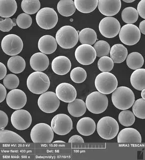
- Plasma atomization produced the metal implant from Ti-6Al-4V Grade 5 powder. d10 = 27 μm; d50 = 47 μm; d90 = 76 μm. The particles are spherical without satellites
- The metal implant can be made using SLM Solutions SLM 280HL machine with parameters set to 99.9% relative density
- Manufactured implants are 200 μm accurate.
- In the figure we can see an AM titanium hip implant production sequence.
Microstructure analysis-
- Leica DMI 5000 light microscopes examined microstructure.
a)X200
- SLM standard samples were machined to ASTM E8/E8M size and tested on Zwick/Roell – Z100 machine.
- Each test point used three specimens.
- ALD MonoTherm vacuum furnaces were used to anneal at 10−3–10−4 mbar.
Sequence of operations used to produce an implant via additive manufacturing.
Results and Discussion of the project work
- By matching the patient's bone size and shape, a 3D hip bone model was created using CT data. SLS 3D-printed the polyamide hip bone model.
- After SLM, Ti-6Al-4V was heat-treated twice. Vacuum annealing parameters such as 950 °C for 1.5 hours and 800 °C for 4 hours; furnace cooling.
- Materialise Magics software can be used to position the implant's CAD model relative to the SLM machine's building plate.
- Support structures were also created to preserve the implant geometry.
- The implant orientation minimised thermal stresses and supports during SLM. Supports were placed on surfaces without special patterns to simplify removal and prevent support residue on the implant.
- Table 1 shows that the material meets the requirements of ASTM F2924–14 (Standard Specification for Additive Manufacturing Titanium-6 Aluminum-4 Vanadium with Powder Bed Fusion) and ISO 5832-3 in terms of its mechanical properties as a whole (Implants for surgery – Metallic materials –Wrought titanium 6-aluminium 4-vanadium alloy).
Conclusion-
- The ability of additive manufacturing technologies to successfully produce a custom-made component of a hip implant endoprosthesis from Ti-6Al-4V alloy was demonstrated.
Because the implant's configuration matches the anatomical features of the patient, the risk of early instability development is reduced, as is the surgery time, blood loss, and the risk of infectious complication development. The ability of SLM technology to create a texturized surface of the implant allows for the potential improvement of the osseointegration process by creating areas with high specific surface. - After annealing, martensitic α′-phase microstructures are partially decomposed into α- and β-phases. This mechanical properties meet ASTM F2924 for additively manufactured Ti-6Al-4V alloy and ISO 5832-3 Implants for surgery from titanium 6-aluminium 4-vanadium alloy. Annealed material has 1080 ± 10 MPa tensile strength and 10% elongation at break.
Sugestions for improvement-
- Future research can concentrate on osseointegration processes and improving mechanical properties through the use of different lattice structures, as well as computer simulation of implant and material characteristics.
Reference-
-
https://www.ncbi.nlm.nih.gov/pmc/articles/PMC6128303/#!po=19.6429
- https://www.ijb.sg/index.php/int-j-bioprinting/article/view/83
- https://www.sciencedirect.com/science/article/pii/S1005030216301335
- https://www.sciencedirect.com/science/article/pii/S2238785419318873
- https://www.sciencedirect.com/science/article/pii/S0143816620300993
- https://www.sciencedirect.com/science/article/pii/S2214860418309035
- https://www.sciencedirect.com/science/article/pii/S2351978919300332