Project 1
PART A
Musculoskeletal Disorders
Clinical condition -Osteoarthritis
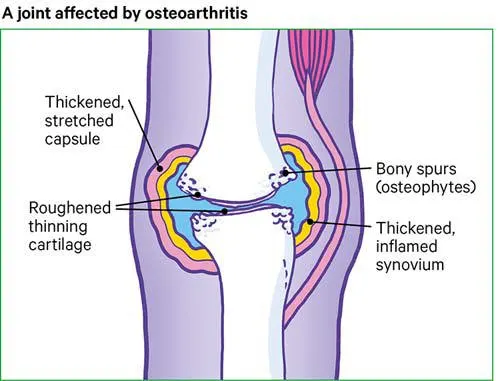
Osteoarthritis is a degenerative joint disease in which joint cartilage is gradually lost. It results from a combination of aging, obesity, irritation of the joints, muscle weakness, and wear and abrasion. Commonly known as “wear-and-tear” arthritis, osteoarthritis is the most common type of arthritis.(Arthritis is a form of rheumatism in which the joints are swollen, stiff, and painful.Rheumatism is any painful disorder of the supporting structures of the body—bones, ligaments, tendons, or muscles—that is not caused by infection or injury.) Osteoarthritis is a progressive disorder of synovial joints, particularly weight-bearing joints. Articular cartilage deteriorates and new bone forms in the subchondral areas and at the margins of the joint. The cartilage slowly degenerates, and as the bone ends become exposed, spurs (small bumps) of new osseous tissue are deposited on them in a misguided effort by the body to protect against increased friction. These spurs decrease the space of the joint cavity and restrict joint movement. Unlike rheumatoid arthritis, osteoarthritis affects mainly the articular cartilage, although the synovial membrane often becomes inflamed late in the disease. Two major distinctions between osteoarthritis and rheumatoid arthritis are that osteoarthritis first afflicts the larger joints (knees, hips) and is due to wear and tear, whereas rheumatoid arthritis first strikes smaller joints and is an active attack on the cartilage. Osteoarthritis is the most common reason for hip- and knee-replacement surgery.
KNEE JOINT
GROSS ANATOMY OF ORGAN
KNEE
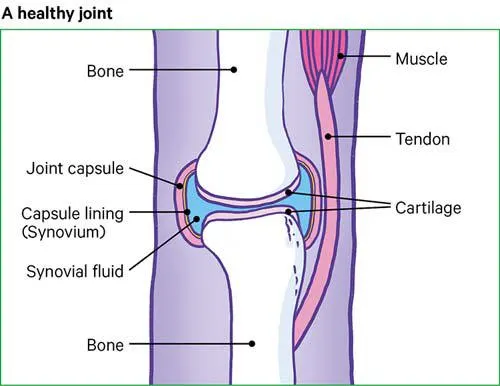
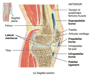
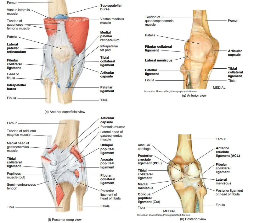
Blood supply of knee joint
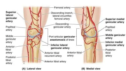
common disorders associated with knee
- ACL, PCL, MCL, LCL injuries (all ligament injuries)
- Cartilage injuries
- Arthritis of the knee
- Chondromalacia - softening of the knee's cartilage cap
- Dislocated kneecaps
- Gout
- Knee dislocation
- Knee pain
- Meniscus injuries
- Osteochondritis
- Osteoarthritis
Diagnostic techniques for osteoarthritis
The diagnostic process involves taking the patient’s medical history and a physical examination, including evaluations of joint function and flexibility. Medical imaging, such as x-rays, and lab tests may also be ordered.
Medical Imaging
Medical imaging provides views of inside the body. Two types of medical imaging commonly used to diagnose osteoarthritis are x-rays and magnetic resonance imaging (MRI):
- X-rays can show how much space is between a joint’s bones, indicating whether there has been a loss of cartilage. An x-ray can also show bone spurs, a sign that the bones have tried to compensate for cartilage loss with extra bone growth.
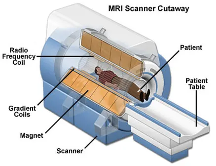
- MRIs provide a detailed look at soft tissues, such as cartilage, ligaments, tendons, and muscle, as well as bone. While not required for an osteoarthritis diagnosis, an MRI may be ordered if to assess the joint’s soft tissues and/or rule out other possible diagnoses. MRIs take longer and are more expensive than x-rays.
- Ultrasound can be useful for evaluating the soft tissue structures that surround the affected joint, such as tendons. Ultrasound can also be used to identify changes to the delicate membrane that encapsulates the joint (synovial lining) or an abnormal increase in joint fluid (synovial fluid).
A physician may use ultrasound along with an injection of local anesthetic (intraarticular injection) to try to identify the exact source of pain.
Many individuals have signs of osteoarthritis that can be seen on medical imaging, even if they do not experience symptoms. Medical imaging is just one diagnostic tool and must be used in conjunction with the patient interview and physical exam.
Currently, there is no standard lab test to verify the presence of osteoarthritis. Lab tests may be used to rule out other problems, such as rheumatoid arthritis, which also causes joint pain. Lab tests may require drawing fluid from the joint (arthrocentesis) or taking a blood sample. In the future, labs may be able to test blood or joint fluid for biomarkers that predict or identify osteoarthritis. The above combination of medical history, physical exam, and possibly diagnostic testing, all inform the diagnosis of osteoarthritis.
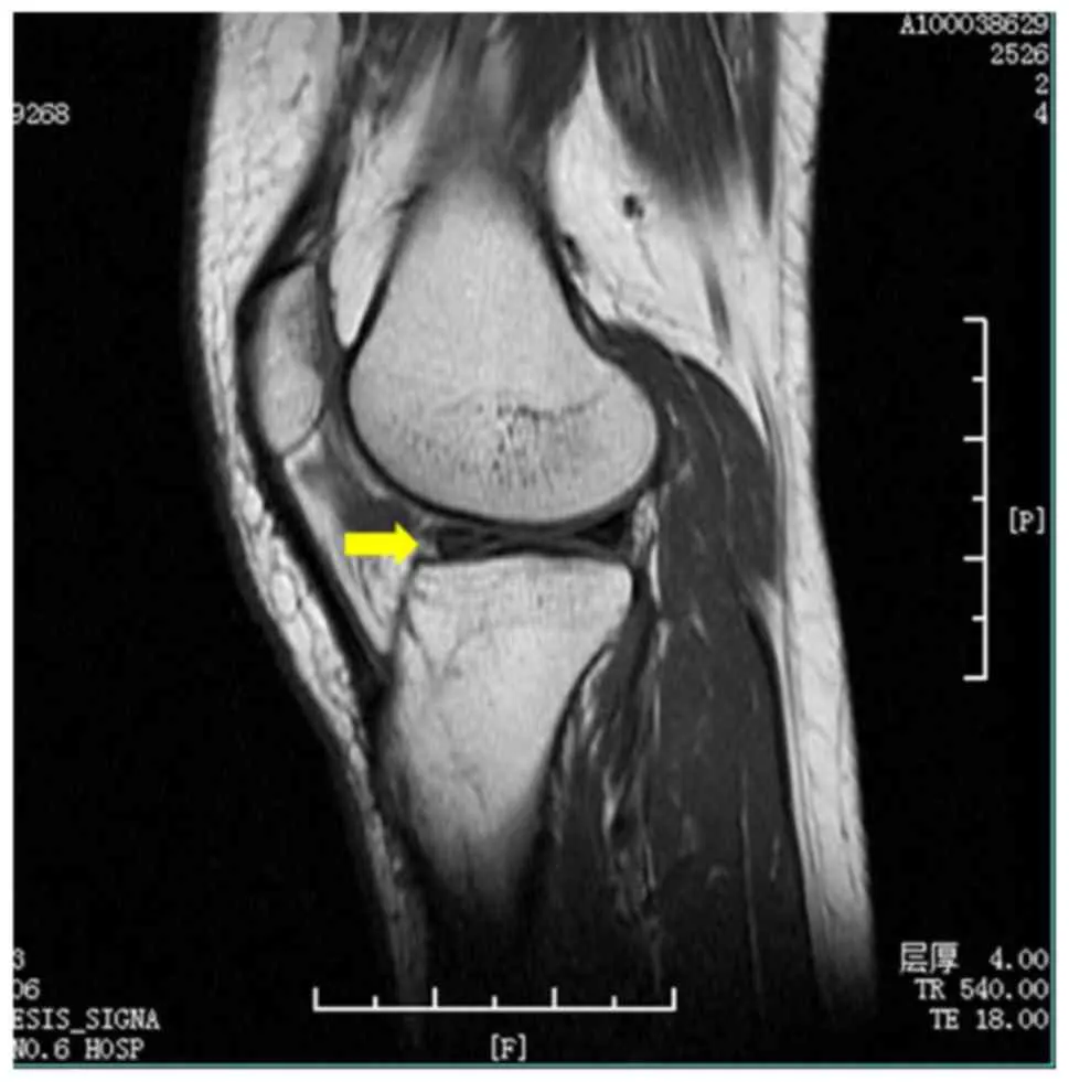
Figure - MRI image of MI (male, 32 years). The arrow shows the meniscus injury in the knee joint. MRI, magnetic resonance imaging; MI, meniscus injury.
Treatment
Osteoarthritis can't be reversed, but treatments can reduce pain and help you move better.
Medications
Medications that can help relieve osteoarthritis symptoms, primarily pain, include:
- Acetaminophen. Acetaminophen (Tylenol, others) has been shown to help some people with osteoarthritis who have mild to moderate pain. Taking more than the recommended dose of acetaminophen can cause liver damage.
-
Nonsteroidal anti-inflammatory drugs (NSAIDs). Over-the-counter NSAIDs, such as ibuprofen (Advil, Motrin IB, others) and naproxen sodium (Aleve), taken at the recommended doses, typically relieve osteoarthritis pain. Stronger NSAIDs are available by prescription.
NSAIDs can cause stomach upset, cardiovascular problems, bleeding problems, and liver and kidney damage. NSAIDs as gels, applied to the skin over the affected joint, have fewer side effects and may relieve pain just as well.
- Duloxetine (Cymbalta). Normally used as an antidepressant, this medication is also approved to treat chronic pain, including osteoarthritis pain.
Therapy
- Physical therapy. A physical therapist can show you exercises to strengthen the muscles around your joint, increase your flexibility and reduce pain. Regular gentle exercise that you do on your own, such as swimming or walking, can be equally effective.
- Occupational therapy. An occupational therapist can help you discover ways to do everyday tasks without putting extra stress on your already painful joint. For instance, a toothbrush with a large grip could make brushing your teeth easier if you have osteoarthritis in your hands. A bench in your shower could help relieve the pain of standing if you have knee osteoarthritis.
- Transcutaneous electrical nerve stimulation (TENS). This uses a low-voltage electrical current to relieve pain. It provides short-term relief for some people with knee and hip osteoarthritis.
Surgical and other procedures
If conservative treatments don't help, you might want to consider procedures such as:
- Cortisone injections. Injections of a corticosteroid into your joint might relieve pain for a few weeks. Your doctor numbs the area around your joint, then places a needle into the space within your joint and injects medication. The number of cortisone injections you can receive each year is generally limited to three or four, because the medication can worsen joint damage over time.
- Lubrication injections. Injections of hyaluronic acid might relieve pain by providing some cushioning in your knee, though some research suggests that these injections offer no more relief than a placebo. Hyaluronic acid is similar to a component normally found in your joint fluid.
- Realigning bones. If osteoarthritis has damaged one side of your knee more than the other, an osteotomy might be helpful. In a knee osteotomy, a surgeon cuts across the bone either above or below the knee, and then removes or adds a wedge of bone. This shifts your body weight away from the worn-out part of your knee.
- Joint replacement. In joint replacement surgery, your surgeon removes your damaged joint surfaces and replaces them with plastic and metal parts. Surgical risks include infections and blood clots. Artificial joints can wear out or come loose and might eventually need to be replaced.
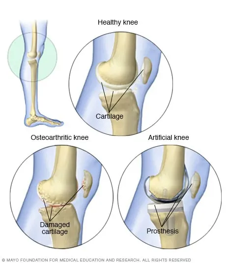
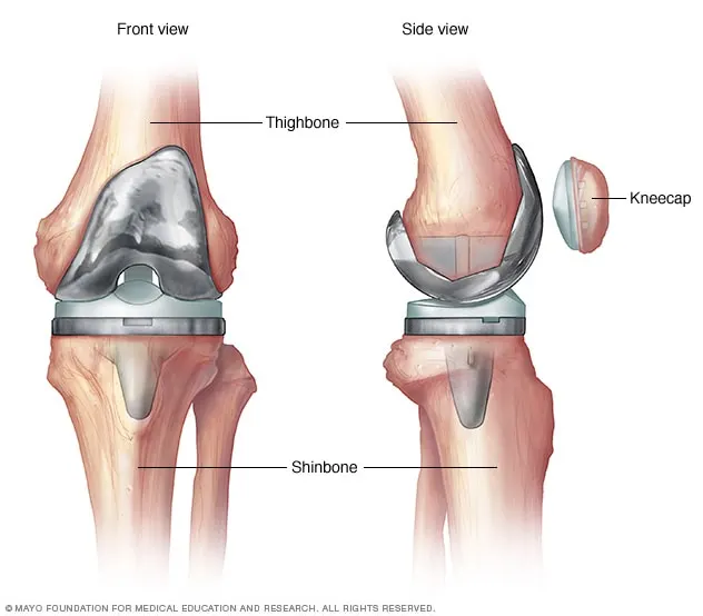
One of the most common reasons for knee replacement surgery is severe pain from joint damage caused by wear and tear arthritis (osteoarthritis). Osteoarthritis can erode the slick cartilage that helps your knee joint move smoothly. An artificial knee joint has metal alloy caps for your thighbone and shinbone, and high-density plastic to replace damaged cartilage.
Surgical treatments for knee OA include arthroscopy, cartilage repair, osteotomy, and knee arthroplasty. Determining which of these procedures is most appropriate depends on several factors, including the location, stage of OA, comorbidities on the one side and patients suffering on the other side.
Knee replacement surgery
Knee replacement, also called knee arthroplasty or total knee replacement, is a surgical procedure to resurface a knee damaged.Metal and plastic parts are used to cap the ends of the bones that form the knee joint, along with the kneecap.
During the procedure
Knee replacement requires a stay in a hospital. Procedures may vary depending on your condition and your doctor's practices.
Knee replacement surgery is most often performed while you are asleep under general anesthesia. Your anesthesiologist will discuss this with you in advance.
Generally, knee replacement surgery follows this process:
-
Patient will be asked to remove clothing and will be given a gown to wear.
-
An intravenous (IV) line may be started in patient's arm or hand.
-
Patient will be positioned on the operating table.
-
A urinary catheter may be inserted.
-
If there is excessive hair at the surgical site, it may be clipped off.
-
The anesthesiologist will continuously monitor patient's heart rate, blood pressure, breathing, and blood oxygen level during the surgery.
-
The skin over the surgical site will be cleansed with an antiseptic solution.
-
The doctor will make an incision in the knee area.
-
The doctor will remove the damaged surfaces of the knee joint and resurface the knee joint with the prosthesis. The knee prosthesis is made up of metal and plastic. The most common type of artificial knee prosthesis is a cemented prosthesis. Uncemented prostheses are not commonly used anymore. A cemented prosthesis attaches to the bone with surgical cement. An uncemented prosthesis attaches to the bone with a porous surface onto which the bone grows to attach to the prosthesis. Sometimes, a combination of the 2 types is used to replace a knee.
The prosthesis is generally comprised of 3 components: the tibial component (to resurface the top of the tibia, or shin bone); the femoral [thigh bone] component (to resurface the end of the thighbone; and the patellar component (to resurface the bottom of the kneecap that rubs against the thighbone). -
The incision will be closed with stitches or surgical staples.
-
A drain may be placed in the incision site to remove fluid.
-
A sterile bandage or dressing will be applied.
After the procedure
In the hospital
After the surgery the patient will be taken to the recovery room for observation. Once blood pressure, pulse, and breathing are stable and the patient is alert, he will be taken to the hospital room. Knee replacement surgery usually requires an in-hospital stay of several days.
It is important to begin moving the new joint after surgery. A physical therapist will meet with patient soon after surgery and plan an exercise program for him. A continuous passive motion (CPM) machine may be used to begin the physical therapy. This machine moves the new knee joint through its range of motion while he is resting in bed. Pain will be controlled with medication so that he can participate in the exercise. He will be given an exercise plan to follow both in the hospital and after discharge.
The patient will be discharged home or to a rehabilitation center. In either case, doctor will arrange for continuation of physical therapy until the patient regain muscle strength and good range of motion.
At home
Once the patient is home, it is important to keep the surgical area clean and dry. Doctor will give patient specific bathing instructions. The stitches or surgical staples will be removed during a follow-up office visit.
To help reduce swelling, patient may be asked to elevate leg or apply ice to the knee.
Take a pain reliever for soreness as recommended by doctor. Aspirin or certain other pain medications may increase the chance of bleeding. Be sure to take only recommended medications.
Notify doctor to report any of the following:
-
Fever
-
Redness, swelling, bleeding, or other drainage from the incision site
-
Increased pain around the incision site
Recent advances
- ACI has been shown to improve the symptoms of osteoarthritis, including pain and mobility. It can also slow or stop osteoarthritis developing, delaying or preventing the need for joint replacement surgery. Autologous Chondrocyte Implantation (ACI) involves the removal of a small sample of tissue from an individual’s joints. This tissue is then used to grow a fresh supply of cartilage cells in a laboratory. Around three weeks later, these cells are returned to the damaged area of the joint in another operation, where they form new cartilage.
- Proprioceptive exercise, Tia-chi exercise, Aquatic resisted exercise, Neuromuscular exercise
- Injections of a natural “energy” molecule prompted regrowth of almost half of the cartilage lost with aging in knees, a new study in rodents shows.(“Our latest study shows that replenishing adenosine stores by injection works well as a treatment for osteoarthritis in animal models of the disease, and with no apparent side effects,” says lead study author Carmen Corciulo, PhD, a postdoctoral fellow at NYU Langone.)
- DNA- or Gene-Based Therapy, Noncoding RNA-Based Therapy, MiRNA, LncRNA, CircRNA, Protein-Based Therapy, Exosomes.
Digestive Disorders
Clinical condition - Gallstones
Gallstones are hardened deposits of digestive fluid that can form in the gallbladder. The gallbladder is a small, pear-shaped organ on the right side of the abdomen, just beneath the liver. The gallbladder holds a digestive fluid called bile that's released into the small intestine.Gallstones range in size from as small as a grain of sand to as large as a golf ball. Some people develop just one gallstone, while others develop many gallstones at the same time.
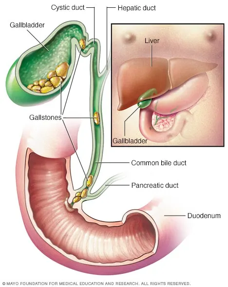
GROSS ANATOMY OF ORGAN
Gall bladder
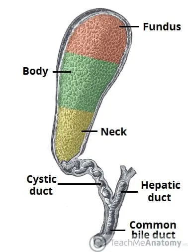
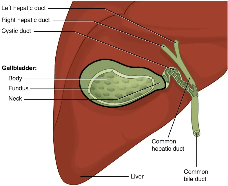
Blood supply of knee joint
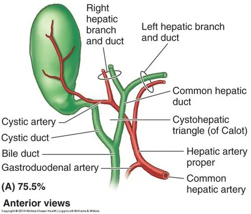
common disorders associated with gallbladder
- Cholecystitis (inflammation of the gallbladder)
- Gallstones.
- Chronic acalculous gallbladder disease (in which the natural movements needed to empty the gallbladder do not work well)
- Gangrene or abscesses.
- Growths of tissue in the gallbladder.
- Congenital defects of the gallbladder.
Diagnostic techniques for gallstones
Gallstone Diagnosis
Doctor will do a physical exam and might order tests including:
Blood thests: These check for signs of infection or blockage, and rule out other conditions.
This makes images of the inside of the body.
CT scan: Specialized X-rays let the doctor see inside the body, including the gallbladder.
Magnetic resonance cholangiopancreatography(MRCP): This test uses a magnetic field and pulses of radio wave energy to make pictures of the inside of the body, including the liver and gallbladder.
Cholescintigraphy (HIDA scan): This test can check whether the gallbladder squeezes correctly. Doctor injects a harmless radioactive material that makes its way to the organ. A technician can then watch its movement.
Endoscopic ultrasound: This test combines ultrasound and endoscopy to look for gallstones.
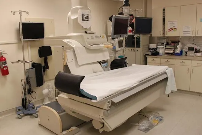
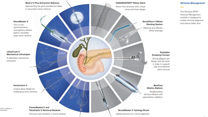
Endoscopic retrograde cholangiopancreatography (ERCP): The doctor runs a tube called an endoscope through mouth down to the small intestine. They inject a dye so they can see bile ducts on a camera in the endoscope. They can often take out any gallstones that have moved into the ducts.
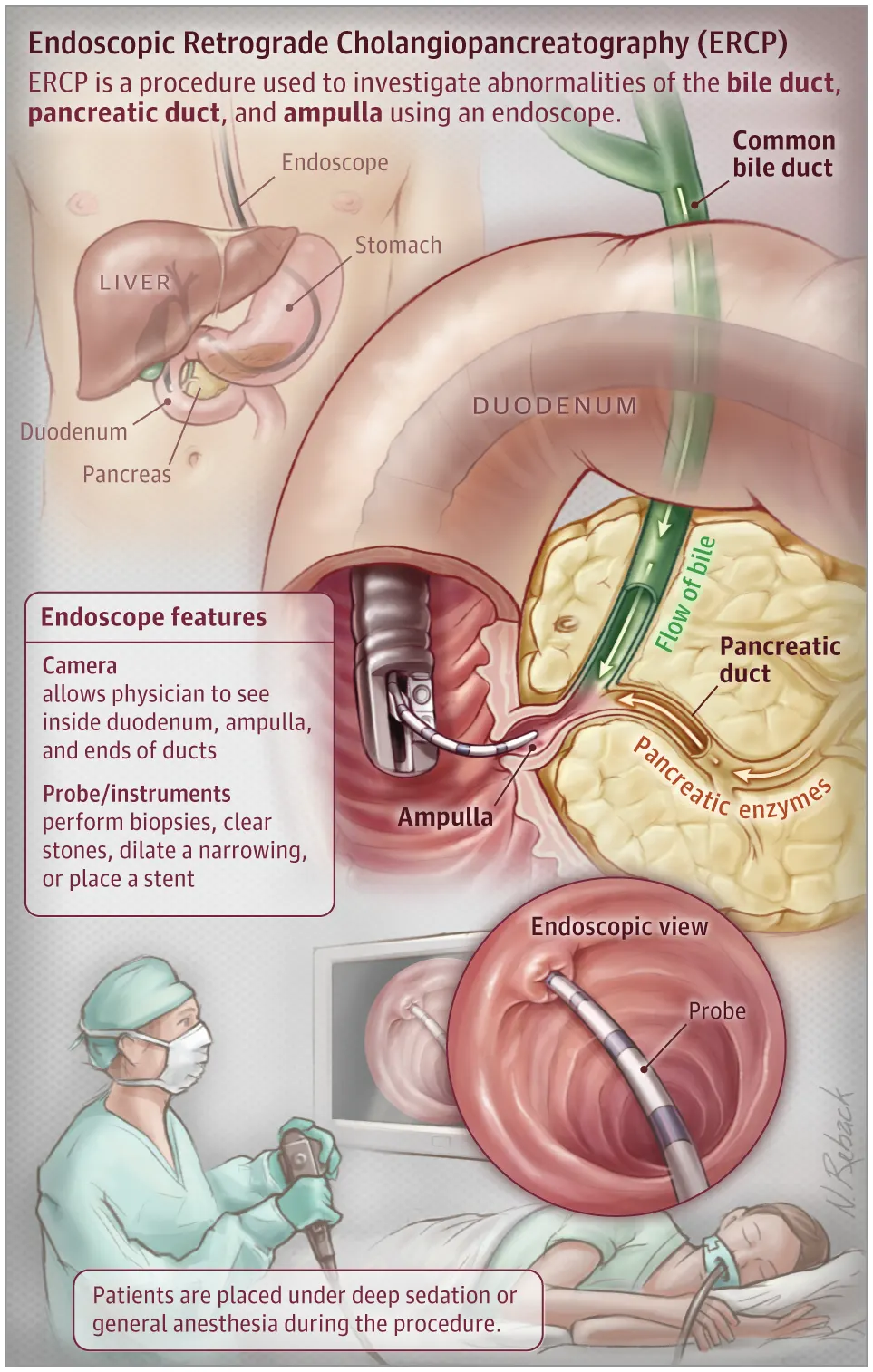
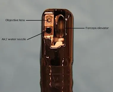
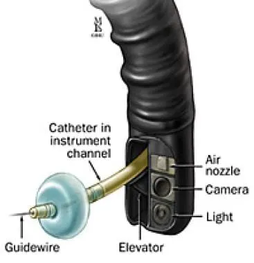
The standard endoscope for endoscopic retrograde cholangiopancreatography (ERCP) is the therapeutic side-viewing duodenoscope, which has a side-viewing objective lens, a tip with four-way angulation capability, a side-positioned air/water nozzle, a 4.2-mm instrument channel, and a forceps elevator adjacent to the instrument channel outlet that allows fine linear instrument position changes (see the images below).
Gallstone Treatment
Treatment is not needed if patients don’t have any symptoms. Some small gallstones can pass through the body on their own.
Most people with gallstones have their gallbladders taken out. They can still digest food without it. Doctor will use one of two procedures.
Laparoscopic cholecystectomy: This is the most common surgery for gallstones. Doctor passes a narrow tube called a laparoscope into the belly through a small cut. It holds instruments, a light, and a camera. They take out the gallbladder through another small cut. The patient will usually go home the same day.
Open cholecystectomy: The doctor makes bigger cuts in the belly to remove gallbladder. The patient will stay in the hospital for a few days afterward.
If gallstones are in the bile ducts, the doctor may use ERCP to find and remove them before or during surgery.
If the patient has another medical condition and the doctor thinks he shouldn't have surgery, they might give him medication instead. Chenodiol and ursodiol dissolve cholesterol stones. They can cause mild diarrhea.
A cholecystectomy is a surgical procedure to remove gallbladder — a pear-shaped organ that sits just below liver on the upper right side of your abdomen.
To prepare for a cholecystectomy, surgeon may ask to:
- Eat nothing the night before your surgery: Patient may drink a sip of water with medications, but avoid eating and drinking at least four hours before surgery.
- Stop taking certain medications and supplements: Tell doctor about all the medications and supplements patient take. Continue taking most medications as prescribed. Doctor may ask to stop taking certain medications and supplements because they may increase risk of bleeding.
Before the procedure
A cholecystectomy is performed using general anesthesia, so the patient won't be aware during the procedure. Anesthesia drugs are given through a vein in the arm. Once the drugs take effect, health care team will insert a tube down the throat to help the patient breathe. The surgeon then performs the cholecystectomy using either a laparoscopic or open procedure.
During the procedure
Depending on patient's situation, surgeon will recommend one of two surgical approaches:
Minimally invasive (laparoscopic) cholecystectomy
During a laparoscopic cholecystectomy, the surgeon makes four small incisions in abdomen. A tube with a tiny video camera is inserted into the abdomen through one of the incisions. Surgeon watches a video monitor in the operating room while using surgical tools inserted through the other incisions in the abdomen to remove the gallbladder.
Next the patient may undergo an imaging test, such as an X-ray or ultrasound, if the surgeon is concerned about possible gallstones or other problems in the bile duct. Then patient incisions are sutured, and he is taken to a recovery area. A laparoscopic cholecystectomy takes one or two hours.
A laparoscopic cholecystectomy isn't appropriate for everyone. In some cases surgeon may begin with a laparoscopic approach and find it necessary to make a larger incision because of scar tissue from previous operations or complications.
Traditional (open) cholecystectomy
During an open cholecystectomy, the surgeon makes a 6-inch (15-centimeter) incision in abdomen below ribs on right side. The muscle and tissue are pulled back to reveal liver and gallbladder. Surgeon then removes the gallbladder.
The incision is sutured, and the patient is taken to a recovery area. An open cholecystectomy takes one or two hours.
After the procedure
The patient will be taken to a recovery area as the anesthesia drugs wear off. Then he will be taken to a hospital room to continue recovery. Recovery varies depending on the procedure:
- Laparoscopic cholecystectomy. People are often able to go home the same day as their surgery, though sometimes a one-night stay in the hospital is needed. In general, you can expect to go home once you're able to eat and drink without pain and are able to walk unaided. It takes about a week to fully recover.
- Open cholecystectomy. Expect to spend two or three days in the hospital recovering. Once at home, it may take four to six weeks to fully recover.
Recent advances
- Statins
- Ezetimibe
- n-3 polyunsaturated fatty acids
- Androgen receptors agonist
- Liraglutide
- GPER antagonist
-
The robotic-assisted laparoscopic cholecystectomy appears to be an interesting and emerging technology
Respiratory Disorders
Clinical condition -Asthma
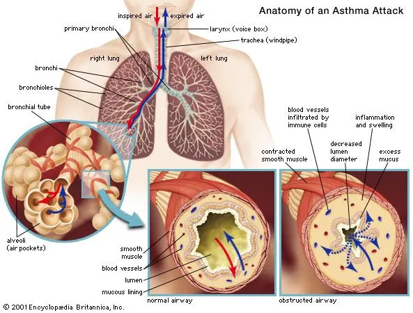
Asthma is a condition in which your airways narrow and swell and may produce extra mucus. This can make breathing difficult and trigger coughing, a whistling sound (wheezing) when you breathe out and shortness of breath.
For some people, asthma is a minor nuisance. For others, it can be a major problem that interferes with daily activities and may lead to a life-threatening asthma attack.
Asthma can't be cured, but its symptoms can be controlled. Because asthma often changes over time, it's important that you work with your doctor to track your signs and symptoms and adjust your treatment as needed.
Blood supply to lungs
Most common lung disorders
- Asthma.
- Collapse of part or all of the lung (pneumothorax or atelectasis)
- Swelling and inflammation in the main passages (bronchial tubes) that carry air to the lungs (bronchitis)
- COPD.
- Lung cancer.
- Lung infection (pneumonia)
- Abnormal buildup of fluid in the lungs (pulmonary edema)
The main tests used to help diagnose asthma are:
- FeNO test – you breathe into a machine that measures the level of nitric oxide in your breath, which is a sign of inflammation in your lungs
- Spirometry – you blow into a machine that measures how fast you can breathe out and how much air you can hold in your lungs
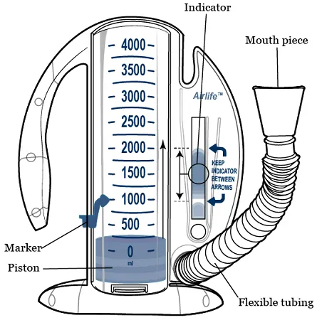
- Peak flow test – you blow into a handheld device that measures how fast you can breathe out, and this may be done several times over a few weeks to see if it changes over time
Other tests to diagnose asthma include:
- Methacholine challenge. Methacholine is a known asthma trigger. When inhaled, it will cause your airways to narrow slightly. If you react to the methacholine, you likely have asthma. This test may be used even if your initial lung function test is normal.
- Imaging tests. A chest X-ray can help identify any structural abnormalities or diseases (such as infection) that can cause or aggravate breathing problems.
- Allergy testing. Allergy tests can be performed by a skin test or blood test. They tell you if you're allergic to pets, dust, mold or pollen. If allergy triggers are identified, your doctor may recommend allergy shots.
- Nitric oxide test. This test measures the amount of the gas nitric oxide in your breath. When your airways are inflamed — a sign of asthma — you may have higher than normal nitric oxide levels. This test isn't widely available.
- Sputum eosinophils. This test looks for certain white blood cells (eosinophils) in the mixture of saliva and mucus (sputum) you discharge during coughing. Eosinophils are present when symptoms develop and become visible when stained with a rose-colored dye.
- Provocative testing for exercise and cold-induced asthma. In these tests, your doctor measures your airway obstruction before and after you perform vigorous physical activity or take several breaths of cold air.
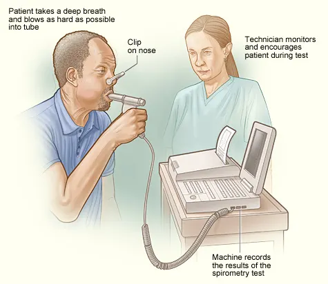
Parts of Spirometry machine
Treatment
Prevention and long-term control are key to stopping asthma attacks before they start. Treatment usually involves learning to recognize your triggers, taking steps to avoid triggers and tracking your breathing to make sure your medications are keeping symptoms under control. In case of an asthma flare-up, you may need to use a quick-relief inhaler.
Medications
The right medications for you depend on a number of things — your age, symptoms, asthma triggers and what works best to keep your asthma under control.
Preventive, long-term control medications reduce the swelling (inflammation) in your airways that leads to symptoms. Quick-relief inhalers (bronchodilators) quickly open swollen airways that are limiting breathing. In some cases, allergy medications are necessary.
Long-term asthma control medications, generally taken daily, are the cornerstone of asthma treatment. These medications keep asthma under control on a day-to-day basis and make it less likely you'll have an asthma attack. Types of long-term control medications include:
-
Inhaled corticosteroids. These medications include fluticasone propionate (Flovent HFA, Flovent Diskus, Xhance), budesonide (Pulmicort Flexhaler, Pulmicort Respules, Rhinocort), ciclesonide (Alvesco), beclomethasone (Qvar Redihaler), mometasone (Asmanex HFA, Asmanex Twisthaler) and fluticasone furoate (Arnuity Ellipta).
You may need to use these medications for several days to weeks before they reach their maximum benefit. Unlike oral corticosteroids, inhaled corticosteroids have a relatively low risk of serious side effects.
-
Leukotriene modifiers. These oral medications — including montelukast (Singulair), zafirlukast (Accolate) and zileuton (Zyflo) — help relieve asthma symptoms.
Montelukast has been linked to psychological reactions, such as agitation, aggression, hallucinations, depression and suicidal thinking. Seek medical advice right away if you experience any of these reactions.
- Combination inhalers. These medications — such as fluticasone-salmeterol (Advair HFA, Airduo Digihaler, others), budesonide-formoterol (Symbicort), formoterol-mometasone (Dulera) and fluticasone furoate-vilanterol (Breo Ellipta) — contain a long-acting beta agonist along with a corticosteroid.
- Theophylline. Theophylline (Theo-24, Elixophyllin, Theochron) is a daily pill that helps keep the airways open by relaxing the muscles around the airways. It's not used as often as other asthma medications and requires regular blood tests.
Quick-relief (rescue) medications are used as needed for rapid, short-term symptom relief during an asthma attack. They may also be used before exercise if your doctor recommends it. Types of quick-relief medications include:
-
Short-acting beta agonists. These inhaled, quick-relief bronchodilators act within minutes to rapidly ease symptoms during an asthma attack. They include albuterol (ProAir HFA, Ventolin HFA, others) and levalbuterol (Xopenex, Xopenex HFA).
Short-acting beta agonists can be taken using a portable, hand-held inhaler or a nebulizer, a machine that converts asthma medications to a fine mist. They're inhaled through a face mask or mouthpiece.
- Anticholinergic agents. Like other bronchodilators, ipratropium (Atrovent HFA) and tiotropium (Spiriva, Spiriva Respimat) act quickly to immediately relax your airways, making it easier to breathe. They're mostly used for emphysema and chronic bronchitis, but can be used to treat asthma.
- Oral and intravenous corticosteroids. These medications — which include prednisone (Prednisone Intensol, Rayos) and methylprednisolone (Medrol, Depo-Medrol, Solu-Medrol) — relieve airway inflammation caused by severe asthma. They can cause serious side effects when used long term, so these drugs are used only on a short-term basis to treat severe asthma symptoms.
If you have an asthma flare-up, a quick-relief inhaler can ease your symptoms right away. But you shouldn't need to use your quick-relief inhaler very often if your long-term control medications are working properly.
Keep a record of how many puffs you use each week. If you need to use your quick-relief inhaler more often than your doctor recommends, see your doctor. You probably need to adjust your long-term control medication.
Allergy medications may help if your asthma is triggered or worsened by allergies. These include:
- Allergy shots (immunotherapy). Over time, allergy shots gradually reduce your immune system reaction to specific allergens. You generally receive shots once a week for a few months, then once a month for a period of three to five years.
- Biologics. These medications — which include omalizumab (Xolair), mepolizumab (Nucala), dupilumab (Dupixent), reslizumab (Cinqair) and benralizumab (Fasenra) — are specifically for people who have severe asthma.
Bronchial thermoplasty
This treatment is used for severe asthma that doesn't improve with inhaled corticosteroids or other long-term asthma medications. It isn't widely available nor right for everyone.
During bronchial thermoplasty, your doctor heats the insides of the airways in the lungs with an electrode. The heat reduces the smooth muscle inside the airways. This limits the ability of the airways to tighten, making breathing easier and possibly reducing asthma attacks. The therapy is generally done over three outpatient visits.
Treat by severity for better control: A stepwise approach
Your treatment should be flexible and based on changes in your symptoms. Your doctor should ask about your symptoms at each visit. Based on your signs and symptoms, your doctor can adjust your treatment accordingly.
For example, if your asthma is well controlled, your doctor may prescribe less medication. If your asthma isn't well controlled or is getting worse, your doctor may increase your medication and recommend more-frequent visits.
Methods of surgical treatment of bronchial asthma (BA) are classified as first generation (glomectomy, gangliectomy, vagotomy, pulmonary roots denervation), second generation (implantation of neurostimulators of sinocarotid, diaphragmal nerves, vagus, sympathic trunks), third generation (implants of programmed microchips).
Recent advances
- Mepolizumab (also known as Nucala) is one of the first in a new line of monoclonal antibody treatments designed to treat a specific type of asthma called severe eosinophilic asthma. This type of asthma is one of the most debilitating forms of the condition, involving an inflammation of the airways linked to a particular type of white blood cell (eosinophils).
- The U.S. Food and Drug Administration has approved an injectable drug called Tezspire, which would be administered every four weeks by a health care professional.The medication is considered an add-on treatment meant to improve severe asthma in a person whose condition isn’t controlled by their current medications. People who are prescribed Tezspire would continue to use other asthma treatments.
- Precision medicine, which is fitting very well asthma’s heterogeneity and complex pathogenesis, is becoming an overarching medical discipline that requires a good understanding of biomarkers, phenotypes, endotypes, genotypes, regiotypes, and theratypes of diseases. ICS have been the foundation for asthma treatment; however, inhaled or systemic corticosteroids can be ineffective in many patients with asthma. Few treatment options exist for patients with steroid-resistant asthma. The recent development of a new class of biological agents that target airway type 2 inflammation has provided an opportunity for treating patients with corticosteroid refractory asthma
- New long-acting β-agonists (LABA) have been developed for chronic obstructive pulmonary disease (COPD) and, a priori, these might also reasonably be expected to be effective against asthma. The recently developed once-daily, very long-acting LABA (indacaterol, vilanterol, olodaterol and others) potentially offer better bronchodilation patterns relevant to treating severe asthma with persistent airflow obstruction. Whether these drugs will also synergistically improve asthma control and reduce exacerbation rates is unknown at present.
PART B
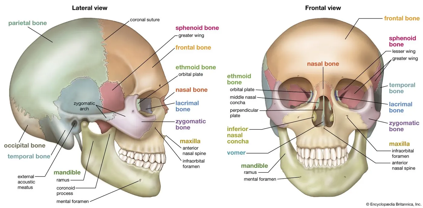
Cranioplasty is the surgical repair of a bone defect in the skull resulting from a previous operation or injury. There are different kinds of cranioplasties, but most involve lifting the scalp and restoring the contour of the skull with the original skull piece or a custom contoured graft made from material such as:
- Titanium (plate or mesh).
- Synthetic bone substitute (in liquid form).
- Solid biomaterial (prefabricated customized implant to match the exact contour and shape of the skull).
Cranioplasty might be performed for any of the following reasons:
- Protection: In certain places, a cranial defect can leave the brain vulnerable to damage.
- Function: Cranioplasty may improve neurological function for some patients. In some instances, a customized cranial implant is designed ahead of time to help the surgeon obtain an ideal shape and outcome, as well as to house embedded neuro technologies.
- Aesthetics: A noticeable skull defect can affect a patient’s appearance and confidence.
- Headaches: Cranioplasty can reduce headaches due to previous surgery or injury.
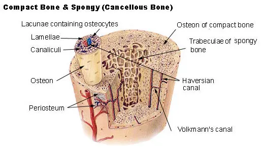
Microscopic anatomy of bone
Schematic of the blood supply to the brain
A cranioplasty is performed in a hospital setting with the patient under general anesthetic. A portion of the scalp will be shaved and a topical cleanser is applied to prevent infection. Doctor then makes an incision in the skin to access the skull bones. If the existing bone is intact and undamaged, Doctor can reshape and reposition it and use special discs as well as titanium plates and screws to secure it in place.
It is generally considered optimal to use the patient’s original bone if it remains functional. However, in cases where the bone has been badly damaged or removed, there are several options available. Prior to the surgery, it will be determined exactly what substance will be used to replace the bone. Depending on a number of factors, the surgeon may choose to graft.
Possible graft materials include:
- A piece of bone from your own skull that has been removed and replaced
- A piece of bone from your own rib or pelvis
- Mesh or plate titanium
- Synthetic bone-like material
- Acrylic
All of these are attached to the nearby bones with titanium plates and screws.
Once the bone or grafting material has been properly affixed, Doctor will close the scalp incision using either sutures or surgical staples.
Cranioplastic surgery following decompressive craniectomy (DC) is a well-established neurosurgical intervention. The operation not only restores the integrity of the skull but also seems to have positive effects on cerebral blood flow and cerebrovascular reserve capacity. There is evidence that the rehabilitation potential as well as the neurological outcome of patients improves after cranioplasty. In addition, the procedure helps to achieve a cosmetically satisfactory result.
The restoration of the integrity of the skull can be achieved either by reimplantation of the autologous bone or by allogenic material such as bone cement (polymethyl methacrylate (PMMA)) or a previously manufactured patient-specific implant (PSI) made of titanium or polyether ether ketone (PEEK).
The literature regarding a potential correlation between graft material and infection rate is inconsistent. Matsuno et al. describe a significant infection rate for titanium mesh compared to autologous bone flaps and PMMA . However, while the implantation of the autologous bone is associated with significantly lower costs than the manufacturing of a PSI, a particular disadvantage remains: the occurrence of aseptic bone resorption (ABR) is a specific and not to be underestimated complication of this method that leads to a considerable number of patients requiring further surgery.
This resorption leads to structural instability and consecutive loss of protective function in addition to the already existing risk of infection.
The rate of ABR varies in the literature with incidences around 20%. Bone flap fragmentation, shunt-dependent hydrocephalus, and especially, young age have been discussed lately as risk factors leading to ABR. Furthermore, some authors suggest a relationship between the method of storage, preparation of the implant, time interval between DC and reimplantation (“freezer time”), or the size of the defect. A possible contamination and subsequent low-grade infection with Propionibacterium acnes has been discussed as a potential risk factor as well. Schütz et al. were further able to show that hypertensive patients treated with ACE inhibitors had a lower ABR rate than patients treated with other antihypertensive medication or patients who do not suffer from arterial hypertension.
Despite these frequent descriptions of ABR in the literature, we still lack a thorough understanding of the physiology of bony reintegration at the skull and thus the underlying pathology leading to the pathological process of resorption.
Macroscopically, the typical bone structure of cortical and cancellous bone was no longer present in some areas. The graft had thin ends in the marginal areasMicroscopically, a bone remodeling with a build-up of new, vital bone tissue was observed in the entire area of the bone flaps. The remodeling occurred not only from the edge of the transplants, but almost simultaneously in more central sections.
The new bone formations were visible in the form of additions on the original graft bone. In the osteocyte cavities of the new bone formations, the vital cells were clearly visible, whereas no living cells were found in the old graft bone, but empty osteocyte cavities. The avital graft bone seemed to serve as guidance for new bone formation by osteoblasts. Bone resorbing osteoclasts were also found.
The remaining marrow spaces were predominantly fibrotic or necrotic (avital). In areas with marrow cavity fibrosis, hardly any new bone tissue was found in the adjacent bone, while there were increased signs of osteoclastic resorption.
In general, there was more resorption on the outer side of the transplants facing away from the brain than on the inner side facing the brain. Here, new, elongated, parallel bone lamellae dominated.
Taking into account freeze storage, both control samples showed a normal bone structure. The marrow spaces were intact and hematopoiesis activity appeared normal. Neither control showed signs of fibrosis or necrosis.
With increasing time interval between reimplantation and explantation, the bone volume in the marginal areas of the graft bone increased.
Compared to the controls, the grafts showed an altered structure, the typical layering of cortical and cancellous bone was mainly eliminated. The measurements showed that the grafts were less thick and had distinctly thinned edges. Histologically, new bone formations were distributed over the entire graft area.
The shown resorption processes indicate that the measured thickness decrease of the grafts mainly originates from the outer side as this side showed increased osteoclastic resorption. On the side facing the brain, more elongated parallel bone appositions were found. However, this could not be shown in all samples.
Despite the relatively small control group, the reference values measured in the two control cases for the skull thickness in the peripheral and central areas corresponded to the values in the literature.
Fatty bone marrow may be responsible for insufficiency of bone reintegration. We postulate a possible barrier-function of the remaining marrow cavities, since these areas were predominantly fibrotic and showed hardly any adjacent new bone tissue but increased resorption. Thorough rinsing of the implant to remove the remaining bone marrow could be considered. However, a recommendation for this procedure cannot be derived from our study. Further studies to investigate this idea are necessary.
Some risk factors for ABR include fragmentation of the bone flap, shunt-dependent hydrocephalus, and young age.
One option under discussion is the initial implantation of artificial grafts in pediatric patients and fragmented bone flaps, although it should be noted that this involves higher costs which might be of relevance in developing countries.
The histological examinations performed in this work provide a new insight into the underlying processes in ABR of autologous cranial grafts.
As endocrinological serum parameters did not differ significantly between patients in whom ABR had occurred and the controls, no systemic serological endocrine predictors could be identified for aseptic bone necrosis in our population.
Since there is no comparable study to date in which results of histological examinations of explanted autologous grafts of the skull have been published, a direct comparison of the own findings with other studies is hardly possible. However, some histological examinations of grafts from other skeletal regions exist. A peripheral bridge-like reintegration starting from the vital bone was observed in homologous transplants from the acetabulum region. In contrast, the skull transplants examined here showed new bone deposits in the entire area, not only in the marginal area of a few millimeters as described for homologous hip transplants.
Both the homologous grafts of the hip and the examined autologous grafts of the skull had been cryopreserved. It is therefore to be discussed whether this alters the marrow cavities in such a way that the body’s own cells are no longer able to colonize these areas, resulting in scar-like fibrosis. Most of the existing studies on the influence of cryopreservation of bone have been performed in animal models. While freezing appears to preserve the morphological state of the mineralized bone tissue and the mineral content, the cellular components are affected more severely. Chan et al. could not detect vital osteoblasts in any of the 18 cryopreserved human bone flaps examined, with a minimum storage time of 4 months in this study.
Limitations of this study include the inhomogeneous patient population. The samples were derived from male and female patients of different ages, so that differences in bone metabolism must be suspected. However, endocrinological serum parameters did not differ significantly between patients with ABR and controls. Additionally, the time interval between DC and reimplantation (“freezer time”) varied from 1 to 6 months.
The heterogeneous sample collective and missing samples with time intervals between DC and explantation of less than 1 year do not allow for reliable conclusions about the timing of changes in the transplants. However, the measurements carried out on bone volume indicate that increased resorption of the graft by osteoclasts may occur first, followed by densification of the graft by new bone formation.
It should further be noted that an adverse selection occurred in the samples, as only cases of graft failure could be investigated. With only a limited number of samples, a further statistical evaluation of the measurement results was considered to be of limited use, so that the focus of this work was based on a descriptive approach.
Recovery from Cranioplasty
After undergoing a cranioplasty procedure, patients are typically required to stay in the hospital for a period of five to seven days. Drains may be needed initially to help the body remove any excess fluid that has accumulated in the area. Most patients will experience some headaches in the days following the surgery, but these respond well to pain relievers that will be prescribed by doctor. Fatigue is also a common side effect that should subside within the first few weeks. Some activities may be limited for a period of time after your cranioplasty, and Doctor will provide patient with information on any restrictions that he will be expected to adhere to and for how long.
Cranioplasty Risks
As in the case of any surgery, there are risks, including (but not limited to) the following:
- Infection (which may need to be treated with antibiotics).
- Postoperative blood clot requiring drainage.
- Stroke.
- Seizure.
- Clot in the legs (which rarely can travel to the lungs).
- Complication not related directly to the surgery:
- Pneumonia.
- Heart attack.
- Urinary infection.