Project 2
Abstract
Robot-assisted kidney transplantation (RAKT) has recently been introduced to reduce the morbidity of open kidney transplantation (KT). Robot-assisted surgery has been able to overcome many of the limitations of classical laparoscopy, certainly in complex and technically demanding procedures, such as vascular and ureteral anastomosis. Since the first RAKT in 2010, this technique has been standardized and evaluated in highly experienced robot and KT centers around the world. In Europe, the European Association of Urology Robotic Urology Section (ERUS) created an RAKT working group in 2016 in order to prospectively follow the outcomes of RAKT. When performed by surgeons with both robotic and KT experience, RAKT has been proven to be safe and reproducible in selected cases and yield excellent graft function with a low complication rate. Multiple institutions have now adopted RAKT, and its use will likely increase in the near future. However, structured training and proctoring will be mandatory for those embarking on RAKT in order to help them negotiate the learning curve and avoid technical mistakes. This chapter will describe RAKT from living and deceased donors and its application in kidney autotransplantation (KAT).
1. Introduction
Kidney transplantation (KT) is considered the preferred treatment for patients with end-stage renal disease (ESRD) owing to the greater survival rate and better quality of life in comparison to hemodialysis. To date, the open approach has been the gold standard in KT, despite its invasiveness and high morbidity. Minimally invasive surgery may be a good alternative to reduce the morbidity associated with the open approach, especially in immunocompromised and fragile KT patients and even more in obese recipients who are known to have a higher complication rate.
Minimally invasive surgery (using a pure laparoscopic or a robot-assisted approach) offers significant benefits to patients compared to open surgery, including improved peri- and postoperative outcomes, such as shorter hospital stay, less postoperative pain, shorter convalescence period, fewer wound infections, and better cosmetic results. Robot-assisted surgery has been able to overcome many of the limitations of classical laparoscopy, especially in complex and technically demanding surgical procedures, such as reconstruction or vascular and ureteral anastomosis. This has been attributed to the superb three-dimensional vision, magnification (12×), control of the camera by the surgeon, elimination of hand tremor, and seven degrees of freedom of movement.
This chapter will describe the history, technique and results of robot-assisted kidney transplantation (RAKT) from living donors and deceased donors, and also its application in kidney autotransplantation (KAT).
2. Robot-assisted kidney transplantation (RAKT): living donor
2.1 History and background
2.1.1 Living donor nephrectomy
Open donor nephrectomies were carried out for nearly 50 years until the introduction of laparoscopy in 1995 by Ratner et al. Since its first description in 1995, the laparoscopic approach for donor nephrectomy has demonstrated to improve peri- and postoperative outcomes such as blood loss, pain, hospital stay, as well as cosmetic results, when compared to open surgery. In 2001, the first series of robot-assisted laparoscopic donor nephrectomy, using the da Vinci® Surgical System (Intuitive Surgical, Sunnyvale, CA, USA), was reported by the Group of the University of Illinois (Chicago). They demonstrated that robot-assisted nephrectomy is feasible, safe, and reproducible, providing similar results in comparison to the laparoscopic approach. Nowadays, around 40% of all KT in the USA and around 20% of all KT in Europe are performed with living donors. Every year, the ratio of “emotionally related” living donors to genetically related living donors increases slightly, with most of the living donors currently being family members. In comparison with KT from deceased donors, KT from living donors provides several advantages in terms of long-term patient survival, earlier graft function, longer graft survival, less aggressive immunosuppressive regimens, and reduced waiting lists. When a living donor has two equally functioning kidneys, the left kidney is preferred for donation as the left renal vein is longer compared to the right renal vein. When the kidney function of both kidneys is different, the lesser functioning kidney is used for donation, in order to limit the risks for the donor. Many concerns have been raised regarding the use of the right kidney for living donation, but literature suggests that right laparoscopic donor nephrectomy is feasible and results in good graft function.
2.1.2 Living donor kidney transplantation
RAKT has recently been introduced to reduce the morbidity of open KT. Since the first RAKT in 2010 by Giulianotti et al. in the USA, this technique has been standardized and evaluated in highly experienced robot and KT centers around the world. In 2014 Menon et al. standardized the technique with the transperitoneal approach and regional hypothermia known as the Vattikuti-Medanta technique. The authors highlighted that RAKT is a safe technique with possible advantages such as low intra- and postoperative complications, better cosmetic results, and superlative vision that could result in better quality of the vascular and ureteral anastomoses. The first two European pure RAKTs were performed in July 2015 by Breda et al. and Doumerc et al. In 2016, the European Association of Urology (EAU) formed the EAU Robotic Urology Section (ERUS) RAKT working group in order to prospectively follow the outcomes of RAKT. Breda et al. published the largest multicenter series of RAKT to date (120 patients). Angelo Territo et al. addressed the functional results of RAKT from living donors at 1-year follow-up, and Vignolini et al. developed a RAKT program with grafts from deceased donors. Siena et al. described the technique for RAKT in grafts with multiple vessels. The feasibility of RAKT in children was described by the Ghent University group by Spinoit et al.
2.2 Operative technique
2.2.1 Living donor nephrectomy
To date, several techniques are described for living donor nephrectomy, including open surgery, pure laparoscopy, hand-assisted laparoscopy, and robot-assisted surgery. The most commonly used technique is the minimally invasive transperitoneal laparoscopic approach. According to the literature, laparoscopic surgery for living donor nephrectomy achieves similar functional results compared to open and robot-assisted living donor nephrectomy, being equally safe for the donor. Robot-assisted surgery offers clear advantages over conventional laparoscopy, thanks to the use of EndoWrist instruments, three-dimensional view, enhanced visualization of the operative field (12x), and, possibly, a shorter learning curve. Open nephrectomy for donation may offer an advantage in challenging cases such as grafts with multiple vessels and/or vascular anomalies and prior abdominal surgery. Furthermore, the open approach may be preferred in centers with low experience in laparoscopy and/or a low case volume of living donor nephrectomies (Figure 1).
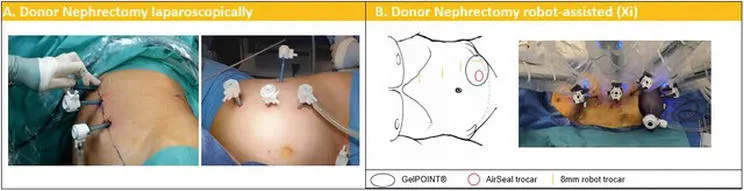
Figure 1.
Trocar placement and patient positioning for nephrectomy during RAKT on living donors. (A) Laparoscopically: patient positioned in lateral decubitus, linear port configuration along the pararectal line, with the camera placed at the most cephalic position (at the 12th rib level). (B) Robot-assisted: patient positioned in lateral decubitus; GelPOINT® device at the level of the ipsilateral fossa through a 6 cm Pfannenstiel incision; a 15-mm AirSeal trocar is placed in the device to introduce endovascular stapler and the 15-mm EndoCatch bag for organ extraction. An additional trocar is used to raise the kidney during the section of the vessels.
2.2.2 Back table preparation
After robot-assisted/laparoscopic living donor nephrectomy, the preparation of the kidney is performed at the back table close to the operating bed. First, the graft is placed in a basin with slushed ice and perfused with 1 liter of storage solution (Celsior®, or Custodiol®, or Institut Georges Lopez-1®). Next, the graft vessels are carefully dissected. If the donor kidney has multiple arteries or veins, a vascular reconstruction can be performed. Siena et al. demonstrated that RAKT is possible using grafts with multiple vessels. The ureter can be pre-stented with a double-J if preferred. Subsequently, the kidney is wrapped in a gauze filled with slushed ice, with the artery and vein brought out through an opening in the gauze (Figure 2). The aim is to keep the donor kidney at a constant low temperature after insertion in the abdominal cavity, until the vascular anastomoses are finished, and the kidney is reperfused. In addition, the gauze can prevent potential graft injury from manipulation with the robot arms. To keep the graft temperature below 20°C intracorporeally, ice is added through the GelPOINT® (Applied Medical, Rancho Santa Margarita, CA, USA) every 15 min.
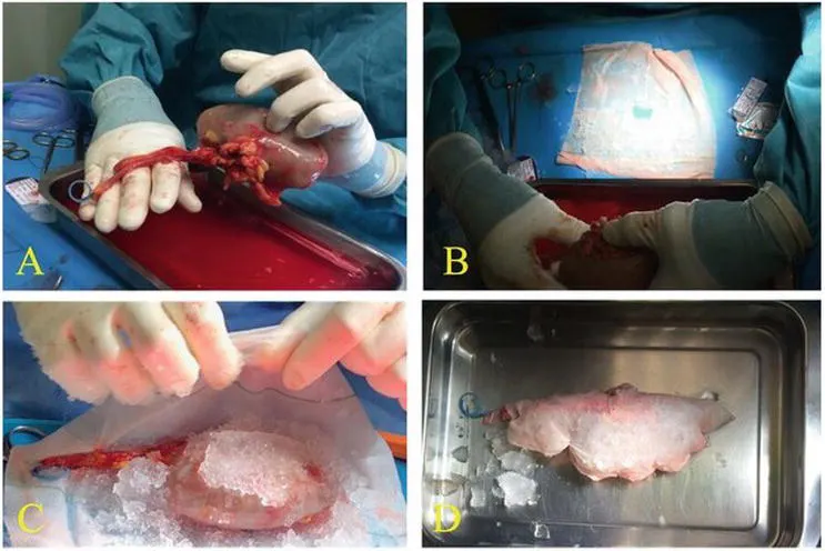
Figure 2.
Preparation of the kidney graft after nephrectomy during RAKT from living donors. (A) Ureteral double J stent is placed in the graft. (B) A central hole in the gauze from which the artery and vein are outside. (C and D) The graft is wrapped in a gauze jacket filled with ice slush.
2.2.3 Robot-assisted living donor kidney transplantation
2.2.3.1 Patient and trocar positioning
When using the da Vinci Si® or X® system, the patient is positioned in lithotomy position according to the Vattikuti-Medanta technique. When the da Vinci Xi® system is used, the patient is positioned in dorsal decubitus. A 20–30° Trendelenburg position is recommended. The required robotic instruments are monopolar scissors, Potts scissors, bipolar forceps, prograsp forceps, large needle driver, black diamond micro-forceps, and bulldog clamps. A 12 mm camera port is inserted in the supra-umbilical area, and a pneumoperitoneum is created. Veress needle puncture, optical trocar access, or the Hasson technique can also be used for access to the abdomen and creation of a pneumoperitoneum. The open approach (Hasson technique) has been reported to result in fewer complications. Three extra robotic 8 mm ports are placed under vision, and the robot is docked. Minimal changes in port placement are related according to the robotic system used (Figure 3). A GelPOINT® device replaces the camera trocar through a 6–8 cm (four fingers) periumbilical incision (Figure 4) once the transplant bed preparation has been performed. Alternatively, the GelPOINT® device can be introduced from the beginning through a 6–8 cm periumbilical incision, containing the camera and an assistant port. This GelPOINT® device is used to introduce the graft in the abdominal cavity (Figure 4) and allows for insertion of slushed ice (± 200 ml) via modified Toomey syringes into the abdominal cavity, surrounding the graft surface with the intent to achieve regional hypothermia (i.e., low constant temperature (<20°C) of the graft). Additionally, GelPOINT® is a useful device for fast hand introduction if needed (i.e., in case of massive bleeding). In selected cases, the graft can be introduced transvaginal as described by few authors. The AirSeal® (Conmed, Utica, NY, USA) system might be used in order to maintain a stable and low-pressure pneumoperitoneum at 8 mmHg.
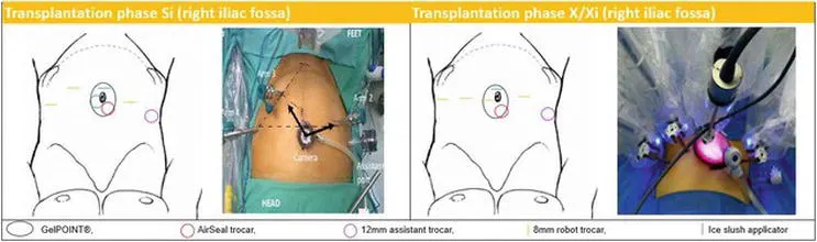
Figure 3.
Trocar placement and patient positioning for RAKT for Si/X/Xi in the right iliac fossa. Patient repositioned in dorsal decubitus, legs in Allen stirrups, table in 20–30° Trendelenburg; GelPOINT® device at the level of the umbilicus through a 6–8 cm vertical peri-umbilical incision; camera trocar and ice applicator in the GelPOINT® (eventually with 12 mm AirSeal® port); 3–8 mm robotic trocars in the lower abdomen, 2 in the left fossa and 1 in the right iliac fossa.
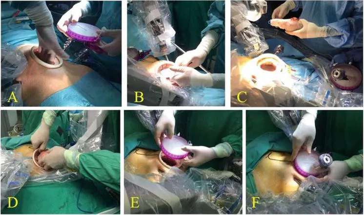
Figure 4.
Introduction of the kidney and ice through the Gel-POINT®. (A) The GelPOINT® device is placed through a 6 cm (four fingers) incision. (B) Ice slush is introduced in the abdominal cavity using modified Toomey syringes. (C and D) The graft is introduced into the abdominal cavity. (E and F) Once the graft is inside, Gel-POINT® cup is inserted to close the abdomen.
2.2.3.2 Transplant bed preparation
Accurate dissection of the external iliac vessels is performed. Subsequently, the bladder is prepared for ureteral reimplantation. A retroperitoneal pouch is created by incision of the peritoneum following a transverse line above the level of the appendix and mobilization of the peritoneal flaps. These will be used to cover (retroperitonealize) the graft once the vascular anastomosis is completed. Although RAKT is a transperitoneal approach, retroperitonealization of the kidney is performed to avoid pedicle torsion and to enable future graft biopsies.
2.2.3.3 Venous and arterial anastomosis
After clamping of the external iliac vein with robotic bulldog clamps and the distal clamp followed by the proximal clamp, a longitudinal venotomy using cold scissors is performed. An end-to-side anastomosis between the graft renal vein and the external iliac vein is created, using a 6/0 Gore-Tex® CV-6 TTc-9 or THc-12 needle (W.L. Gore and Associates Inc., Flagstaff, AZ, USA) continuous suture. At the proximal angle, the suture is tied to secure the posterior wall of the anastomosis watertight and to avoid stenosis, and then the continuous suture is completed until the distal angle. Prior to finishing the anastomosis, the lumen is flushed with heparinized solution using a 4.8 Fr ureteric catheter. The catheter may be pulled out by the assistant from outside the abdomen while the surgeon tightens the knot to secure the anastomosis (Figure 5). Next, the graft vein is clamped, and the bulldog clamps are removed from the external iliac vein and positioned on the external iliac artery, first proximally and then distally. The artery may be incised with the cold scissors or a scalpel at the 1–2 o’clock position. Arteriotomy may be completed using a laparoscopic aortic punch to transform the linear arteriotomy into a circular one. In both arterial and venous anastomosis, the anastomosis is started by passing the needle in the external iliac vessel in an outside-inside direction and then inside-outside through the graft vessel (Figure 6). For the venous anastomosis, the knot is tied now, and the needle is then passed outside-inside through the renal vein to start the running suture. For the arterial anastomosis, the suture is not tied yet (as for the venous anastomosis), and the needle is passed through the graft artery outside-inside before tying the suture to a loop that is left outside. This is done to prevent a difficult first needle passing in a small arterial lumen. After completing the arterial anastomosis, a clamp is positioned on the graft artery while the external iliac artery is declamped. If no sign of leakage (bleeding) is observed, the graft vein and artery are declamped. The evaluation of the graft perfusion is primarily visual: pink colorization, a pulsatile graft artery, filling of the renal vein, and small bleedings from the renal capsule and urine output are signs of perfusion. Doppler ultrasound evaluation (drop-in ultrasound probe linked to TilePro™ function of the da Vinci Surgical System) is recommended to verify adequate perfusion of the graft.
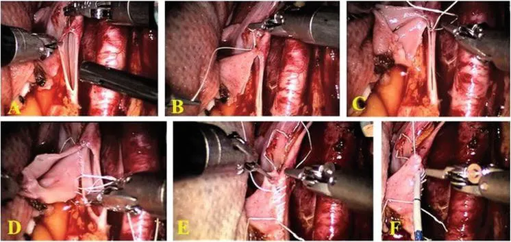
Figure 5.
Overview of the main steps for venous anastomosis during RAKT from living donors. (A) The graft renal vein is anastomosed in an end-to-side continuous fashion to the external iliac vein using a 6/0 Gore-Tex®. (B and C) At cranial angle, the suture is knotted to fix the posterior wall of the anastomosis. (D and E) The running suture is completed at the caudal angle. (F) Before completing the anastomosis, the lumen is flushed with heparinized solution using a 4.8 Fr ureteral catheter.
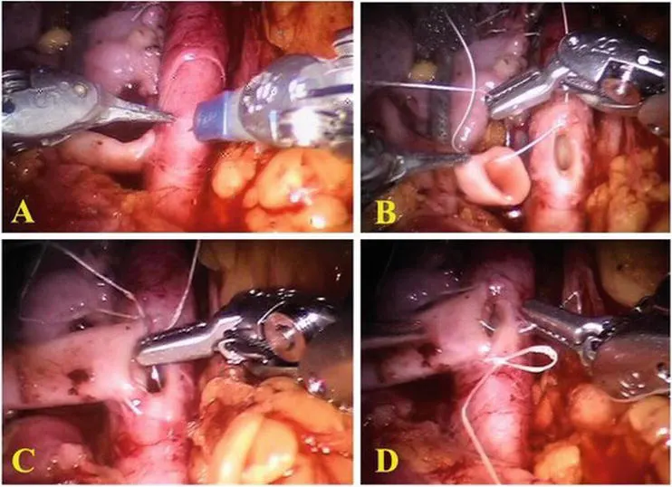
Figure 6.
Overview of the main steps for arterial anastomosis during RAKT from living donors. (A) The robotic scalpel is used to make a linear incision on the iliac artery, converting it in circular hole with a laparoscopic vascular punch. (B) The running suture is carried out using a 6/0 Gore-Tex®; particularly in the caudal tying of an arterial anastomosis, the needle is passed in the external iliac vessel in an outside-inside direction, then outside-inside through the graft vessel. (C and D) The running suture is completed at the caudal angle.
2.2.3.4 Ureteroneocystostomy
After flipping the kidney on the psoas and retroperitonealization of the graft, the ureteroneocystostomy is performed according to the Lich-Gregoire technique using a Monocryl or PDS 5/0 (Ethicon Inc., Cincinnati, OH, USA) continuous suture (Figure 7). Care is taken to construct an adequate detrusor tunnel as anti-reflux mechanism. A double-J stent is inserted to protect the anastomosis. The stent can be removed after 3 weeks.
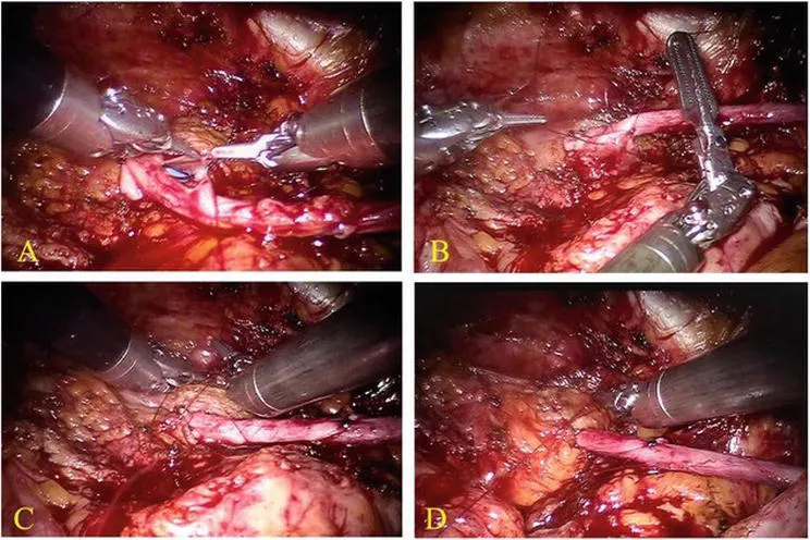
Figure 7.
Ureteroneocystostomy performed according to the Lich-Gregoir technique. In (A) and (B) running suture between ureteral and bladder mucosa using 5-0 Monocryl. In (C) and (D) the details of the anti-reflux tunnel.
2.3 Results
After reporting a single center experience on 17 cases of RAKT from living donation, the European Experience in RAKT was published in 2018. One hundred twenty cases were prospectively collected in eight centers across Europe. The authors demonstrated the low complication rate (at 1 month follow-up) while maintaining excellent graft function (median eGFR at 30 days was 58 ml/min) and cosmetic results (Figure 8). Three cases of graft loss due to arterial thrombosis during the first postoperative week were reported in these series (2.5%). This complication might be associated with technical errors during the learning curve. Territo et al. demonstrated that the functional results at 1-year follow-up were not statistically different from the functional results at 1-month follow-up. The complication rate remained low. To date, there are no studies available comparing RAKT with the conventional open approach. However, an increasing body of evidence confirms that RAKT is at least non-inferior to open KT regarding both patient and graft survival.
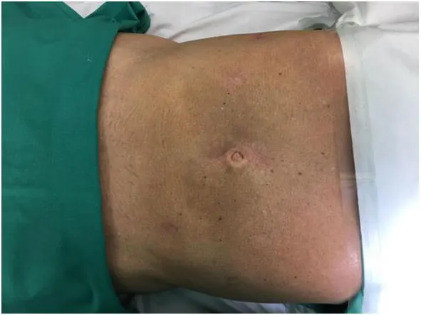
Figure 8.
Esthetic results of RAKT 3 months later.
3. Robot-assisted kidney transplantation (RAKT): deceased donor
3.1 Background
The vast majority of RAKT worldwide has been performed from living donors, raising concerns regarding the generalizability of RAKT outcomes in the broader and more challenging scenario of deceased donors.
While it has been shown that the learning curve for elective RAKT (i.e., living donor RAKT) may be minimal for surgeons with extensive experience in robotic surgery (regardless of their background in open KT) , RAKT from deceased donors has unique technical and logistical challenges. Indeed, due to the timeframe of organ preservation, it can be considered an unforeseeable “emergency” robotic procedure, which requires a structured multidisciplinary framework.
3.2 Development of a structured RAKT program from deceased donors
To fill this gap and move the field forward, the University of Florence group has recently developed a RAKT program from deceased donors aiming to safely and progressively increase the pool of patients who may benefit from minimally invasive KT.
The cornerstones of this program are as follows: (a) an extensive experience in open KT (n > 1100 KTs from 1991) and robotic urologic surgery (>4000 procedures from 2010, including radical prostatectomy, radical nephrectomy, partial nephrectomy, radical cystectomy, dismembered pyeloplasty, and ureteral reconstructive surgery); (b) a codified technique for RAKT; (c) a structured modular training in RAKT, including e-learning, simulation, and dry lab and wet lab training on animal models; (d) the availability of a multidisciplinary team (composed by urologists, anesthesiologists, nephrologists, radiologists, as well as operating room support staff and nurses) with experience in KT and robotic surgery; and (e) the opportunity to perform RAKT at nighttime and/or during the weekend in a dedicated operating room.
3.3 Selection criteria for RAKT from deceased donors
Following the University of Florence, recipient’s exclusion criteria for RAKT from deceased donors include the following: (a) age < 18 years; (b) severe comorbidities with contraindication for robotic surgery; (c) significant atherosclerotic plaques at the level of external iliac vessels; (d) highly complex vascular graft anatomy (likely to require multiple anastomoses); (e) multiple previous abdominal surgeries; and (f) previous KT. A key element of the RAKT program from deceased donor is also represented by the decision-making process aiming to assess the feasibility of RAKT in light of the patient-, graft-, and robotic team-related factors (Figure 9). As such, the decision to proceed with an “emergency” RAKT from a deceased donor relies on a careful balance of the potential advantages of robotic surgery (for both the patient and the surgeon) and the logistical challenges of setting up the operating room and the robotic surgical team in a fixed timeframe, respecting the recipient’s selection criteria and the maximal thresholds of cold ischemia time. Specifically, planning RAKT from deceased donors follows a pre-specified decision-making process. First, after the alert of a kidney offer is addressed to our transplant team by the Regional Allocation Centre, the surgeon in charge considers the opportunity to perform RAKT according to his/her personal experience and the availability of an expert bedside assistant as well as an expert robotic operating room nursing staff. Then, after the recipient is admitted to the nephrology unit for the pretransplant clinical work-up (which systematically includes a computed tomography angiogram of the abdomen to check for severe atherosclerotic plaques at the external iliac vessels), the surgeon checks that the recipient’s inclusion criteria are met. Subsequently, the robotic operating room is checked for availability (even during weekends, at nighttime, and potentially during daytime, if elective non-oncologic robotic procedures can be safely rescheduled), ensuring that the bench surgery starts within 16 hours from the beginning of cold storage of the kidney. Finally, at the time of bench surgery, the graft is carefully inspected to ensure no exclusion criteria for RAKT is present (i.e., highly complex vascular anatomy requiring multiple vascular anastomoses).
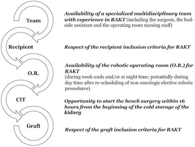
Figure 9.
Flow-chart depicting the decision-making process for RAKT from deceased donors.
3.4 The University of Florence technique for RAKT from deceased donors
A step-by-step overview of our surgical technique for RAKT from deceased donors is shown in Figures 10–12.
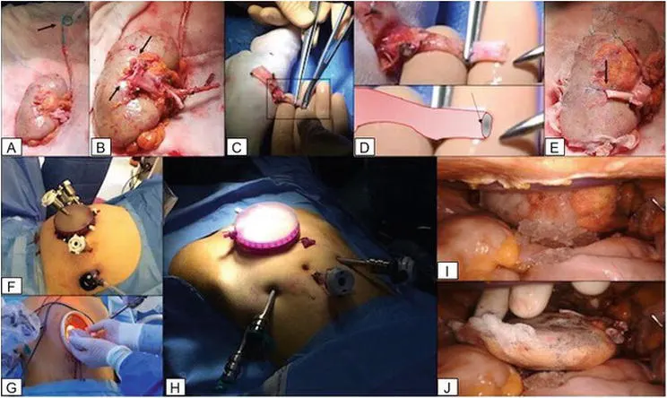
Figure 10.
Overview of the main steps for bench surgery (A-E), port placement (F-H) and insertion of the graft into the abdominal cavity (I, J) during RAKT from deceased donors.
All procedures were performed with the da Vinci Si® or Xi® robotic platform in a four-arm configuration with a zero-degree lens and one assistant port, by a single surgeon (G.V.) with extensive experience in robotic urologic surgery and open KT. The pneumoperitoneum pressure was set at 8 mm using the AirSeal® system, while the Trendelenburg was tilted at 20° in all cases.
The robotic instruments employed for RAKT included the following: (a) large needle driver; (b) black diamond micro-forceps; (c) monopolar curved scissors; (d) Maryland bipolar forceps; (e) prograsp forceps; and (f) robotic bulldog clamps. Laparoscopic instruments included a needle driver, the suction device, laparoscopic scissors, Hem-o-lok (Weck Surgical Instruments, Teleflex Medical, Durham, NC) clips and clip applier, Johan grasping forceps, and the drop-in ultrasound probe.
For insertion of the graft inside the abdominal cavity, we employ the GelPOINT® or the Alexis® wound retractor (Applied Medical, Rancho Santa Margarita, CA) through a Pfannenstiel incision (Figure 10). Regional hypothermia is achieved by cooling the graft with ice slush introduced through the GelPOINT® via modified Toomey syringes, as previously described.
Intraoperative indocyanine green (ICG) fluorescence videography (IFV) (i.e., FireFly® technology) is used to assess the graft and ureteral reperfusion.
Abdominal organ procurement is performed according to established principles for donors after brain death (DBD), while specific protocols were followed in the case of donors after circulatory death (DCD). In this setting, the kidneys are preserved in a hypothermic machine perfusion device (LifePort Kidney Transporter® (Organ Recovery Systems Inc. Chicago, USA)) before transplantation.
During bench surgery (Figure 10), the graft is perfused with Celsior® solution and inserted in a gauze jacket filled with ice (and closed with 4–5 stiches) after placement of a 6F, 14 cm double-J stent. Two landmark stiches are placed on the upper and lower sides of the graft vein, while one landmark stich is placed on the upper side of the graft artery, to facilitate the subsequent orientation of the graft inside the abdominal cavity. Finally, the graft artery is modeled to facilitate subsequent intracorporeal arterial anastomosis.
RAKT from deceased donors at our Institution follows the principles of the Vattikuti-Medanta technique, adopted by all other centers included in the ERUS-RAKT group.
Of note, this technique allows performance of a transperitoneal RAKT with final retroperitonealization of the graft after kidney reperfusion.
After induction of general anesthesia, port placement, and docking of the da Vinci Si/Xi robotic platform, the iliac vessels are dissected and prepared for subsequent anastomoses. Then, an extraperitoneal pouch is created, and access is gained into the Retzius space in order to prepare the bladder for subsequent ureteroneocystostomy.
Then, a Pfannenstiel incision is performed, and the GelPOINT® device or the Alexis® wound retractor is set in place. The graft is subsequently introduced into the abdominal cavity and the robot is redocked. After achievement of regional hypothermia, a venotomy is performed, the lumen of the external iliac vein is flushed with heparinized saline, and the venous anastomosis is completed in an end-to-side fashion using a 6/0 Gore-Tex suture on a CV-6 TTc-9 needle (Figure 11). After additional graft cooling, a liner arteriotomy is performed and converted into a circular arteriotomy with cold scissors. The renal artery is then anastomosed in an end-to-side fashion to the external iliac artery using a 5/0 Gore-Tex suture on a CV-6 TTc-9 needle with two running sutures (Figure 12).
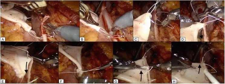
Figure 11.
Overview of the main steps for venous anastomosis during RAKT from deceased donors. (A) A 1–2 cm venotomy is performed on the external iliac vein. (B) The lumen of the external iliac vein is flushed with heparinized saline. (C–F) The graft renal vein is anastomosed in an end-to-side continuous fashion to the external iliac vein using a 6/0 Gore-Tex®. (G and H) The running suture is completed at the caudal angle.
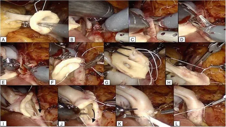
Figure 12.
Overview of the main steps for arterial anastomosis during RAKT from deceased donors. (A-E) A linear arteriotomy is performed and converted into a circular arteriotomy with cold scissors. (F-L) The renal artery is anastomosed in an end-to-side fashion to the external iliac artery using a 5/0 Gore-Tex suture on a CV-6 TTc-9 needle with two running sutures.
After the integrity of the anastomoses is tested, the graft is revascularized and inspected for color and turgor. Intraoperative Doppler ultrasound with the TilePro® feature is employed to check for graft reperfusion in case the da Vinci Si robotic platform is used. On the contrary, if the da Vinci Xi® platform is available, graft and ureteral reperfusions are assessed using intraoperative indocyanine green (ICG) fluorescence videography (IFV). Then, the graft is allocated in the extraperitoneal pouch, which is partially closed by reapproximating the previously prepared peritoneal flaps with a V-lock running suture or single Hem-o-lok clips. This step avoids graft torsion, facilitates postoperative graft monitoring, and allows convenient access for ultrasound-guided transplant renal biopsy and/or placement of nephrostomy tubes, if needed. Interestingly, we recorded a laminar perirenal fluid collection in almost all patients at ultrasound examinations during the first postoperative period (i.e., postoperative days 1 and 7 and at hospital discharge). However, subsequent follow-up evaluations showed a progressive reduction of this fluid collection until complete reabsorption within 20–30 days after surgery, confirming the complete retroperitonealization of the graft.
The last step of the procedure is the ureterovesical anastomosis, which is performed according to a modified Lich-Gregoire technique over the double-J stent. The ureter is anastomosed to the bladder mucosa with two 4.0 Monocryl running sutures; then the detrusor muscle is closed creating an anti-reflux mechanism.
The technique for RAKT from deceased donors evolved over time throughout the learning curve. In particular, few technical nuances have been introduced, according to the surgeon’s preference and skills, with the aim to improve the esthetic result and the cost-effectiveness of RAKT, as well as to optimize critical steps such as the assessment of graft and ureteral reperfusion before ureterovesical anastomosis.
These technical modifications include insertion of the graft into the abdominal cavity through a Pfannenstiel incision (rather than a periumbilical incision), to improve the esthetic result of RAKT and facilitate the positioning of the graft in the pelvis close to the iliac vessels.
Second, the placement of the GelPOINT® device is achieved after preparation of iliac vessels, bladder, and extraperitoneal pouch, to make sure not to injure the bladder while positioning the GelPOINT® device through the Pfannenstiel incision. Of note, in the case of recipients with autosomal dominant polycystic kidney disease (ADPKD), all ports are positioned approximately 3 centimeters downward on the same lines to obtain an increased working space in the right iliac fossa far from the enlarged polycystic kidneys.
While mirroring the Vattikuti-Medanta technique, the steps of venous and arterial anastomoses differ slightly from those originally described by the ERUS-RAKT group. In all cases of right-sided grafts, an inferior vena cava (IVC) cuff is used during bench surgery to increase the length of the graft renal vein to facilitate the subsequent venous anastomosis. However, according to our experience in RAKT using right-sided grafts from living donors, the use of an IVC cuff does not appear to be mandatory, as the robotic platform facilitates the performance of a tension-free venous anastomosis even in case of short renal veins.
The venotomy is performed using the standard curved scissors (rather than the Potts scissors), and the venous anastomosis is completed in a two-step fashion: first, the posterior plate is closed using a running suture from 12 to 6 o’clock position; then, a knot is tied, and the anterior plate is completed with a second running suture with the same thread from 6 to 12 o’clock position (Figure 11). Tying the knot at 6 o’clock position after completion of the posterior plate may be key to reduce the tension on the anterior plate of the anastomosis, tailoring it according to the graft vein shape and length.
Similarly, the technique for arterial anastomosis follows the principles of the Vattikuti-Medanta technique with key technical nuances in case of deceased donors. First, the arteriotomy is achieved using standard cold scissors (rather than using the aortic punch). Second, due to the increased risk of atherosclerotic plaques and/or calcifications at the level of the external iliac arteries, the arterial anastomosis is performed using two different threads (Figure 12). First, the posterior plate of the anastomosis is closed using a running suture from 12 to 6 o’clock position, without tying a knot at 6 o’clock position. Then, the anterior plate is closed in a similar fashion with a running suture from 12 to 6 o’clock position using a second thread. Finally, the two free ends of the threads are tied together at 6 o’clock position, after checking the integrity of the anastomosis. This technique allows to tailor the tension of the anastomosis according to the stiffness and characteristics of both the renal and iliac vessels.
Third, the arterial anastomosis during RAKT from deceased donors is performed using a slightly thicker thread (Gore-Tex 5/0 instead of 6/0).
A specific challenge in case of RAKT from deceased donors is represented by the management of the Carrel’s patch for arterial anastomosis. At the beginning of our robotic program for deceased donor RAKT, after careful bench surgery showing no significant atherosclerotic plaques at the level of the renal artery ostium, we performed the anastomosis using the Carrel’s patch (Figure 12). However, with increasing experience the anastomosis is increasingly being performed without the Carrel’s patch. While in few cases at the beginning of the program, the surgeon had to remove the patch due to the presence of significant atherosclerotic plaques, in the following cases, the Carrel’s patch was removed intentionally during bench surgery, and the anastomosis was performed mirroring the technique adopted for living donor RAKT. From a technical point of view, and thanks to the robotic platform, removing the Carrel’s patch may provide significant advantages for the surgeon. These include (a) the opportunity to perform a shorter arteriotomy; (b) a more anatomic anastomosis, thanks to the similar caliber of the graft renal artery and external iliac artery; and (c) the reduced risk of atherosclerotic plaques at the level of the graft renal artery (as compared to Carrel’s aortic patch).
Finally, a specific technical modification proposed by our group for RAKT from deceased donors is represented by the use of intraoperative indocyanine green (ICG) fluorescence videography (IFV) to assess graft and ureteral reperfusion. The tips and tricks for the use of this technique during RAKT from deceased donors are discussed in the previous publications. In brief, a bolus of 0.3 mg/kg of ICG dissolved in 5% glucose (2 mg/ml) is administered intravenously after completion of vascular anastomoses to allow performance of a real-time qualitative and potentially quantitative assessment of graft and ureteral fluorescence signal. Forty seconds after ICG injection, the camera is switched to reveal fluorescence at the level of vascular anastomoses and the renal parenchyma and ureter to check for their reperfusion, allowing to adapt the ureteral length according to the fluorescence signal to reduce the chance of postoperative ureteral strictures.
3.5 RAKT from deceased donors: preliminary results
Overall, 32 RAKTs have been performed at the University of Florence from January 2017 (22.4% of all KT in this period).
The first RAKT from a deceased donor was performed in August 2017. Of the 32 RAKT performed so far, 19 (59.4%) were from deceased donors (n = 5 [26%] from DCD donors; n = 14 [74%] from DBD donors).
Interestingly, thanks to the structured RAKT program developed at the University of Florence, the proportion of RAKT increased over time in the last 3 years, being 18.3%, 26.4%, and 24% of all KT in 2017, 2018, and 2019, respectively. Similarly, the proportion of RAKT from deceased donors increased over time, being 11.1% in 2017, 19.6% in 2018, and 14.3% in 2019 of all KT from deceased donors.
The University of Florence group very recently published their results on their first 17 RAKTs from deceased donors. They now performed 19 cases, all successfully completed without need of open conversion.
The da Vinci Si and Xi robotic platform was used in 15 (79%) and 4 (21%) cases, respectively. In the vast majority of cases (n = 17), RAKT was performed in the right iliac fossa.
The graft was introduced using the GelPOINT® device (or the Alexis® port) in 14 (73.7%) cases through a Pfannenstiel incision, while in 5 (26.3%) through a periumbilical incision.
They recorded one case of intraoperative complication (intraoperative bleeding not requiring transfusion) that required positioning of an additional 5 mm port to increase exposure and help aspiration. Three patients (16%) suffered a high-grade (Clavien-Dindo grade 3) complication (transplant renal artery stenosis requiring percutaneous angioplasty in one patient; percutaneous placement of a nephrostomy tube for hydronephrosis in one patient and transplant renal artery thrombosis requiring graft nephrectomy).
A progressive improvement of renal function was recorded at all time points during the postoperative period. Median eGFR at hospital discharge (median 12 days post-op) was 47.2 ml/min/1.73m2 (IQR 28.9–59.4), while median eGFR at a median follow-up of 15 months was 58.6 ml/min/1.73m2 (IQR 40.0–80.4).
Overall, five (26%) patients required dialysis during the first postoperative week. Of these, one patient due to primary nonfunction after RAKT from an uncontrolled DCD donor; one patient due to graft nephrectomy due to arterial thrombosis; one patient for suspected acute rejection treated with intravenous corticosteroids; and two patients for DGF.
At a median follow-up of 15 months, all patients are alive, and two patients are still on dialysis.
3.6 RAKT from deceased donors: future perspectives
We developed the first structured program of RAKT from deceased donors worldwide.
Despite being preliminary, our experience confirms the feasibility of RAKT in this donation setting in centers with a solid background in open KT and robotic surgery and after proper standardized modular training.
From a logistical perspective, RAKT from deceased donors poses specific challenges, which require a comprehensive, multidisciplinary effort to redesign the management strategy of the institution’s transplantation pathway. In this view, a highly trained and committed surgical team, as well as the opportunity of a dedicated flexible robotic operating room, are key elements for the success of the program.
The perioperative and functional data in our preliminary series are promising but warrant further investigation. In particular, larger studies with longer follow-up are needed to confirm the safety of RAKT from deceased donors (especially DCD donors), to evaluate the impact of learning curve on patient outcomes, and to identify the predictors of adverse perioperative events (i.e., major surgical complications, PNF and DGF) to refine patient selection.
Yet, extending the number of robotic transplantations performed by centers experienced in robotic surgery using grafts from deceased donors would be key to increase the pool of patients who may benefit from minimally invasive surgery. Notably, this is a critical step toward the definition of evidence-based indications for RAKT in this clinical scenario also from a Guideline’s perspective.
We entirely share Dr. Alcaraz and colleagues’ perspective that “although major improvements in outcomes for transplant patients are likely to come from the field of immunology, small, measurable improvements may yet be possible via technical advances”. As such, we believe RAKT, which is likely to be increasingly performed by referral transplant centers worldwide, may provide unique opportunities for both patients and surgeons, allowing to improve the precision and reduce the morbidity of this procedure. To move the field forward, implementing RAKT programs from deceased donors is the most compelling clinical unmet need for the transplant and urological community.
4. Robot-assisted kidney autotransplantation (RAKAT)
4.1 History and background
In 1902 Ullmann and Carrel experimentally achieved the first successful animal kidney autotransplantation (KAT), moving the kidney of a dog from its lumbar fossa to its neck, in this way laying the foundations for future kidney (auto)transplantation. In 1956, the Brazilian Campos Freire attempted KAT for the first time on a man with a renal artery aneurysm, although an early thrombosis forced him to perform a nephrectomy. In 1961, Shackman and Dempster repeated this in a case of unilateral renal artery stenosis but with unsatisfactory results. It was not until 1963 when JD Hardy et al. achieved the first successful KAT in human in a case of high ureteral stricture after a large aortic aneurysm repair. McLaughlin et al. demonstrated its utility in the management of complex renal lesions. Following these pioneering surgeries, KAT was adopted as a method to perform renal artery angioplasty in treating hypertension caused by severe renal artery stenoses. In these vascular cases, the ureter was usually not transected; the vascular pedicle is reconstructed in or close to the abdominal incision with subsequent vascular reimplantation on the iliac vessels, making this actually a renal transposition. Only when the indication concerns primary ureteral pathology or when the kidney needs complex surgery on the bench table (and both the vascular pedicle and the ureter are sectioned and subsequently reimplanted), the term KAT is correct. Nowadays, most renovascular problems are treated with endovascular methods, and the indications for KAT have been shifted more toward managing complex ureteral strictures or malignant pathologies for which endoluminal or in vivo repair is impossible or contraindicated. Less frequent indications include loin pain hematuria syndrome, retroperitoneal fibrosis, and metabolic stone disease.
KAT is an important surgical last resort technique in order to spare the kidney in select cases. It allows ex vivo management of complex renal or ureteral pathology, which would not be treatable with conventional techniques, without resulting in important kidney function loss. For this purpose, the harvested kidney is cooled on ice slush and flushed with an ice-cold preservation solution during bench surgery, thus reducing tissue oxygen requirement.
Traditionally, KAT has been performed through a large midline laparotomy, a large paramedian extraperitoneal incision or the combination of a lumbotomy for the nephrectomy phase with a lower abdominal incision for the transplantation phase; thus being a very invasive procedure, and, albeit having a low mortality rate (1.3%), postoperative morbidity may be as high as 46.2%. Due to the historic invasiveness of the procedure, and the unfamiliarity of many urologists with the field of KT, KAT has never gained much popularity and often these kidneys were sacrificed. However, since the introduction of laparoscopy in urology, attempts have been made to reduce the morbidity of open KAT. Fabrizio et al. described in 2000 the first laparoscopic nephrectomy for KAT; however, the patient still required a periumbilical incision for extraction of the graft and a pelvic incision for the transplantation. Today, the most accepted approach is to perform laparoscopic nephrectomy, via a three-port transperitoneal access, and open KAT, using the classical Gibson incision for both extraction and introduction of the graft, resulting in possible low complication rate and excellent long-term autograft function.
In recent years, due to the success of RAKT, interest is rising to reevaluate KAT as an ultimate nephron-sparing option in complex cases. In 2014, Gordon et al. published the first case of robot-assisted kidney autotransplantation (RAKAT) to treat extensive ureteral loss after complicated ureterorenoscopy for ureterolithiasis. They used a completely intracorporeal technique, establishing in vivo hypothermic renal perfusion by continuous renal artery irrigation. The robot was redocked between nephrectomy and transplantation phase, and the table was repositioned rather than the patient. The complete intracorporeal technique was successfully repeated by Lee et al. in 2015. Despite these promising results, the totally intracorporeal technique is not suitable in patients where ex vivo bench repair is required (e.g., multiple vessel grafts, complex oncological cases, lithiasis treatment) or where the surgeon would prefer to transplant the autograft in the contralateral iliac fossa (e.g., disturbed pelvic vascular anatomy or excessive fibrosis). In the same period, Sood et al. theoretically described a technique for RAKAT using extracorporeal tabletop graft reconstruction, using a GelPOINT device, in order to broaden indications of RAKAT. In 2017, Araki et al. published a case using this extracorporeal RAKAT technique in a patient with proximal ureteral stenosis. Also in 2017, Decaestecker et al. performed the first extracorporeal RAKAT in Europe and to date have the largest series of RAKAT worldwide. This European series also adopted a totally intracorporeal technique.
4.2 Operative technique
Decaestecker et al. published the largest series of RAKAT worldwide, all for non-oncological cases. Their technique is based on the experience within the ERUS-RAKT working group, as described by Breda et al., and on the theoretical report by Sood et al. We will describe the extracorporeal bench work technique and the totally intracorporeal technique with the da Vinci Si® and Xi® robotic platform.
4.2.1 Extracorporeal bench work technique (video)
4.2.1.1 Trocar and patient position
Figures 13 and 14 describe the patient and trocar positioning for Si and Xi, right or left, during nephrectomy and transplantation phase.
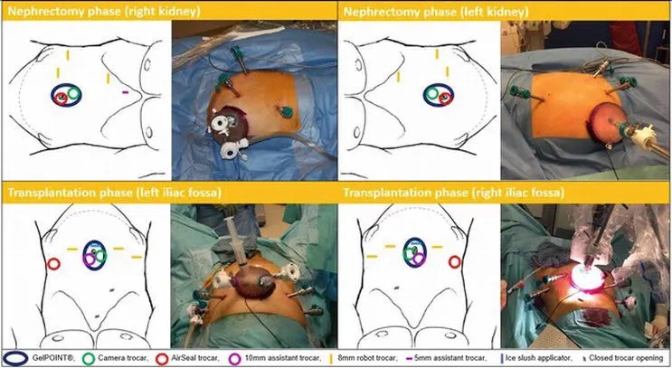
Figure 13.
Trocar and patient positioning for extracorporeal RAKAT using Si. Nephrectomy phase: patient in lateral decubitus; GelPOINT® through a 6 cm vertical peri-umbilical incision containing camera and assistant trocar; three 8 mm robot trocars in hemiabdomen, one subcostal and 2 in the iliac fossa. Transplantation phase: patient repositioned in lithotomy position, legs in Allen stirrups, table in 20–30° Trendelenburg; three 8 mm robotic trocars in the lower abdomen, reusing the 2 former iliac fossa trocar sites. Low-pressure pneumoperitoneum realized with the AirSeal®System.
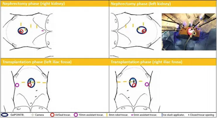
Figure 14.
Trocar and patient positioning for extracorporeal RAKAT using Xi. Nephrectomy phase: patient in lateral decubitus; GelPOINT® through a 6 cm vertical peri-umbilical incision containing robot and assistant trocar; three 8 mm robot trocars in hemiabdomen on a slightly oblique line, 2 subcostal and 1 in the iliac fossa. Transplantation phase: patient repositioned in lithotomy position, table in 20–30° Trendelenburg; three 8 mm robotic trocars in the lower abdomen, reusing 1 former iliac fossa trocar site. Low-pressure pneumoperitoneum realised with the AirSeal®System.
4.2.1.2 “Donor” nephrectomy
A “donor” nephrectomy is performed with the patient in the lateral decubitus position, maximizing the renal vessel length and transecting the ureter just proximal of the strictured segment or at the level of the crossing with the iliac vessels. After administering 2500–5000 units of heparin intravenously according to the weight of the patient, the renal vessels are transected after securing the vessels with either a laparoscopic/robotic vascular stapler or double clipping the vessels with Click’aV plus® clips (Grena, Amsterdam, the Netherlands, Europe), followed by a transfixing ligation of the clipped vascular stump with Prolene® 5/0 (Ethicon Inc., Johnson & Johnson Corp, Cincinnati, OH, USA) to prevent clip slipping. The latter is done robotically after the kidney is exteriorized by the table-side assistant through the GelPOINT®.
4.2.1.3 Bench work
Upon retrieval, the graft is immediately perfused on the bench with 4° Celsius preservation solution (Institut Georges Lopez-1®). If necessary, bench vascular reconstruction can be performed for duplicated renal arteries (end-to-side reconstruction of the lower pole to main artery or pantaloon reconstruction of two central arteries). During cold storage, tabletop flexible ureterorenoscopy or nephroscopy can be performed to extract nephro- or ureterolithiasis. The kidney is wrapped in an ice gauze jacket with a central hole exposing the renal hilum (Figure 15).
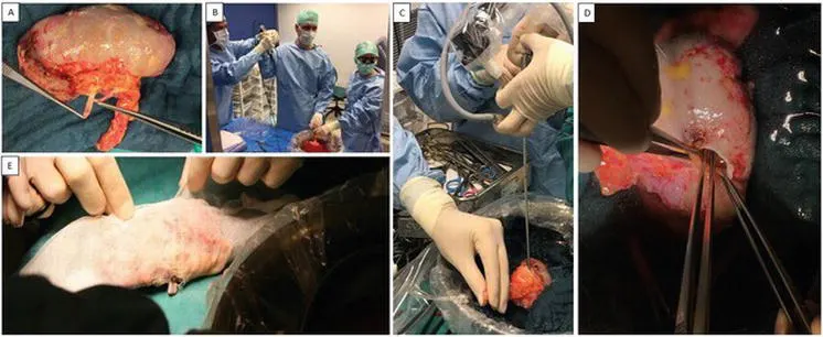
Figure 15.
Bench work in extracorporeal RAKAT technique. (A) End-to-side anastomosis of lower pole artery on main renal artery. (B) Extraction of nephrolithiasis. (C) Nephroscopy. (D) Extraction of lithiasis from calyceal diverticulum and closing diverticulum infundibulum. (E) Wrapping of the kidney in ice gauze jacket, leaving opening for the structures.
4.2.1.4 Transplantation phase
During cold storage, the robot is undocked, the incisions are temporarily closed, and the patient is repositioned in lithotomy position (Si/X) or dorsal decubitus (Xi). For the transplantation phase, the robot is redocked, and RAKT is performed following the Vattikuti-Medanta technique as previously described and adopted by the ERUS-RAKT group. With the extracorporeal technique, the kidney is usually transplanted to the contralateral iliac fossa unless the proximal ureter of the graft is absent or very short and urges a ureteropyelostomy (native distal ureter to transplant pyelum) or uretero-ureterostomy (native distal ureter to transplant ureter). The iliac vessels can be very hard to dissect due to excessive fibrosis caused by previous surgery and/or radiotherapy.
4.2.2 Totally intracorporeal technique
4.2.2.1 Selection of patients
The totally intracorporeal technique is not suitable in patients where ex vivo bench repair is required (e.g., multiple vessel grafts, complex oncological cases, lithiasis treatment) and might be less optimal in cases where the surgeon would prefer to transplant the autograft in the contralateral iliac fossa (e.g., disturbed pelvic vascular anatomy or excessive fibrosis).
4.2.2.2 Trocar and patient position
Figures 16 and 17 describe the patient and trocar positioning for Si and Xi, right or left, during nephrectomy and transplantation phase. Note that the GelPOINT® device is not used and as much trocars as possible are reused, although some need to be exchanged for each other. For the transplantation phase, the patient is positioned on the PinkPad® (Kebomed, Apeldoorn, the Netherlands, Europe) in a modified lateral flank position using inflatable pressure bags. Together with maximal side tilting of the table (15°), the patient is in a 65° flank position which makes nephrectomy feasible. After nephrectomy and intracorporeal flushing, the robot needs to be undocked, and the table is repositioned rather than the patient (from maximal side tilt to contralateral maximal side tilt, partially) deflating the pressure bags to bring the patient more or less horizontal and adding 20–30° of Trendelenburg). Using the Si/X system, the robot needs to be repositioned from flank-docking tot side-docking; using the Xi system, only the boom needs to be rotated 90° before redocking. In case of expected pelvic fibrosis, it is wise to prepare the iliac vessels before clamping the renal hilum in order to reduce intracorporeal “cold” ischemia time as well as the amount of intracorporeal cold fluid that could potentially lower central body temperature. This means that three phases with two repositions are necessary. The Xi system with easy docking and repositioning of the boom and integrated table motion is a real advantage for these challenging cases.
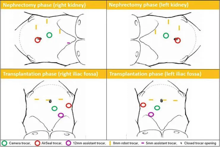
Figure 16.
Trocar and patient positioning for RAKAT intracorporeal technique Si. No use of GelPOINT® device. Nephrectomy phase: patient positioned in lateral decubitus; camera trocar supra-umbilical, three 8 mm robot trocars in hemiabdomen, AirSeal trocar as assistant trocar infra-umbilical. Transplantation phase: robot undocked, table repositioned rather than the patient, and robot redocked (side-docking next to left leg); three 8 mm robotic trocars in the lower abdomen, reusing 1 former caudal robot trocar site and AirSeal trocar site; AirSeal in flank position reusing former robot trocar site; assistant trocar subcostal reusing former robot trocar site.
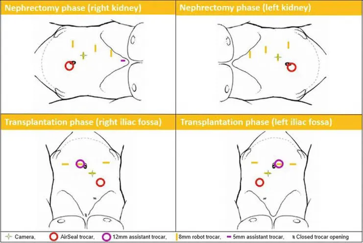
Figure 17.
Trocar and patient positioning for RAKAT intracorporeal technique Xi. No use of GelPOINT® device. Nephrectomy phase: patient positioned in lateral decubitus; camera trocar supra-umbilical, three 8 mm robot trocars in hemiabdomen, AirSeal trocar as assistant trocar infra-umbilical. Transplantation phase: robot undocked, table repositioned rather than the patient, and robot redocked (side-docking next to left leg); three 8 mm robotic trocars in the lower abdomen, reusing 1 former caudal robot trocar site and AirSeal trocar site; AirSeal in flank position reusing former robot trocar site; assistant trocar subcostal reusing former robot trocar site.
4.2.2.3 “Donor” nephrectomy
During nephrectomy, more fat is left on the lower pole of the kidney in order to be able to manipulate the kidney intracorporeally. Care should also be taken to skeletonize the renal vessels and gain adequate length as would normally be performed on the bench. After clamping the renal vessels, they are not transected, but incised to introduce a 5–7 Fr Fogarty catheter with open tip to flush the kidney intracorporeal with 4°C physiologic NaCL solution until the effluent is clear. Note that any solution containing potassium should not be used in order to prevent hyperkalemia and possible arrhythmias by peritoneal reabsorption. After flushing, the renal vessels are transected, and the Fogarty is blocked in the renal artery lumen with inflating the balloon with 0.5–1 cc. The kidney is now continuously flushed at a low flow rate with the intention of intravascular cooling but preventing lowering body temperature by excessive flushing. Effective flushing can be confirmed continuously by looking for renal vein effluent. The kidney is repositioned from flank to pelvis, and the robot is redocked for table repositioning.
4.2.2.4 Transplantation phase
The venous anastomosis is performed in the same way as for the extracorporeal technique. Flushing (and graft cooling) through the renal artery is continued until the venous anastomosis is complete. The Fogarty is removed, and the arterial anastomosis is performed, resulting in only 10––15 min of rewarming time. With the intracorporeal technique, the kidney is usually transplanted to the ipsilateral iliac fossa.
4.2.3 Results
Decaestecker et al. published their initial experience on 7 patients, updated their results of the first 10 cases at EAU 2019 congress, and now performed 15 cases, making this the largest reported series worldwide. Thirteen cases were performed extracorporeally and two intracorporeally. Ureteral stricture disease was the main indication (12/15). All 15 RAKATs were successfully completed without intraoperative complications needing open conversion. Compared to pre-op, there was a significant decrease in mean serum creatinine (p = 0.027) and a nonsignificant increase of mean overall and autotransplant GFR estimated by nuclear DMSA and Cr-EDTA scans at 3 months post-op. All patients were free from indwelling catheters or nephrostomy tube, recurrent urinary tract infections, debilitating stent symptoms, flank pain, or macroscopic hematuria at the last follow-up. Short hospital stay and early recovery confirmed the minimal invasiveness of the approach although 3/15 patients experienced a high-grade complication (pulmonary embolism, wound dehiscence, and lower limb compartment syndrome without lasting disability). Of note is that the high-grade complications occurred in former cancer patients that had the combination of multiple surgeries and radiotherapy.
5. Learning curve and training
The learning curve for RAKT can be relatively short for surgeons experienced in robotic surgery and kidney transplant surgery. It cannot be stressed enough that a high level of robotic experience is recommended before starting this kind of high-stake surgery. Training the technique on dry and wet lab models is mandatory. The structured RAKT course provided at ORSI Academy is recommended as well as an experienced RAKT proctor supporting first cases. In this way a safe introduction of this new technique is possible.
Conclusions
RAKT has been proven to be an advanced application for KT. The technique is now standardized, and surgical data show that RAKT is safe, feasible, and reproducible when performed by surgeons with experience in both robotic and KT surgery. Most experience has been gained with grafts from living donors, but RAKT from deceased donors is feasible taking into account careful selection and optimizing logistics. Finally, RAKAT is an extension of the RAKT technique and is a minimal invasive way to salvage kidneys with complex renal, vascular, or ureteral pathology, which cannot be treated with conventional in situ techniques. Although the ultimate goal could be intracorporeal RAKAT, the extracorporeal technique is more versatile for complex cases. Training and proctoring are key to safely introduce this advanced robotic technique and give the transplant patient population the advantages of a minimal invasive approach for KT.
Nephrectomy for living kidney donation has been performed laparoscopically for more than two decades. Minimally invasive surgical techniques have substantially contributed to the future development of transplant programs and among others significantly increased the number of potential living donors. This evolution was fueled by the fact that minimally invasive operative techniques generate significant patients’ benefits such as reduction in postoperative pain, decreased length of hospital stay, better cosmetic results and lower morbidity rates.
In this context, robotic assisted nephrectomy for living kidney donation has become a well-established procedure, competing for supremacy with laparoscopic and hand assisted minimally invasive approaches. Unlike in other surgical interventions, living kidney donation involves healthy altruistic individuals, who are willing to undergo major surgery for the wellbeing of another human being. Minimization of any potential risk factors thus maximizing donor safety is even more highest priority.
Hemorrhagic complications resulting from inadequate renal artery closure in living kidney donation surgery have been shown to jeopardize donor life and health. Opinions about the safety of specific techniques of renal vascular closure are heterogeneous and predominantly investigated in open and laparoscopic procedures of living kidney donation. In general, transfixing techniques using various vascular stapling devices and surgical sutures are considered superior when compared with non-transfixing techniques like clips and ligatures. A survey among surgeon-members of the American Society of Transplant Surgeons (ASTS) however, also revealed that multiple locking clips are still commonly used in laparoscopic cases of living kidney donation.
From a technical perspective robotic assisted surgery provides the surgeon with enhanced 3D visualization of the operative field and the use of wristed instrumentation allows tremor free articulation with seven degrees of freedom. Surgeons intraoperative comfort while using the robotic unit has been described to be extremely high, resulting in continuous steep reproducibility rates of various major surgical interventions.
The first series of robotic assisted laparoscopic donor nephrectomies were reported in 2001. Recent data suggest that robot assisted nephrectomies account for a fraction of 3,8% of all minimally invasive donor nephrectomies, numbers increasing. Published data suggest that renal vascular closure in robotic assisted nephrectomy for living donation (RANLD) has been prevalently performed safely with vascular staplers.
The da Vinci Surgical System® (Intuitive Surgical, Sunnyvale, CA) is currently the most used device with more than 3,500 operating platforms worldwide. As the company’s name suggests, the design and handling of the robot is meant to be intuitive for a first-time user, which allows for a steep learning curve for novices.
Here we describe the experience of our first 40 consecutive robotic assisted donor nephrectomies with regard to pre- and intraoperative strategies and postoperative outcome. In contrast to previous studies we used multiple locking clips or a combination of clips and suture ligatures for renal vascular closure.
Methods
Patient evaluation
According to the German law, living donors can be either directly related to the recipient or altruistic. In any case, a close relationship without a direct degree of kindship must be evident and will be reviewed by an ethics committee of the German medical association (Bundesärztekammer).
Donor selection was based on a medical and psychological evaluation as recommended by the KDIGO Clinical Practice Guidelines of the Transplantation Society. Table 1 gives an overview of the standard parameters investigated during the assessment process. Older age and comorbidities such as moderate hypertension and a BMI >30 kg/m2 are no contraindications at our center, since previous studies have proven safe results for these types of donors. Preoperative CT or MRI angiography of the kidneys was performed with special emphasis on vascular supply. Split renal function was determined by MAG3 kidney scintigraphy. Presuming that isotope clearance of the organ selected for donation did not differ more than 5% of the total isotope clearance, the kidney with the fewer artery supply was chosen for donation. The institutional review board composed of a multidisciplinary team of transplant surgeons, nephrologists and anesthesiologists approved every case before schedule. Informed consent from all parties involved into the transplant process was obtained before the procedure.
Table 1
| Renal function: GFR CKD EPI (mL/min/1.73 m2), isotope clearance of each side, albuminuria, hematuria, kidney stone disease |
| Cardio vascular risk factors: hypertonia, myocardial infarction, pAVK |
| Metabolic risk factors: obesity, glucose intolerance, dyslipidemia, nicotine abuse |
| Viral serology: HIV, HCV, HBV, CMV, EBV |
| Donor anatomy and renal vascular supply: CT, MRI, ultrasound |
| Anamnesis of family tumor diseases: family anamnesis, gastroscopy, colonoscopy, mammography, PSA |
| Anamnesis of family genetic diseases: autosomal dominant polycystic kidney disease, APOL1 risk allele |
RANLD, robotic assisted nephrectomy for living kidney donation; GFR, glomerular filtration rate; CKD, chronic kidney disease.
Surgical technique
All robotic assisted donor nephrectomies were performed by two board certified urologic and transplant surgeon specialists using the da Vinci® Si Surgical System. As described earlier, the patient was placed in a left or right 45° lateral decubitus position, depending on the nephrectomy side. Following the team-timeout, under general anesthesia and sterile conditions, the 12mm camera trocar was placed on the hemiclavear line, next to the umbilicus. Capnoperitoneum was maintained at 12 mmHg. The 3 trocars (8 mm) for the robotic arms were subsequently placed in an imaginary semicircular line equidistant to the camera. Another 12 mm trocar used by the assistant for suction-irrigation, introduction and removal of suture materials and clip application was placed in the lower lateral abdomen. The “docking maneuver”, which virtually means connecting the robotic arms with all trocars, was performed after the robot was positioned lateral at the backside of the donor. Standard instruments used for all operations comprised hot shears (MSC), fenestrated bipolar forceps, Cadiere forceps and a large SutureCut needle driver (Endowrist®, Intuitive Surgical).
In short, for left donor nephrectomy (LDN) the descending colon was mobilized and displaced medially to allow opening of the Gerota’s fascia. In a first step the ureter was identified at the level of the iliac axis and dissected free up to the lower pole of the kidney preserving the periureteral tissue commonly known as “golden triangle”. After dissection of the lateral and posterior region of the kidney the renal vein was dissected free until its outlet into the inferior vena cava, and its branches (lumbar, gonadal and adrenal) are clipped and transected free. In a subsequent step the renal artery was identified and dissected free up to the level of its aortic origin. When the kidney was completely mobilized the ureter got clipped with one hemlock clip and was divided leaving the clip in situ. Subsequently the kidney was placed into an endobag for extraction. Before removal of the wrapped kidney through a Pfannenstiel or Rutherford-Morrison incision, the renal artery and renal vein was either double clipped with locking hemlock clips or addressed by one locking hemlock clip and one non-transfixing ligature.
For right donor nephrectomy (RDN), in a similar fashion the ascending colon was mobilized and displaced medially to allow access to the Gerota’s fascia and perirenal fat. In contrast to the left side the renal vascular stalk, especially the right renal vein is shorter. Ligation and clipping close to the inferior vena cava is even more crucial for a simple and successful subsequent transplant procedure. Again, the kidney was extracted manually in an endo-bag and afterwards flushed with 200 mL of heparinized ice-cold perfusion solution (Custodiol®). Before wound closure the renal bed and vascular stumps were inspected for bleeding. All trocars were removed under direct vision during evacuation of the pneumoperitoneum and occasionally a 12 Chr. Robinson drain was inserted into the wound bed depending on the surgeon’s decision.
Data analysis
Data collection was performed retrospectively in context with quality inspection for all 40 consecutive RANLD. Evaluated donor parameters included, relationship to the recipient, gender, age, BMI, preoperative creatinine and GFR CDK EPI as well as vascular supply of the organ selected for donation and transplantation. Data related to the surgical procedure included operation time (operation time was defined as time between skin incision and placement of the last skin suture), warm ischemia time during the donation operation(warm ischemia time was defined as the time from renal artery clipping to the beginning of back-table perfusion), intra- and postoperative complications in terms of Clavien-Dindo classification as well as the pre- and postoperative laboratory parameters including serum creatinine (µmol/L) and glomerulus filtration rate (mL/min/1.73 m2) which were estimated using the CDK-EPI-formula. Laboratory parameters were evaluated preoperatively and at date of discharge. Furthermore, days of intermediate care unit (ICU) and length of hospital stay (LOS) were assessed.
Data analysis was performed using descriptive statistics. Continuous variables were reported as mean values ± standard deviation (SD). SD was calculated using Microsoft® Excel (Redmond, WA, USA). Categorical variables were presented as the absolute values and/or percentage of the group. Statistical analysis was performed using Graph Pad Software (La Jolla, CA, USA) and statistical significance was calculated using Student’s t test, with significance defined as P<0.05.
Results
From November 2013 till August 2018, forty consecutive RANLD were performed at our center. Within this time period an exponential increase of donation surgeries could be detected (Figure 1). A number of 13 cases alone was performed within the last 8 months, which represents 32.5% of the total patient volume. Sex distribution among donors was n=21 (52.5%) women and n=19 (47.5%) men, with a donor’s average age of 53.075±11.68 years (range, 28–70) and an average body mass index of 25.99±3.58 kg/m2 (range, 19.9–35.3 kg/m2). All donors were Caucasian. Relation to the recipient was distributed in parent (n=18, 45%), sibling (n=5, 12.5%), spouse (n=9, 22.5%) and children (n=1, 2.5%) for direct relation as well as other (n=7, 17.5%) for a very close relationship. Preoperative laboratory parameters showed average creatinine values of 72.7±11.95 µmol/L and a mean glomerular filtration rate of 92.48±12.46 mL/min/1.73 m2 (see all Table 2).
Number of performed RANLDs per year beginning from 11/2013 to 08/2018, total n=40. (reference date: August 31st 2018).
Table 2
| Variables | Number |
|---|---|
| Gender | |
| Female | 21 |
| Male | 19 |
| Relation to recipient | |
| Parent | 18 |
| Sibling | 5 |
| Spouse | 9 |
| Child | 1 |
| Other | 7 |
| Age (years) | 53 [28–70] |
| BMI (kg/m2) | 25.99 [19.9–35.3] |
| Creatinine (µmol/L) | 72.7 [52–99] |
| GFR CKD EPI (mL/min/1.73 m2) | 92.48 [71–123] |
Continues variables are depicted as median [range]. Categorical variables are depicted as absolute values. BMI, body mass index; GFR, glomerular filtration rate; CKD, chronic kidney disease.
A total of n=21 (52.5%) left and n=19 (47.5%) right kidneys were used for donation. Hereof n=31 (77.5%) kidneys had a single arterial vascular supply and n=9 (22.5%) a multiple (seven donors with 2 arteries and two with 3 arteries). Venous vascular supply was as follows: n=37 (92.5%) single and n=3 (7.5%) multiple (maximum of two veins) (see all Table 3). Eighteen cases, (including all cases with multiple vessel supply) were operated with double clipping of renal vessels using locking hemlock clips and in 22 cases vessels were addressed with one locking hemlock clip and one Vicryl ligature (Vicryl 3-0). No significant difference in warm ischemia time (WIT; defined as time from renal artery closure to cold perfusion) was evident in both groups (WIT double clipping: 3.9±1.2 min vs. clip + ligature: 3.8±1.5 min, P=n.s.).
Table 3
| Variables | Kidney | Vascular supply | |||
|---|---|---|---|---|---|
| Left | Right | Single | Multiple | ||
| N | 21 | 19 | Arterial 31 (venous 37) | Arterial 9 (venous 3) | |
| Operative time [range], min | 160.95±27.93 [119–232] | 139±22 [110–198] | 150.9±26.99 [110–232] | 150.27±28.99 [114–198] | |
Overall mean operative time was 150.75±27.30 min with no individual difference between the two surgeon specialists (surgeon A with n=18 RANLD and a mean operation time of 153.11±30.92 min, surgeon B with n=22 RANLD with a mean operative time of 148.8±24.53 min, P=0.636). Also, the mean operative time for patients with single 150.9±26.99 min or multiple 150.27±28.99 min vascular supply (arterial and or venous) showed no significant difference (P=0.949) (no figure provided).
Interestingly a significant difference in operation time could be detected when looking at the donation side. Right donor nephrectomy (139±22 min) was performed significantly faster than left (160.95±27.93 min) (P=0.01) (Figure 2).
Total operative time in minutes per case for right and left kidney.
Total operation time gradually decrease with the numbers of robotic donor nephrectomies performed (Figure 3). There was also a significant decrease in operation time when comparing the first 20 RANLD (161±29.16 min) with the last 20 cases (140.5±21.38 min) (P=0.016).
Chronologically arranged total operative time in minutes per case (dot). Dashed line indicates interpolation with decreasing time trend. Average total operative time was 150.75 minutes (range, 110–232 minutes).
Average warm ischemia time (WIT) was 2.25±0.63 min. 30 patients received an intraoperative drainage (Redon, 16 Charr), placed in the nephrectomy loge. First eight Patients were transferred to the ICU postoperatively for safety reasons. The mean length of stay was 1.25±0.54 days (range, 0–2). Average length of hospital stay was 7.25±3.59 days (range, 4–21). Mean value of postoperative serum creatinine at day of discharge was 114.05±18.42 µmol/L and average GFR was 55.89±11.17 mL/min/1.73 m2.
No conversion to hand assisted, laparoscopic or total open surgery was necessary. There were no intraoperative complications. A number of five (12.5%) postoperative complications were identified. Clavien-Dindo grade I complications occurred in n=2 (5%), lymphatic fistula, urinary tract infection), grade IIIa complications occurred in n=1 (2.5%) (abscess of the trocar insertion site) and grad IIIb complications occurred in n=2 (5%) n=1 fascial dehiscence and n=1incisional hernia). Grad IV and V complications did not occur (see all Table 4).
Table 4
| Postoperative complications* | Type of complication | Hospital stay (days) | Creatinine (µmol/L) | GFR CKD EPI (mL/min/1.73 m2) |
|---|---|---|---|---|
| n=5 | 7.25 [4–21] | 114.05 [77–150] | 55.86 [38–88] | |
| I (n=2) | Lymphatic fistula, urinary tract infection | 8, 8 | ||
| II (n=0) | – | – | ||
| IIIa (n=1) | Abscess of the trocar insertion site | Post-stationary | ||
| IIIb (n=2) | Fascial dehiscence, incisional hernia | 21, post-stationary |
Length of hospital stay and creatinine and GFR values at the day of discharge. Continues variables are depicted as median (range). Categorical variables are depicted as absolute values [RANLD (n=40)]. RANLD, robotic assisted nephrectomy for living kidney donation; GFR, glomerular filtration rate.
Similar to results of operative time, there is a small difference for complication rate between the first 20 (n=3, 7.5%) and the second 20 (n=2, 5%) cases. Donor mortality was 0%. Thirteen kidneys (32.5%) were transplanted across the AB0 barrier, requiring therapeutic apheresis as well as T and B cell directed immunosuppressive therapy for the recipient.
Discussion
Kidney transplantation is the treatment of choice for patients with end-stage renal disease. In particular, live kidney donation has been shown to be superior to deceased donor kidney transplantation mainly in terms of long-term benefits and better graft survival. In Germany, living donor kidney transplantation is currently available in 37 transplant centers, and 39% of all kidney transplants in 2018 were living donor related.
As surgical robots begin to diversify into all different fields of surgery worldwide, innovations in robotic surgical procedures continuously strive to improve patients’ outcome, including the field of transplant surgery.
Our data support the evidence of a limited number of articles highlighting the feasibility and safety of robotic assisted living kidney donation. The majority of European, American and Asian transplant centers performing living donor kidney transplantation offer their patients a minimally invasive donor organ procurement strategy. The elective type surgery allows transplantation of kidney grafts with excellent function to recipients under optimal conditions. Living kidney donation furthermore allows to reduce wait time to transplantation and time on dialysis and should therefore additionally be propagated in the field of transplant surgery. As described earlier, older age and comorbidities such as moderate hypertension and a BMI >30 kg/m2 are no contraindications at our center and have again proven safe outcomes for the donor.
Interestingly the introduction of robotic assisted donor surgery has furthermore propelled the living kidney donation program in our center. A similar effect has already been described by Oberholzer et al. for the kidney transplantation program at the University of Illinois at Chicago. In 2001 for the first time the number of living kidney donors has exceeded the number of transplantations with organs from deceased donors in several specialized transplant centers. In 2018 almost 39% (n=638) of all kidneys transplanted (n=1,653) in Germany were living donor related. This mirrors our numbers of living donations which have almost quintupled within the last 5 years since our debut of robotic assisted donor surgery.
Renal scintigraphy and volumetry for split renal function assessment are key decision parameters for donor side selection, which generally falls for the side with inferior function. Accurate preoperative evaluation of renal vascular anatomy using CT angiography or MRI imaging is vital for successful and safe postoperative outcomes. As a principle we selected the kidney with inferior function and smaller number of vessels. If split renal function was well balanced, we generally selected the left kidney for anatomical reasons which are mainly the longer renal vein. We share the idea that in contrast to early publications, kidney grafts with multiple vessels are no longer considered as a relative contraindication for living donation. In this context our cohort included an amount of 27.5% donor organs with multiple (n=2 or n=3) renal arteries, all originating from the aorta. In case of a significant difference of split renal function (more than 5% difference) we did not hesitate to select the qualitatively inferior kidney for donation, even if the number of vessels was increased.
Although uncommon, major vascular complications during minimally invasive donor nephrectomy can be potentially fatal. Based on data from laparoscopic donor nephrectomies, it has been postulated that vascular clips alone are inadequate for safe closure of the main renal artery and vascular staplers would be saver due to transfixion of the vessel wall. In 2006 the US Food and Drug administration (FDA) and the manufacturer of the hemlock clip, Teleflex, as a reaction to two death of living kidney donors, announced a recall stating that the use of hemlock clips in laparoscopic donor nephrectomy is contraindicated. In addition, three live kidney donors, two form the United States and one from India reportedly died after the release of the statement and several other donors obviously suffered severe hemorrhagic events from clipping failures during laparoscopic kidney donation. This is a substantial dilemma, and we strongly believe that the incidence of fatal vascular complications in living kidney donation is even under-reported. By now, from all vessel sealing devices and techniques that were used for laparoscopic kidney donation [intracorporal knot tying, bipolar vascular sealing devices ultrasonic shears staplers and clips still remain the prevalent techniques used for vascular control. The reason why staplers may not have entirely replaced clips in this surgical scenario may be the fact that stapler use is itself is associated with a considerable amount of malfunction. In addition to malfunction stapler use in laparoscopic kidney donation is furthermore associated with donor vessel length shortening and increased operative costs.
In our patients, we either used two locking hemlock clips, or a combination of one locking hemlock clip and one Vicryl 3-0 ligature for renal artery closure. Informed consent on this technique was obtained by all patients. Our strategy is supported by a recent metanalysis on vessel controlling devices for renal pedicle ligation in laparoscopic live donor nephrectomy, which revealed no significant differences between hemlock clips and staplers with regard to device failure rate, death rate and severe hemorrhage rate. Our strategy utilizes the full capacity of the robot, namely enhanced 3-D vision and when indicated, the ability of delicate intracorporal tying. This method does not waste significant millimeters of vessel length when compared to vascular stapling and seems to be cost effective. When compared to the robotic clip applicator, the conventional re-usable laparoscopic applicator proved sufficient, allowing for even more cost savings in our setup.
Furthermore, the simultaneous use of two laparoscopic clip applicators, one used for clipping and another one ready to be applied might furthermore reduce warm ischemia time, since the repeat loading of one robotic clip applicator might be more time consuming.
In case of multiple vascular supply, the individual vessel diameter trends to be smaller. Under these circumstances our strategy of double clipping proved to be save. Our indigenous technique furthermore does not significantly delay the operation time or increase warm ischemia time. We believe that vessel length might be of crucial importance in case of multiple artery supply, requiring microvascular reconstruction before transplantation. Reduction of material costs in terms of saving expenses for stapling devices, might in our situation furthermore compensate for the current global higher costs associated with robotic surgery.
In general, robotic operations are predominantly performed by two surgeons, one handling the robotic console and the second one scrubbed in the operating field for assistance. In contrast, fully laparoscopic, laparoscopic hand assisted, or open donor nephrectomies may require up to three surgeons depending on the strategy and technique.
In comparison to previous studies our mean operative time was relatively low. This is probably due to the fact, that both surgeons have a work experience with the robotic platform of more than 10 years, having completed more than 400 complex robotic assisted kidney surgeries each. Still a learning curve was evident after the first 10 operations for each surgeon, resulting in a decrease of operation time and patient morbidity. In parallel, steep learning curves have also been reported for both minimally invasive retroperitoneoscopic and laparoscopic donor nephrectomies. 3-D vision which is not only exclusively restricted to robotic assisted operations, but also broadly available for laparoscopic procedures in general seem to reduce operation time and warm ischemia times when compared to 2-D techniques.
Interestingly multiple vessel supply did not have a significant impact on operation time or warm ischemia time, nor did it have an impact on graft survival or morbidity after transplantation (data not shown). This supports our strategy of vessel occlusion, using a combination of both ligatures and clips.
A new surgical technique must always be compared to the gold standard. RALDN has been reported to be feasible and save, although operation time and warm ischemia time in some studies may be slightly longer when compared to laparoscopic kidney donation. A recent study however, revealed that skin to skin time of RALDN approached that of laparoscopic which each subsequent procedure, and the standard operation time of the laparoscopic kidney donation procedure could be reached at the 22nd case. Longer operation times might hypothetically be explained with an initial cautious and slower approach of the surgeon to an unfamiliarly procedure.
Our study presented here is not devoid of limitations, which are mainly a small sample size and the retrospective character, which has to be taken into consideration. We used a total of six trocars for the robotic procedure which is two more than in the average laparoscopic procedure. In addition to the 4 robotic trocars (three working trocars and one camera trocar) our additional two trocars are necessary for simultaneous suction irrigation and clip application. Admittedly we also have a fairly high complication rate, especially when it comes to fascia dehiscence and early hernia development. This might be directly related to the Rutherford Morrison incision, since no complications developed when a Pfannenstiel incision was used for kidney extraction.
However, to date no data using explicitly hemlock clips or ligatures for renal vascular control in robotic assisted living kidney donation have been published. Further prospectively randomized studies involving robotic surgery in living kidney donation are in demand.
We consider the robotic assisted nephrectomy for living kidney donation as the method of choice for the experienced robotic surgeon which incorporates lowest donor risks and highest reproducibility rates. Vascular closure with two locking clips or a combination of one locking clip and one ligature have been proven save and feasible using the robotic platform. Laparoscopic or hand-assisted laparoscopic techniques may always be taken into consideration for selected cases or at institutions where a robotic device is currently not available.