Project 2
1. PROJECT (a)
1. Pathological Condition : Kidney failure in urinary system
a.
Healthy kidneys do many important jobs. They keep your whole body in balance by:
- removing waste products and extra water from your body
- helping make red blood cells
- helping control blood pressure
b. Pathology stage: Acute kidney failure occurs when your kidneys suddenly become unable to filter waste products from your blood. When your kidneys lose their filtering ability, dangerous levels of wastes may accumulate, and your blood's chemical makeup may get out of balance.Acute kidney failure — also called acute renal failure or acute kidney injury — develops rapidly, usually in less than a few days.
c. Reasons: Acute kidney failure can occur when:
- You have a condition that slows blood flow to your kidneys
- You experience direct damage to your kidneys
- Your kidneys' urine drainage tubes (ureters) become blocked and wastes can't leave your body through your urine
d. Homeostasis change:Kidney failure causes a very serious and possibly fatal disruption of homeostasis in the body. Complications include weakness, shortness of breath, widespread swelling (edema), metabolic acidosis, and heart arrhythmias.
2. Diagnostic method:
a.
- Urine output measurements. Measuring how much you urinate in 24 hours may help your doctor determine the cause of your kidney failure.
- Urine tests. Analyzing a sample of your urine (urinalysis) may reveal abnormalities that suggest kidney failure.
- Blood tests. A sample of your blood may reveal rapidly rising levels of urea and creatinine — two substances used to measure kidney function.
- Imaging tests. Imaging tests such as ultrasound and computerized tomography may be used to help your doctor see your kidneys.
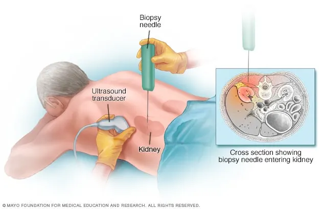
- Removing a sample of kidney tissue for testing. In some situations, your doctor may recommend a kidney biopsy to remove a small sample of kidney tissue for lab testing. Your doctor inserts a needle through your skin and into your kidney to remove the sample.
b. Data - By referring the images from above ct, ultrasound and tests.
c. Doctor identifies the problem by checking the above images.The main test for kidney disease is a blood test. The test measures the levels of a waste product called creatinine in your blood. Your doctor uses your blood test results, plus your age, size, gender and ethnic group to calculate how many millilitres of waste your kidneys should be able to filter in a minute.
d. Medication - Your kidney numbers include 2 tests: ACR (Albumin to Creatinine Ratio) and GFR (glomerular filtration rate). GFR is a measure of kidney function and is performed through a blood test. Your GFR will determine what stage of kidney disease you have – there are 5 stages. Know your stage.
3. Medication - ACE inhibitors and ARBs are two types of blood pressure medicine that may slow the loss of kidney function and delay kidney failure. Surgery - Kidney transplant
4. Surgical Solution :
a.Kidney Transplant
b.The patient is placed in the supine position. The legs are secured to the table with a strap with slight laxity. The lower extremities should be exposed enough to allow surgical access to the infrainguinal femoral vessels for the rare case where arterial reconstruction is necessary.
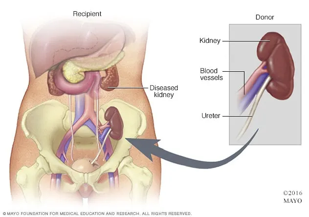
c. Kidney transplantation involves placing a healthy kidney into your body, where it can perform all of the functions that a failing kidney cannot.Your new kidney is placed on the lower right or left side of your abdomen where it’s surgically connected to nearby blood vessels. Placing the kidney in this position allows it to be easily connected to blood vessels and your bladder. The vein and artery of your new kidney are attached to your vein and artery. The new kidney's ureter is attached to your bladder to allow urine to pass out of your body.
A kidney transplant requires a stay in a hospital. Procedures may vary depending on your condition and your healthcare provider's practices.
Generally, a kidney transplant follows this process:
-
You will remove your clothing and put on a hospital gown.
-
An intravenous (IV) line will be started in your arm or hand. More catheters may be put in your neck and wrist to monitor the status of your heart and blood pressure, and to take blood samples. Other sites for catheters include under the collarbone area and the groin blood vessels.
-
If there is too much hair at the surgical site, it may be shaved off.
-
A urinary catheter will be inserted into your bladder.
-
You will be positioned on the operating table, lying on your back.
-
Kidney transplant surgery will be done while you are asleep under general anesthesia. A tube will be inserted through your mouth into your lungs. The tube will be attached to a ventilator that will breathe for you during the procedure.
-
The anesthesiologist will closely watch your heart rate, blood pressure, breathing, and blood oxygen level during the surgery.
-
The skin over the surgical site will be cleansed with an antiseptic solution.
-
The healthcare provider will make a long incision into the lower abdomen on one side. The healthcare provider will visually inspect the donor kidney before implanting it.
-
The donor kidney will be placed into the belly. A left donor kidney will be implanted on your right side; a right donor kidney will be implanted on your left side. This allows the ureter to be accessed easily for connection to your bladder.
-
The renal artery and vein of the donor kidney will be sewn to the external iliac artery and vein.
-
After the artery and vein are attached, the blood flow through these vessels will be checked for bleeding at the suture lines.
-
The donor ureter (the tube that drains urine from the kidney) will be connected to your bladder.
-
The incision will be closed with stitches or surgical staples.
-
A drain may be placed in the incision site to reduce swelling.
-
A sterile bandage or dressing will be applied.
Talk with your healthcare provider about what you will go through during your kidney transplant.
d
.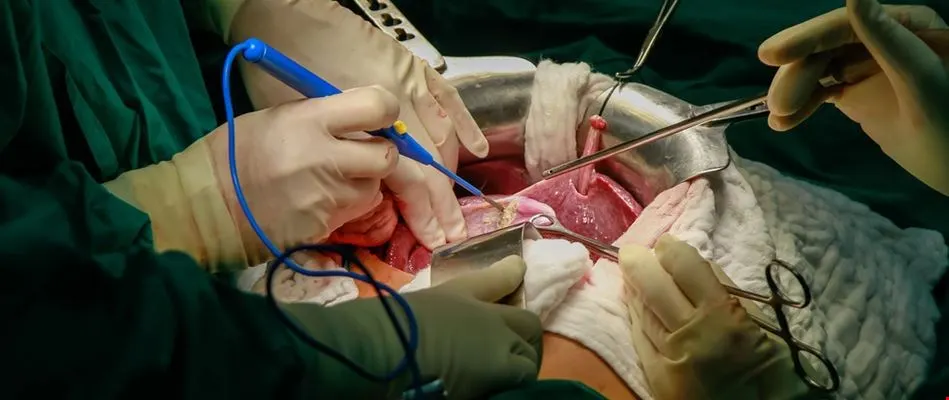
When you wake up after surgery, you will have intravenous lines (IVs) in your neck and arm(s). This allows us to give you enough fluids to keep up with the large amounts of urine usually made by the new kidney. You will have a catheter in your bladder to drain the urine, which prevents pressure on the surgical connection between the ureter and bladder. You may notice blood in the urine. This is from the surgical connection in the bladder. Sometimes clots can form and block the catheter tube. If this happens, the nurse will need to flush it.
e.https://youtu.be/fELn4Fe9Ccc
5. After a kidney transplant, you can expect to:
-
Spend several days to a week in the hospital. Doctors and nurses monitor your condition in the hospital's transplant recovery area to watch for signs of complications.
Your new kidney will make urine like your own kidneys did when they were healthy. Often this starts immediately. In other people, it may take several days, and you may need temporary dialysis until your new kidneys begin to function properly.
Expect soreness or pain around the incision site while you're healing. Most kidney transplant recipients can return to work and other normal activities within eight weeks after transplant. Avoid lifting objects weighing more than 10 pounds or exercising other than walking until the wound has healed (usually about six weeks after surgery).
-
Have frequent checkups as you continue recovering. After you leave the hospital, close monitoring is necessary for a few weeks to check how well your new kidney is working and to make sure your body is not rejecting it.
You may need to have blood tests several times a week and have your medications adjusted in the weeks following your transplant. During this time, if you live in another town, you may need to make arrangements to stay near the transplant center.
- Take medications the rest of your life. You'll take a number of medications after your kidney transplant. Immunosuppressant drugs help keep your immune system from attacking and rejecting your new kidney. Additional drugs help reduce the risk of other complications, such as infection, after your transplant.
6. intravenous (IV)- This allows us to give you enough fluids to keep up with the large amounts of urine usually made by the new kidney. ,urinary catheter-drain the urine.
2. PROJECT (b)
1. Pathological Condition : Pulmonary hypertension
a.The main function of the lungs is the process of gas exchange called respiration (or breathing). In respiration, oxygen from incoming air enters the blood, and carbon dioxide, a waste gas from the metabolism, leaves the blood. A reduced lung function means that the ability of lungs to exchange gases is reduced.
b.Pulmonary hypertension is high blood pressure in your pulmonary arteries, which carry oxygen-poor blood from your heart to your lungs.
c.
Some common underlying causes of pulmonary hypertension include high blood pressure in the lungs’ arteries due to some types of congenital heart disease, connective tissue disease, coronary artery disease, high blood pressure, liver disease (cirrhosis), blood clots to the lungs, and chronic lung diseases like emphysema. Genetics also play a role.
Pulmonary hypertension can happen in association with many other diseases, such as lung disease and heart disease. Heart failure is common in pulmonary hypertension.
d.Pulmonary hypertension (PH) is characterized by high blood pressure in the lungs and affects the circulatory system between the heart and lungs.
2.
Pulmonary hypertension is hard to diagnose early because it's not often detected during a routine physical exam. Even when pulmonary hypertension is more advanced, its signs and symptoms are similar to those of other heart and lung conditions.
To diagnose pulmonary hypertension, a health care provider will perform a physical exam and review your symptoms. You'll likely be asked questions about your medical and family history.
Blood and imaging tests done to help diagnose pulmonary hypertension may include:
- Blood tests. Blood tests can help determine the cause of pulmonary hypertension or detect signs of complications.
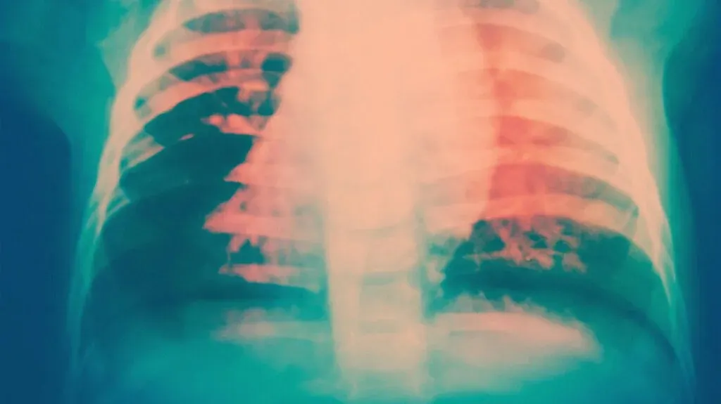
- Chest X-ray. A chest X-ray creates pictures of the heart, lungs and chest. A chest X-ray may be used to check for other lung conditions that can cause pulmonary hypertension.
- Electrocardiogram (ECG). This simple test records the electrical activity of the heart. It can detect changes in the heartbeat. Patterns on an ECG may reveal signs of right ventricle enlargement or strain.
-
Echocardiogram. Sound waves are used to create moving images of the beating heart. An echocardiogram shows blood flow through the heart. This test may be done to help diagnose pulmonary hypertension or to determine how well treatments are working.
Sometimes, an echocardiogram is done while exercising on a stationary bike or treadmill to learn how activity affects the heart. If you have this test, you may be asked to wear a mask that checks how well the heart and lungs use oxygen and carbon dioxide.
-
Right heart catheterization. If an echocardiogram reveals pulmonary hypertension, you'll likely have a right heart catheterization to confirm the diagnosis.
During this procedure, a cardiologist places a thin, flexible tube (catheter) into a blood vessel, usually in the groin. The catheter is gently guided into the right lower heart chamber (right ventricle) and pulmonary artery. A cardiologist can then measure blood pressure in the main pulmonary arteries and the right ventricle.
Other tests may be done to check the condition of the lungs and pulmonary arteries and further determine the cause of pulmonary hypertension:
-
Computerized tomography (CT). This imaging test takes a series of X-rays to create cross-sectional pictures of the bones, blood vessels and soft tissues inside the body. Dye (contrast) may be injected into a vein to help the blood vessels show up more clearly on the images.
A heart (cardiac) CT scan can show the size of the heart and any blockages in the pulmonary arteries. It can help diagnose lung diseases that might lead to pulmonary hypertension such as COPD or pulmonary fibrosis.
- Magnetic resonance imaging (MRI). This test uses magnetic fields and radio waves to create detailed images of the heart. It can show blood flow in the pulmonary arteries and determine how well the lower right heart chamber (right ventricle) is working.
- Lung (pulmonary) function test. This noninvasive test measures how much air the lungs can hold and the airflow in and out of the lungs. The test involves blowing into an instrument called a spirometer.
- Sleep study (polysomnogram). A sleep study measures brain activity, heart rate, blood pressure, oxygen levels and other factors during sleep. A sleep study can help diagnose obstructive sleep apnea, which can cause pulmonary hypertension.
- Ventilation/perfusion (V/Q) scan. In this test, a radioactive tracer is given by IV. The tracer shows blood flow and airflow to the lungs. A V/Q scan can determine whether blood clots are causing symptoms of pulmonary hypertension.
- Open-lung biopsy. Rarely, an open-lung biopsy may be done to check for a possible cause of pulmonary hypertension. An open-lung biopsy is a type of surgery in which a small sample of tissue is removed from the lungs.
b. Data - By referring the images from above ct, mri and tests.
c.Normal pulmonary artery pressure is 8-20 mm Hg at rest. If the pressure in the pulmonary artery is greater than 25 mm Hg at rest or 30 mmHg during physical activity, it is abnormally high and is called pulmonary hypertension.
d.
There is no cure for pulmonary hypertension. However, there are many different types of treatments, including
- Inhaled medicine
- Medicine given through the veins under the skin
- Medicine to reduce swelling in the feet (diuretics)
- Oxygen therapy
3.
a.Medications
Medications are available to help improve symptoms of pulmonary hypertension and to slow the progression of the disease.
Medications that may be prescribed to treat pulmonary hypertension symptoms or complications include:
-
Blood vessel dilators (vasodilators). This type of medicine relaxes and opens narrowed blood vessels, improving blood flow. Vasodilators may be taken by mouth, inhaled, injected or given by IV infusion. A commonly prescribed vasodilator for pulmonary hypertension is epoprostenol (Flolan, Veletri).
This drug continuously flows through an IV attached to a small pump, which is worn in a pack on the belt or shoulder. Potential side effects of epoprostenol include jaw pain, nausea, diarrhea, leg cramps, and pain and infection at the IV site.
Other types of vasodilators, including treprostinil (Tyvaso, Remodulin, Orenitram), can be inhaled, injected or taken by mouth. The drug iloprost (Ventavis) is given while breathing in through a nebulizer, a machine that vaporizes the medication.
Side effects associated with treprostinil include chest pain, often with headache and nausea, and breathlessness. Possible side effects of iloprost include headache, nausea and diarrhea.
- Guanylate cyclase (GSC) stimulators. This type of medicine increases nitric oxide in the body, which relaxes the pulmonary arteries and lowers pressure in the lungs. GSC stimulators include riociguat (Adempas). Side effects include nausea, dizziness and fainting. You should not take GSC stimulators if you're pregnant.
- Medications to widen blood vessels. Medications called endothelin receptor antagonists reverse the effect of a substance in the walls of blood vessels that causes them to narrow. Such drugs include bosentan (Tracleer), macitentan (Opsumit) and ambrisentan (Letairis). They may improve energy level and symptoms. However, they can damage the liver. Monthly blood tests may be done to check liver function. Don't take these drugs during pregnancy.
- Medications to increase blood flow. Drugs called phosphodiesterase 5 (PDE5) inhibitors may be used to increase blood flow through the lungs. These medicines are also used to treat erectile dysfunction. They include sildenafil (Revatio, Viagra) and tadalafil (Adcirca, Cialis, Alyq).
- High-dose calcium channel blockers. These drugs help relax the muscles in the walls of blood vessels. They include amlodipine (Norvasc), diltiazem (Cardizem, Tiazac, others) and nifedipine (Procardia). Although calcium channel blockers can be effective, only a small number of people with pulmonary hypertension improve while taking them.
-
Blood thinners (anticoagulants). Warfarin (Jantoven) may be prescribed to help prevent blood clots. Blood thinners increase the risk of bleeding, especially in those who are having surgery or an invasive procedure. If you take blood thinners, you'll need occasional blood tests to see if the medicine is working as it should. Don't stop taking blood thinners without first talking to your care provider.
Many other drugs, herbal supplements and foods can interact with warfarin. Always tell your provider about your diet and all the medications you take, including those bought without a prescription.
- Digoxin (Lanoxin). This medicine helps the heart beat stronger and pump more blood. It can help control irregular heartbeats (arrhythmias).
- Water pills (diuretics). These medications help the kidneys remove excess fluid from the body. This reduces the amount of work the heart has to do. Diuretics may also be used to reduce fluid buildup in the lungs, legs and abdomen.
- Oxygen therapy. Breathing pure oxygen is sometimes recommended as a treatment for pulmonary hypertension, especially for those who live at a high altitude or have sleep apnea. Continuous oxygen therapy may be needed.
b.Surgery and other procedures
If medications do not help control the signs and symptoms of pulmonary hypertension, surgery may be recommended. Surgeries to treat pulmonary hypertension include:
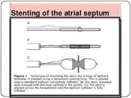
- Atrial septostomy. This open-heart surgery may be recommended if medications don't control pulmonary hypertension signs and symptoms. In an atrial septostomy, a surgeon creates an opening between the upper left and right chambers of the heart (atria) to relieve the pressure on the right side of the heart. Potential complications include heart rhythm problems (arrhythmias).
-
Lung or heart-lung transplant. Sometimes, a lung or heart-lung transplant may be recommended, especially for younger people who have idiopathic pulmonary arterial hypertension.
Major risks of any type of transplantation include rejection of the transplanted organ and serious infection. Immunosuppressant drugs must be taken for life to help reduce the chance of rejection.
4. a. Surgical solution: Atrial Septostomy Surgery
b.Supine position
c. A surgeon puts a catheter with a balloon on the tip through the wall between the upper chambers (atria) of the heart to make a hole.
This allows blood to pass from the right side of the heart to the left side and bypass the lungs, which have high resistance due to pulmonary hypertension.
This procedure alleviates the pressure on the right ventricle of the heart.
After surgery, usually the oxygen levels in the blood fall, but the heart failure improves.
Atrial septostomy is only done under special circumstances, when doctors believe the benefits outweighs the risks. It only relieves symptoms and is not a permanent solution for pulmonary hypertension.
d. catheter
e. https://youtu.be/hio-h3YRDwA
5.After the procedure, your baby will be allowed to wake up and slowly come off of the breathing machine depending on how strong your baby is breathing.
If the oxygen levels are good, your child’s doctors will try to stop the prostaglandin medicine. This can help with the baby’s breathing and coming off of the breathing machine.
6. catheter, echocardiogram