Project 2 - Clinical Investigation Plan
1.0 Clinical Investigation Plan Overview
- The commercially available Günther Tulip and Cook Celect inferior vena cava filters have been previously evaluated in clinical studies.
- This prospective, multicenter, single-arm clinical study will further evaluate the safety and effectiveness of Cook’s permanent and retrievable inferior vena cava (IVC) filters (specifically, the Günther Tulip and the Cook Celect® filters) in patients in need of temporary or permanent IVC filter placement for the prevention of pulmonary embolism (PE).
- This study will enroll 320 patients in the Celect filter stratum and up to 150 patients in the Günther Tulip stratum at up to 40 clinical sites globally; patients will be stratified based on the type of filter they receive.
- Consistent with current clinical practice and already-completed studies of Cook’s IVC filters, the study will evaluate the performance and safety of filters in patients considered at risk for PE for a variety of clinical reasons (i.e., a broad patient population).
Study assessments will include, but are not limited to rate of technical placement success, rate of freedom from new symptomatic PE, rate of freedom from new PE, rate of clinical perforation, rate of migration, rate of filter fracture, rate of filter embolization,
- rate of IVC thrombotic occlusion, rate of new symptomatic deep vein thrombosis (DVT; total incidence and new incidence), rate of procedure-related complications, time to filter retrieval, number of successful retrievals, and rate of technical retrieval success.
- Patients will be followed until 1 month after successful filter retrieval or until study completion 2 years after filter placement.
- Enrollment is expected to be completed within 2 years of initiating the study.
- The primary study endpoints for the Celect® filter stratum will be compared to performance goals for safety (i.e., freedom from major adverse events) and effectiveness (i.e., technical placement success and freedom from new symptomatic PE while a filter is indwelling).
- Additional study outcomes will be evaluated separately for each stratum (i.e., separately for patients receiving the Günther Tulip® and Celect® filters) and for the combined patient set.
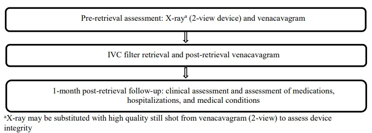
- The study flow diagrams are presented in Figure 1.1 (filter placement and follow-up) and Figure 1.2 (filter retrieval and follow-up).
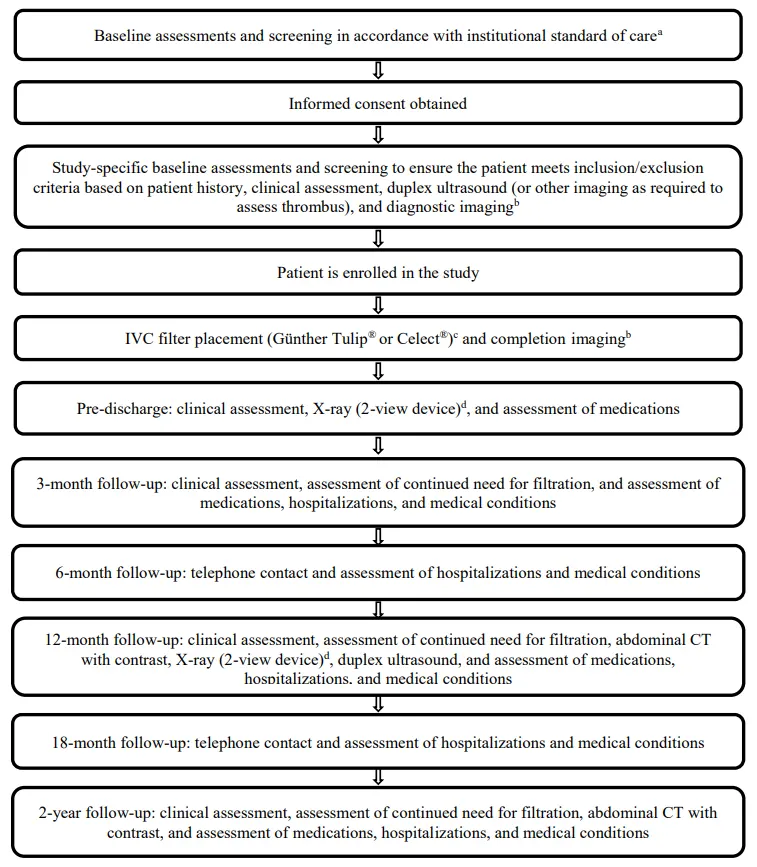
- Imaging performed to document the presence of PE in patients with suspected PE is to be collected
- Imaging performed pre-placement to verify anatomical study criteria and post-placement to verify the position of the filter
- Type of filter is at the discretion of the physician X-ray may be substituted with high quality still shot from venacavagram (2-view) to assess device integrity Figure 1.1. Study flow diagram (filter placement and follow-up schedule)
2.0 Objective of the Clinical Study
The objectives of this study are to further evaluate the safety and effectiveness of Cook’s commercially available IVC filters (specifically, the Günther Tulip® filter and the Cook Celect® filters) in patients in need of temporary or permanent IVC filter placement for the prevention of PE. This study is intended to address FDA’s concerns related to IVC filters.4
3.0 Device Description and Intended Use
3.1 General Device Description
- All Cook vena cava filter sets include an IVC filter intended for prevention of recurrent PE in specific situations, such as chronic, recurrent PE where anticoagulant therapy has failed or is contraindicated.
- All Cook vena cava filter sets are available in a femoral vein access version, with which the filter is introduced through the femoral vein, a jugular vein access version, with which the filter is introduced through the jugular vein, and a universal vein access version, which includes the components for both femoral and jugular vein approaches.
- Each filter set consists of a preloaded filter, a coaxial introducer sheath system with a Check-Flo® valve, a hydrophilically coated pre-dilator, and a threeway stopcock.
- The jugular and femoral vein access versions of all filters are available with the NavAlign introducer system (K090140), with design improvements to the introducer sheath and filter introducer intended to ease the filter placement procedure and reduce the likelihood of procedure-related complications.
- The femoral vein access version of all filters is available with a flexible tip (composed of flexible stainless steel cable tubing covered with polyether block amide (PEBAX) shrink tubing) on the filter introducer, which is intended to make the tip more flexible to improve trackability of the introducer through the venous anatomy (K121057).
- The femoral vein access version of the Günther Tulip® filter and the Celect® filter is also available with added markers along the introducer sheath to improve visibility on procedural imaging (K112119).
- In this set, the femoral introducer sheath is made of polyethylene (as compared to fluorinated ethylene propylene, which is the material component of the femoral introducer sheath for all other femoral vein access sets) and has an attached clip (composed of stainless steel spring wire with polycarbonate, acrylonitrile/butadiene/styrene (PC-ABS) handle) to mark the position of the sheath at the insertion site.
- The Celect® filter is available with added platinum markers on its primary legs to improve visibility on procedural imaging (K121629). The femoral vein access version of the Celect® with platinum markers on its primary legs is only available with a flexible tip, and is not available with markers along its introducer sheath.
- Please reference the manufacturer’s Instructions for Use (IFU) for a complete description of each of these devices.
3.2 Indication for Use
The Günther Tulip filter and the Cook Celect® filters are intended for the prevention of recurrent PE via placement in the vena cava in the following situations:
- Pulmonary thromboembolism when anticoagulant therapy is contraindicated;
- Failure of anticoagulant therapy in thromboembolic diseases;
- Emergency treatment following massive PE where anticipated benefits of conventional therapy are reduced; and
- Chronic, recurrent PE where anticoagulant therapy has failed or is contraindicated.
- In addition to the specific labeled indications, it is well-documented in clinical practice guidelines that IVC filters have additional clinical indications, and are frequently placed in patients with the following: severe trauma, closed head trauma, spinal cord injury,
- multiple long-bone or pelvic fractures, or other conditions putting them at high risk for PE (e.g., prolonged immobilization, pre-/post-operatively).
- 5-8 Outcomes from use in these broad patient populations have been widely reported in the literature1-3,9-22, including those based on results of previous studies for the Cook IVC filters.
- 1-3 Based on available data, patient outcome is not specifically related to indication for filter placement, but may be related to each patient’s overall medical condition, including the patient’s state of hypercoagulability, body mass index, use of anticoagulation, comorbid conditions, etc.
- Moreover, Cook has conducted three previous studies of the performance of its permanent and retrievable IVC filters in a broad patient population,1-3 and the results (or interim results) of these studies are included in product labeling.
- Furthermore, these broad patient populations were included in the indications for use that were approved in the PMA for the Cook Bird’s Nest® Vena Cava Filter (P850049).
3.3 Device Identification and Tracking
- As commercially available devices, the Cook IVC filters will be obtained by each site through normal commercial procurement methods and stored and accounted for following site-specific policies.
- Lot numbers for devices used in study patients will be recorded on the appropriate Case Report Forms (CRFs).
3.4 Instructions for Use
The manufacturer’s IFU for each filter contains the following information:
- Complete instructions including storage and handling requirements, preparation for use, pre-use checks, precautions to be taken after use, and disposal.
- Complete summary of the necessary training and experience required for use of these devices.
- Complete description of the procedures involved in the use of these devices.
4.0 Literature Review
Background
- Venous thromboembolism (VTE) affects nearly 1,000,000 people in the US each year, and includes both DVT and PE.
- Pulmonary emboli commonly result from DVT.
- PE is a major cause of morbidity and mortality in the US, with estimates of 400,000 – 630,000 nonfatal PEs and 50,000 – 200,000 fatal PEs per year.8 Populations at an increased risk
- for VTE include those with an inherited hypercoagulability, those undergoing complex surgery (including orthopedic surgery and bariatric surgery, among others), those with cancer, those with trauma, those with prolonged immobilization, those with a previous episode of VTE, as well as those patients with acute medical situations such as stroke, heart failure, respiratory failure, or infections.
- The typical prevention method for VTE is anticoagulation therapy (e.g., heparin, warfarin).
- However, anticoagulation may be contraindicated in some patients; for instance, those undergoing complex surgery, those with acute or recent bleeding, those with liver disease, and those with trauma.
- Therefore, IVC filter placement via percutaneous access is frequently utilized to mitigate the potential risk of PE, either as an individual prevention modality or in combination with anticoagulation therapy.
- IVC filter placement is a well-established method for PE prophylaxis. The first IVC filter was introduced in the late 1960s as an alternative to surgical ligation, caval plication, and clips.The early filters (Mobin-Uddin Umbrella and Greenfield) required surgical cut-down for placement;
- Cook’s Bird’s Nest® Filter was the first percutaneously placed filter, and was approved under a PMA with expert advisory panel review.
- All early filters (Mobin-Uddin Umbrella, Greenfield, Bird’s Nest) were intended for permanent placement, i.e., the filters could not be retrieved after placement.
- Filter technology has evolved to include a variety of percutaneously placed retrievable filters, which were initially developed in response to the following clinical observations:
- 1) a need for temporary periods of PE prophylaxis and
- 2) potential complications of permanently indwelling filters.
- Retrievable filters may be retrieved or left as permanent devices; treatment is at the discretion of the physician, with specific considerations to the individual medical condition of each patient. Cook’s Günther Tulip filter has the longest history of permanent and retrievable use in the US, and Cook’s Celect filter has also been used as a permanent and retrievable filter for many years.
- Despite the clinical rationale for the development of retrievable filters, in particular in periods of temporary high risk for PE (which includes patients with trauma and those in the perioperative setting, among others), the labeled filter indications do not adequately reflect use in the clinical studies performed to date or in routine clinical practice.
- It should be noted that the expert panel recommended approval of the Bird’s Nest for use in a broad population of patients considered at risk for PE, reflective of the clinical data supporting the filter and more appropriate than the currently labeled indication (the indications were changed following a 1995 letter from FDA mandating that the product indications for use be modified).
- It is well-documented in clinical practice guidelines that IVC filter placement is widely utilized for both patients with proven VTE and patients without proven VTE but considered at high risk for VTE.5-8 Patients with VTE receive filters because
- 1) they are at high risk for PE and have a contraindication to or complication or failure of anticoagulation therapy or
- 2) they are at high risk for PE despite anticoagulation therapy, have increased risks of complications with anticoagulation therapy, or are non-compliant with anticoagulation therapy.
- Patients without proven VTE receive filters when they are considered at risk for developing VTE and cannot receive effective prophylaxis or be monitored for the development of VTE (e.g., trauma patients, patients undergoing certain surgical procedures).
- In each category, filters provide prophylactic protection against the occurrence of PE.
Filter Efficacy
- Two randomized studies on the efficacy of IVC filters have been performed. In the first, Decousus et al.
- randomized 400 patients with proximal DVT to permanent filter placement or no filter (patients were also assigned to two different anticoagulation regimens) and found that the filter group had significantly fewer events of PE (p=0.008), albeit more events of DVT (p=0.042) through 8 years.
- Specifically, the eight-year PE rates were 15.1% in the no-filter group and 6.2% in the filter group.
- More recently, Rajasekhar et al. randomized 34 trauma patients to retrievable filter (Celect®) placement or no filter and observed one PE in the no filter group and no PE in the filter group through 6 months.
- 25 Thus, the randomized data available suggest that filters do provide protection against PE, as intended.
- In addition to these data, a wide body of clinical literature supports filter efficacy in a broad range of patient populations.
- These include data from primarily single and multi-center studies of individual filter types, and some randomized studies comparing outcomes with different filter types.
- Based on Cook’s ongoing analysis of the published literature, IVC filter use in a broad patient population is associated with a low PE rate (an average of 1.7%).
- This rate is much lower than that observed in the PREPIC study; moreover, this rate is lower than the expected PE rate in specific patient populations (for example, a literature review suggests that the PE rate in trauma patients without an IVC filter is approximately 4.9%).
- In general, the data are supportive of the use of filters to prevent PE, though there are important covariates that may impact patient outcomes (e.g., history of VTE, anticoagulation scheme).
The benefit of filter placement in specific patient populations has also been discussed in the literature, including:
- In a recent systematic review of 24 studies describing filter placement in trauma patients, in which the authors concluded that (for the trauma population) filter-related complications are infrequent (and those that do occur tend not to be clinically significant) and that filter placement does reduce the incidence of PE and PE-related mortality.42
- A recent assessment of the impact of IVC filters on in-hospital fatality due to PE based on the Nationwide Inpatient Sample using ICD-9-CM codes to identify patients with PE.
- Results of the analysis suggested that IVC filter placement resulted in significantly fewer in-hospital fatalities in unstable patients with PE (regardless of the use of thrombolytic therapy; 33% vs. 51% in patients without thrombolytic therapy and 7.6% vs. 18% in patients with thrombolytic therapy;
- The incidence of in-hospital fatality in stable patients not receiving thrombolytic therapy was also significantly reduced (7.2% vs. 7.9%; p<0.0001), though this reduction was not as clinically-meaningful as for the other
populations. - Therefore, the clinical body of evidence supports filter placement for the prevention of PE in a broad range of patients.
Filter Safety
- As with any medical implant, IVC filters are associated with a range of potential adverse events; these events are clearly outlined in Cook’s IFUs for its IVC filters.
- Recent clinical practice guidelines report the incidence of adverse events for IVC filters, in general, to be as follows:
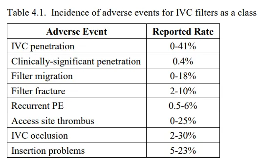
- These event rates are derived from events reported in the clinical literature and are based on studies for various types of IVC filters and with a wide range of sample sizes (which directly impacts the reported event rates). Moreover, definitions for the events vary among publications;
- in particular, they include both those that are clinically significant (i.e., those associated with patient symptoms and/or requiring intervention) and those without clinical consequences. Therefore, the data should be interpreted carefully.
- In 2010, FDA issued a public notice in which it expressed concerns related specifically to the number of adverse event reports for filter migration, filter embolization, filter fracture, and perforation of the IVC; notably, event rates cannot be deduced from the FDA database of reported events.
- In light of these concerns, FDA recommended that filter retrieval be considered as soon as the mechanical protection against PE is no longer warranted.
- FDA’s recommendation is consistent with literature reports which suggest that patients may be at increased risk of adverse events when filters are left indwelling for extended periods of time;
- despite the fact that most filters are retrievable, a large percentage are left in place permanently. Several publications have documented the fact that dedicated patient follow-up, with recurring assessment for continued risk of PE,
results in increased rates of filter retrieval. - These same authors postulate that increased filter retrieval rates may correlate to reduced incidence of filter complications.
- This hypothesis has yet to be verified in clinical studies, but some filter practices (both filter placement decisions and clinical follow-up practices) have changed in light of these safety concerns.
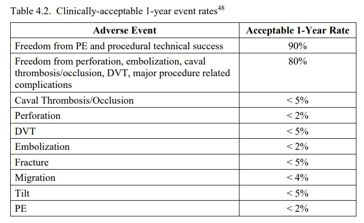
- Recently, the SVS/SIR physician working group presented what they consider to be the clinically-acceptable 1-year event rates for filter-related safety and effectiveness outcomes (Table 4.2).
- As detailed below, Cook’s IVC filter data is favorable in comparison to the rates in Tables 4.1 and 4.2.
- Finally, these rates were utilized in the development of the performance goals and sample size for this study (described in furthermdetail in Sections 8.1 and 8.3).
Cook’s IVC Filter Data
- Cook Incorporated has sponsored three prospective, multi-center, single-arm clinical studies of Cook IVC filters that are currently marketed in the US.
- Briefly, these include the IDE study of 41 US patients treated with the Günther Tulip® filter (G000242;
- results of which supported a retrievable indication, cleared in K032426), a second study of 554 US patients treated with the commercially available Günther Tulip® filter, and a study of 129 OUS patients treated with the Celect® filter (interim results of which supported permanent and retrievable indications).
- Results from all three studies have been published in peer-reviewed journals.
- 1-3 All three studies evaluated filter safety and effectiveness (outcome measures similar to those listed in Tables 4.1 and 4.2) in patients at risk for developing a PE; as such, a variety of reasons for filter placement are captured in the studies.
- Approximately half of the filter placements in these studies were associated with bariatric procedures or trauma cases; specifically, 28/41 patients (68.3%) in the IDE Günther Tulip® study, 329/554 patients (59.4%) in the second Günther Tulip® study, and 53/129 patients (41.1%) in the Celect® study received filters for these types of indications.
- Mean follow-up duration (with range in parentheses) was 70.4 ± 49.3 (0-160) days for the IDE Günther Tulip® study, 124 ± 75.3 (3-494) days in the second Günther Tulip® study, and 277 ± 146 (1- 602) days in the Celect® study (note: the Celect® study included an arm specifically intended to assess permanent filters).
- Mean filter indwell times (for retrieved filters) were 11.0 days in the IDE Günther Tulip® study, 58.9 days in the second Günther Tulip® study, and 179 days in the retrieval arm of the Celect® study.
- The significant safety findings (including PE and those AEs associated with the safety endpoint specified in this clinical investigation plan) in each study are summarized as follows.
- In the Günther Tulip® IDE study, events included one filter misplacement and migration requiring immediate retrieval and placement of a new filter, one filter migration to the right atrium (with successful retrieval), one PE, one patient with a caval thrombosis/occlusion, and four deaths (none were device related);
- in addition, one patient had a vena cava injury following the retrieval procedure (with a normal 3-month postretrieval follow-up examination).
- Events in the second Günther Tulip® study included five PEs, one DVT, one filter fracture noted at retrieval, one vascular access site complication following filter implant, and 13 deaths (two were related to PE and six were within 30 days of filter placement).
- In the Celect® study, related events included two PEs, one DVT, one migration (without clinical sequelae), and 26 deaths (one associated with a PE and eight within 30 days of filter placement); there was no difference in the type of events observed in the permanent and retrievable study arms.
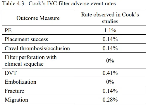
- The incidence of each type of event observed in Cook’s clinical investigations is on the low end of, or below, the rates reported in Tables 4.1 and 4.2 above (Table 4.3).
- Moreover, the overall rate of events associated with the primary endpoints of this clinical investigation (i.e., PE, placement success, caval thrombosis/occlusion, perforation with clinical sequelae, DVT, embolization, fracture, and migration) for the Günther Tulip® filter was 3.7% and the overall rate of events for the Celect® filter was 9.3%.
Conclusion
- Many clinical studies have been conducted on IVC filters, including Cook’s studies of its Günther Tulip® and Celect® filters; Cook’s study results support the safety and effectiveness of its filters.
- In light of FDA’s ongoing concerns regarding the use, performance, and safety of IVC filters, this study is intended to collect additional safety and effectiveness data on Cook’s IVC filters (i.e., the Günther Tulip® and Celect® filters) in patients in need of temporary or permanent IVC filter placement for prevention of PE.
- The study population and endpoints of interest are similar to those investigated in Cook’s previous IVC filter studies, to those of interest in the literature, and to those of interest to FDA4 and several physician society groups.
- Results of this study will be provided to FDA and may be used to support expanded indications for Cook’s IVC filters, and may also be compared to safety and effectiveness results associated with implantation of other filter types to the extent that this protocol is consistent with the PRESERVE study.
5.0 Risk Analysis and Risk Assessment
5.1 Anticipated Benefits
Filter implantation may prevent or reduce the incidence of PE.
5.2 Risks and Foreseeable Adverse Events and Adverse Device Effects
The following foreseeable adverse events, which are captured in the IFUs of these commercially available devices, may be considered as potential risks to patients receiving a Cook IVC filter:
• Damage to the vena cava
• PE
• Filter embolization (including embolization of fractured components)
• Vena cava perforation/penetration
• Vena cava thrombosis or occlusion
• Hemorrhage
• Hematoma at vascular access site
• Infection at vascular access site
• Cardiac tamponade
• Filter malpositioning
• Postphlebitic syndrome
• Death
• DVT
In addition, participation in this clinical study is associated with the risk of additional radiation exposure from study imaging requirements that are above and beyond the standard of care.
5.3 Methods to Minimize Risks
- The Cook IVC filters will be used only by trained healthcare professionals who are experienced in the study procedure/treatment. Patients will be selected in accordance with the inclusion/exclusion criteria outlined in this document.
- The imaging required as part of this clinical investigation has been recommended by FDA, in order to provide additional data for interpreting the occurrence of adverse events. The risk of radiation exposure associated with study imaging has been minimized by collecting imaging data only where essential for the study or otherwise clinically indicated or per institutional standard of care.
- The device design, non-clinical testing, clinical study design, and the IFUs are intended to minimize the risks associated with the use of these devices. The risks of the stud
- have been minimized and the potential benefits outweigh the risks in light of the importance of the knowledge to be gained about the safety and effectiveness of Cook’s IVC filters.
6.0 Design of the Clinical Study
- Type/Design of Study
- This prospective, multicenter, single-arm clinical study will further evaluate the safety and effectiveness of Cook’s commercially available IVC filters (specifically, the Günther Tulip® and the Cook Celect® filters) in patients in need of temporary or permanent IVC filter placement for prevention of PE.
- This study will enroll 320 patients in the Celect® filter stratum and up to 150 patients in the Günther Tulip® filter stratum at up to 40 clinical sites globally. At least half of the study patients in the Celect® filter stratum will be enrolled at US sites. Consistent with current clinical practice, the study is expected to evaluate the performance and safety of filters in a broad range of patients considered at risk for PE (i.e., in patients considered at risk for PE for a variety of clinical reasons).
- This could include patients ranging from those with PE or DVT and a contraindication to, complication of, failure of, or poor compliance with anticoagulation to patients without evidence of PE or DVT, but considered to be at risk for PE for other reasons.
- The results of the study will be compared to safety (i.e., freedom from major adverse events) and effectiveness (i.e., technical placement success and freedom from new symptomatic PE while a filter is indwelling) performance goals.
Inclusion and Exclusion Criteria
Patient eligibility for enrollment shall be based on known information at the time of the procedure. Information obtained at a later date may contradict these criteria, but this will not be considered a violation of the CIP.
Inclusion Criterion
A patient may be suitable for inclusion in the study if he/she requires temporary or permanent IVC filter placement for the prevention of PE
Exclusion Criteria
Patients will be excluded from study enrollment if any of the following are true:
General Exclusion Criteria
- Less than 18 years of age
- Patient or legally authorized representative is unable or unwilling to provide written informed consent prior to initiation of study procedures
- Unable or unwilling to comply with follow-up schedule
- Known hypersensitivity or contraindication to contrast medium that cannot be adequately premedicated
- Known allergy or sensitivity to cobalt, chromium, or nickel
- Pregnant or planning to become pregnant in the next 12 months
- Simultaneously participating in another investigational drug or device study in which the patient has not completed the follow-up phase for that study’s primary endpoint 30 days or more prior to being enrolled in this study
- Patient refuses blood transfusions
Medical Exclusion Criteria
- Unable to tolerate the amount of contrast required by the procedure (in the opinion of the investigator)
- At risk of septic embolism
- Medical condition or disorder that would limit life expectancy to less than 12 months or that may cause noncompliance with the protocol or confound the data analysis
- Existing IVC filter
Anatomical Exclusion Criteria
- Duplicate IVC
- Anatomy that would prevent safe filter placement (e.g., condition of access vessels)
- IVC diameter > 30 mm or < 15 mm
Endpoints
Primary Effectiveness Endpoint
- The primary effectiveness endpoint is the rate of technical placement success and 12- month freedom from new symptomatic PE while a filter is indwelling. Analysis of the primary effectiveness endpoint will be performed separately for each filter type (i.e., each stratum).
- Events of symptomatic PE occurring after filter retrieval (i.e., through the 1-month post- retrieval follow-up period) will be collected; such events will not count against the primary effectiveness endpoint, but will be reported as a secondary measure.
Primary Safety Endpoint
- The primary safety endpoint is the rate of 12-month freedom from major adverse events. Major adverse events are defined as: clinical perforation, clinical migration, clinical fracture, embolization of the filter or filter fragments to the heart or lungs, IVC thrombotic occlusion, new symptomatic DVT while a filter is indwelling, access site complications with clinical sequelae, and procedure-/device-related death.
- Analysis of the primary safety endpoint will be performed separately for each filter type (i.e., each stratum).
- Events occurring after filter retrieval (i.e., through the 1-month post-retrieval follow-up period) will be collected, but only the events of access site complications with clinical sequelae and procedure-related death will count against the primary safety endpoint.
- Other events (i.e., potentially new DVT without symptoms) will be reported as a secondary measure.
Secondary Endpoints
The secondary endpoints include:
- Rate of technical placement success and 12-month freedom from new symptomatic PE while a filter is indwelling for the combined patient population
- Rate of 12-month freedom from major adverse events for the combined patient population
- Rate of 12-month freedom from Grade 2 or Grade 3 filter leg interaction with the IVC, filter migration, filter fracture, and filter embolization for both the individual stratum and the combined patient population
All primary and secondary endpoints will also be reported at 24 months. In addition, analysis of the following secondary measures will be performed separately for each filter type (i.e., each stratum) and for the combined patient population:
- Filter leg interaction with the IVC wall
- Clinical perforation
- Migration > 2 cm (including direction)
- Symptomatic PE (occurring before and after filter retrieval)
- PE (occurring before and after filter retrieval)
- IVC thrombotic occlusion (occurring before and after filter retrieval)
- Symptomatic DVT (occurring before and after filter retrieval)
- DVT (total incidence and new incidence; occurring before and after filter retrieval)
- Retroperitoneal hematoma
- Hematoma at access site
- Infection at access site
- Thrombosis at access site
- Death
- Filter tilt (with measurement)
- Filter fracture
- Filter embolization
- Filter deformation
- Length of procedure (insertion and retrieval)
- Length of ICU stay
- Length of hospital stay
- Technical placement success
- Filter retrieval attempts
- Technical retrieval success
- Failed retrieval attempts, with reason for failed attempt
- Retrieval-related complications
- Adverse events occurring after filter retrieval
- Delivery system performance
Rationale for Endpoints
The endpoints were chosen as applicable measures of device safety and effectiveness because of their similarity to those of interest in the literature and to those of interest to FDA4 and several physician society groups.49
Variables to be Measured to Demonstrate Achievement of Endpoints
Independent core laboratories (to be designated) will be used to provide detailed analysis of imaging data collected.
The clinical data and imaging measurements will be collected on standardized case report forms (CRFs), which may serve as source documents. The schedule for assessments is summarized in Table 6.1.
Table 6.1. Data collection schedule
|
Pre-procedurea |
Placement |
Post-procedureb |
3 months post-placement |
6 months post-placement |
12 months post-placement |
18 months post-placement |
2 years post-placement |
Pre-retrievalc |
Retrieval |
1 month post-retrieval |
|
|
Chest CT or other imaging to document pre-procedure PE in patients with clinical indications |
Xa |
||||||||||
|
Medical history |
Xd |
||||||||||
|
Clinical assessment |
Xd |
X |
X |
X |
X |
X |
|||||
|
Duplex ultrasound |
Xe |
X |
|||||||||
|
Diagnostic imaging |
Xf |
||||||||||
|
Venacavagram |
Xg |
||||||||||
|
Abdominal CT with contrast |
X |
X |
|||||||||
|
X-ray (2-view device) |
Xh,i |
Xi |
Xi |
||||||||
|
Telephone contact |
X |
X |
Measures to be Taken to Avoid or Minimize Bias
This study is designed as a prospective clinical trial to minimize bias in analyzing the results. Specifically, the study will utilize event adjudication by an independent clinical events committee and imaging analysis by a centralized core laboratory to minimize bias in analyzing the results. Patients requiring temporary or permanent IVC filter placement to prevent PE will be screened consecutively to mitigate selection bias. Investigational sites will maintain a screening log of patients; for patients who are excluded from the study, the log will indicate the primary inclusion/exclusion criterion that is violated. A single institution may enroll up to 20% of the patients in each stratum to minimize bias that may be introduced if the majority of patients are enrolled from one or two large clinical sites. Finally, filter choice will be left to the discretion of the physician.
Methods
Patient Consent
Patients who meet the inclusion criterion and none of the exclusion criteria will be invited to participate in this study. All patients eligible for entry into the study will have the study explained to them, as well as potential risks and benefits of their participation in the study. Each patient (or legally authorized representative) who agrees to participate will be required to sign an informed consent document prior to undergoing study-specific testing or the filter placement procedure. If new information is obtained after a patient receives treatment with the device, patients who have not exited the study will be informed about the new information, and will be reconsented at the discretion of the investigators and/or the sites’ IRBs/ECs/REBs.
Point of Enrollment
Patients will be considered enrolled in the study once the filter introducer sheath is inserted below the skin.
Medications
Patients should receive medication(s) according to the institution’s standard of care and/or at the physician’s discretion, based on each patient’s individual medical condition. The use of antiplatelet and anticoagulation medications should be recorded on appropriate CRFs.
Pre-procedure
A pre-procedure clinical assessment will be completed (up to 6 weeks prior to the index procedure) including documentation of medical comorbidities and assessment of medications. Duplex ultrasound of the IVC, bilateral iliac veins, and bilateral lower extremity veins will be performed; presence of DVT will be documented on the appropriate CRF. For patients in whom a CT with contrast was performed as part of routine care within 2 weeks of the filter placement procedure, the CT may be used to assess for thrombus in vein segments visualized on CT. For cases in which a physician feels it is in the best interest of the patient to forego the ultrasound exam so the filter may be placed immediately and/or in which a duplex ultrasound may not be physically possible prior to filter placement (e.g., patients with trauma to the pelvis), the venogram may be used to assess for thrombus. If segment(s) of vein are not assessed before the filter is placed, assessments are to be completed within one day after the placement procedure. Assessments of all vein segments of interest are to be completed, unless a patient’s injuries prevent examination of one or more vein segment(s).
Patients with suspected PE should undergo appropriate assessment for the presence of PE, including appropriate imaging (i.e., pulmonary arteriography, cross-sectional imaging, or ventilation/perfusion lung scan), according to the institution’s standard of care. This imaging will be collected and results of these assessments will be documented on the appropriate CRF.
Filter Placement Procedure
- Diagnostic imaging will be performed prior to study enrollment to verify anatomical study
- Only one Cook IVC filter should be placed in each The Cook IVC filter should be implanted in accordance with the respective IFU (i.e., caudal to the renal veins).
- Completion imaging will be performed to assess filter position (including tilt) and to assess the vena cava for evidence of injury.
- Post-Filter Placement/Pre-Discharge
Post-placement care includes observation and discharge according to institutional standard of care and completion of clinical and medication assessments. In addition, a 2-view device X-ray will be performed prior to hospital discharge; the X-ray should be performed in AP and LAT views. Patients undergoing bedside filter placement in whom a 2-view device X-ray could not be obtained prior to discharge must have a 2-view device X-ray within 45 days post-placement.
Post-Placement Follow-up
Follow-up windows are intended as guidelines only. They are not absolute and are not intended to limit data collection due to scheduling conflicts.
- 3 Months Post-Placement (90 ± 15 days): Office visit including an assessment of the need for continued filtration, and an assessment of medications, hospitalizations, and medical conditions.
- 6 Months Post-Placement (180 ± 30 days): Telephone contact by the clinical site to assess hospitalizations and medical conditions. Up to three attempts should be made to contact the
- 12 Months Post-Placement (365 ± 30 days): Office visit including an assessment of the need for continued filtration, and an assessment of medications, hospitalizations, and medical conditions. An abdominal CT with contrast to assess filter leg interaction with the IVC, a 2-view device X-ray to assess device integrity (the X-ray should be performed in AP and LAT views), and duplex ultrasound (IVC, bilateral pelvic veins, and bilateral lower extremity veins) to assess thrombus/DVT will be
- 18 Months Post-Placement (540 ± 60 days): Telephone contact by the clinical site to assess hospitalizations and medical conditions. Up to three attempts should be made to contact the
- 2 Years Post-Placement (730 ± 60 days): Office visit including an assessment of the need for continued filtration, and an assessment of medications, hospitalizations, and medical conditions. An abdominal CT with contrast to assess filter leg interaction with the IVC will be performed.
- The need for continued filtration will be assessed based on each patient’s overall medical condition. The decision to proceed to a retrieval attempt should also be based on factors such as the patient’s ability and willingness to undergo the retrieval procedure and the physician’s assessment of the risk/benefit ratio associated with the retrieval procedure.
- If at any time during follow-up a patient presents with adverse clinical symptoms potentially related to his/her indwelling filter or the development of DVT/PE, appropriate assessments and treatment should be conducted according to the institution’s standard of care. Clinical and imaging findings will be documented on the appropriate CRFs. For example:
- If the physician suspects a vena cava perforation, an abdominal CT with contrast will be performed to assess filter leg interaction with the
- If the physician suspects DVT, the DVT will be documented by duplex ultrasound, contrast venography, CT, or MR venography.
- If the physician suspects PE, the PE will be documented by pulmonary arteriography, cross-sectional imaging (e.g., CT, MRI), or ventilation/perfusion lung
Filter Retrieval Procedure
- The Cook IVC filters may be retrieved if clinically indicated.
- Before retrieval, the following will be performed:
- Two-view device X-ray to assess device integrity; X-ray should be performed in AP and LAT views; and
- Venacavagram to assess filter position, presence and extent of thrombus in the filter, and IVC wall morphology.
If a 2-view device X-ray was collected within 30 days prior to retrieval, an additional pre- retrieval X-ray would not be required. The venacavagram will be collected procedurally, just prior to filter retrieval.
Filter retrieval should not be attempted in the instance that significant trapped thrombus (i.e., > 25% of the cone volume) is observed in the filter. Filter retrieval should be performed using the Günther Tulip® Retrieval Set and in accordance with the IFU. Following retrieval, the IVC will be assessed by venacavagram.If the filter retrieval attempt is unsuccessful, the patient will continue his/her post- placement follow-up schedule, indexed to the filter placement procedure. Another filter retrieval attempt can be made at a later date, depending on the patient’s medical condition.
Post-Retrieval Follow-up
- If the filter retrieval attempt is successful, the patient will assume the post-retrieval follow-up schedule. Follow-up windows are intended as guidelines only. They are not absolute and are not intended to limit data collection due to scheduling conflicts.
- 1 Month Post-Retrieval (30 ± 15 days): Office visit including an assessment of medications, hospitalizations, and medical conditions.
- Suspected PE post-retrieval must be objectively confirmed by imaging (i.e. pulmonary arteriography, cross-sectional imaging, or ventilation/perfusion lung scan) or at autopsy and will not count against the primary endpoint.
Duration of Study and Patient Participation
Patients will be followed until 1 months after successful filter retrieval or until study completion 2 years after filter placement. Total study duration (from study initiation through patient enrollment and follow-up) is expected to be less than 5 years.
Imaging
An imaging manual will be provided by the sponsor. The imaging criteria specified in the manual should be followed for all imaging required per this CIP. Any imaging performed at pre-procedure, filter placement, filter retrieval, or at any time during follow- up to evaluate potential adverse events will be recorded on the appropriate CRF and submitted to the data coordinating center.
Criteria and Procedures for Withdrawal
- A patient may decide to withdraw from the study at any time without prejudice or loss of care. The patient should notify the investigator of his/her desire to withdraw. The investigator will notify the sponsor. The investigator may also decide to withdraw a
- patient from the study at any time based on medical judgment. In all instances of withdrawal, data collected up to the time of patient withdrawal, including the study exit form, should be submitted to the data coordinating center, and should include the reason why the patient has been withdrawn from the study. Patients withdrawn from the study should be followed according to institutional standard of care.
- In the event a patient cannot be contacted for post-treatment assessments, at least three attempts should be made to contact the patient, and these efforts should be documented on the appropriate CRF. If a patient misses post-treatment assessments and cannot be located, a lost to follow-up entry should be submitted.
Participation Endpoints of the Study
A patient’s participation in the study will end after any of the following:
- Completion of all scheduled clinical and imaging evaluations to 1 month post- retrieval or 2 years post-placement;
- Device not implanted;
- Patient withdrawal or lost to follow-up;
- Closure of the study; or
- Patient
Any data collected on the patient up to a participation endpoint (or until all queries involving the patient are resolved) may be used in the study.
Statistical Considerations
Hypotheses to be Tested
The study hypotheses will be tested only for patients receiving Celect® filters. No formal hypotheses will be tested for patients receiving the Günther Tulip® filter; rather, summary statistics will be presented for patients receiving the Günther Tulip® filter.
Primary Effectiveness Hypothesis
The primary effectiveness endpoint is the rate of technical placement success and 12- month freedom from new symptomatic PE while a filter is indwelling. The analysis requires that the performance goal of 90% be met for technical placement success and freedom from new symptomatic PE at 12 months while a filter is indwelling. The performance goal was derived based on what are considered clinically acceptable rates for technical placement success and new symptomatic PE in the overall population.48 The performance goal will be said to have been met provided the null hypothesis is rejected in favor of the alternative with a one-tailed exact binomial test at the 0.025 level. The hypothesis will be tested only for patients receiving Celect® filters. Given that π is the probability that a randomly selected patient did not experience any technical placement failure or new symptomatic PE within 12 months while a filter is indwelling, the null and alternative hypotheses are as follows.
Null Hypothesis: The rate of technical placement success and 12-month freedom from new symptomatic PE while a filter is indwelling, π, does not meet the performance goal (90%).
H0: π ≤ 90%
Alternative Hypothesis: The rate of technical placement success and 12-month freedom from new symptomatic PE while a filter is indwelling, π, meets the performance goal (90%).
Ha: π > 90%
Primary Safety Hypothesis
The primary safety endpoint is the rate of 12-month freedom from major adverse events. Major adverse events are defined as: clinical perforation, clinical migration, clinical fracture, embolization of the filter or filter fragments to the heart or lungs, IVC thrombotic occlusion, new symptomatic DVT while a filter is indwelling, access site complications with clinical sequelae, and procedure-/device-related death. The analysis requires that the performance goal of 80% be met for freedom from major adverse events at 12 months. The performance goal was derived based on what are considered clinically acceptable rates for each complication in the overall population.48
The null (H0) and alternative (HA) hypotheses are expressed as follows: H0: S(t) ≤ 80%
Ha: S(t) > 80%
where:
- t is time through 12 months
- S(t) is the true rate of freedom from major adverse events at time t.
The hypothesis will be assessed using a Z-statistic. The Z-statistic is given by Z = (𝑆̂(𝑡) – 0.8)/SE
where:
- 𝑆̂(𝑡) = Kaplan-Meier estimate of freedom from major adverse events (S(t)) at 12 months
- SE = Standard Error =V(S(t)) , where 𝑉̂(𝑆̂(𝑡)) is the estimate of the
variance of the Kaplan-Meier estimate using the methods described by Peto et al.50
The performance goal will said to have been met provided the null hypothesis is rejected in favor of the alternative if 𝑆̂(𝑡) − 0.8 > ZαSE , for α = 0.025. The hypothesis will be tested only for patients receiving Celect® filters.
General Statistical Analyses
Statistical analysis will be performed using SAS® for Windows® (release 9.3 or higher) or other widely accepted statistical software. Clinically-relevant baseline variables will be tabulated by each stratum. Continuous variables will be reported as means and standard deviations unless otherwise noted. Categorical variables will be reported as percent.
Unadjusted 95% confidence intervals will be presented for the secondary endpoints, with a footnote specifying that they are unadjusted. Survival analysis techniques such as Kaplan-Meier or Cox Proportional Hazards will be incorporated if censoring of data occurs.
Sample Size
The study is designed to enroll 320 patients in the Celect® filter stratum (to evaluate the safety and effectiveness hypotheses) and up to 150 patients in the Günther Tulip® filter stratum at up to 40 sites globally. At least half of the study patients in the Celect® filter stratum will be enrolled at US sites.
For the primary effectiveness endpoint, based on what are considered clinically acceptable rates for technical placement success and new symptomatic PE in the overall
population, a performance goal of the rate of technical placement success and 12-month freedom from new symptomatic PE while a filter is indwelling was established to be 90%.48 Additionally, based on the data from previous Cook sponsored studies (as summarized in Section 4.0), the rate of technical placement success and 12-month freedom from new symptomatic PE while a filter was indwelling was estimated to be 98.4% for the Celect® filter stratum. A sample size of 70 patients will be required to assess the primary effectiveness hypothesis for the Celect® filter stratum, with a one- sided exact binomial test, at a type I error rate of 0.025, and a power of 0.9. The calculation was performed with the SAS UnifyPow macro.
For the primary safety endpoint, based on what are considered clinically acceptable rates for each complication in the overall population, a performance goal of the rate of 12- month freedom from major adverse events was established to be 80%.48 Based on the data from previous Cook-sponsored studies (as summarized in Section 4.0), the rate of 12-month freedom from major adverse events was estimated to be 92.2% for the Celect® filter stratum. A sample size of 88 patients will be required to assess the primary safety hypothesis for the Celect® filter stratum, with a one-sided exact binomial test, at a type I error rate of 0.025, and a power of 0.9. The calculation was performed with the SAS UnifyPow macro.
Based on sample size calculations, a minimum of 88 patients in the Celect® filter stratum will be required to evaluate the safety and effectiveness endpoints. Cook intends to enroll 320 patients in the Celect® filter stratum; this sample size allows for an attrition rate of 72% (i.e., 60% of filters to be retrieved prior to 12 months and an additional 12% of patients who may die, withdraw, or become lost to follow-up). The study may also enroll up to 150 patients in the Günther Tulip® filter stratum.
Missing Data
If the amount of missing data does not result in a reduction of analyzable patients to a number that is below that which is required for sufficient statistical power of the primary endpoints, case deletion will be the primary method to handle missing data.
However, if missing data results in a reduction of analyzable patients to a number that is lower than what is required for sufficient statistical power of the primary measures, then missing data will be addressed using multiple imputation with best available data, if appropriate. This method will be used to predict missing endpoint and imaging data. Previous clinical trial experience suggests that some portion of the imaging data may not meet the criteria for accurate review by the core laboratory; however, it is recognized that the investigator uses this information to provide the best possible care for the patient.
Therefore, it is reasonable to substitute any missing core laboratory measurements with the corresponding measurements made by the investigator or institutional staff. Statistical imputation strategies originating from Schafer may also be used, supplemented with notes provided by Shafer.51,52 All missing endpoint and covariate data will be imputed with the values simulated from their respective empirical distributions. The computations (with no covariates) will be performed using five imputed datasets and PROC MI and PROC MIANALYZE in SAS version 9.3, or WinBUGS 1.4 or later.
Unless evidence suggests otherwise, missing at random data will be assumed. Additional analyses may also be performed to address missing data, including tipping point analysis.
Site-level Poolability
At the final analysis, poolability of data from multiple sites will be verified by examining the primary safety and effectiveness endpoints using logistic regression models. Site- level poolability will be considered appropriate provided that these measures are similar among sites.
It is expected that some sites may have too few patients to provide reasonable site-level estimates of the primary measures. Each investigative site will be allowed to enroll no more than 20% of the patients in each stratum to ensure the overall results are not biased by the results from a single site. Pooling of this information will be explored based on hospital size (large versus small, determined based on number of beds and annual number of discharges), site enrollment (large versus small), type of hospital (community versus teaching), and other group-wise strategies.
It is recognized that patient baseline characteristics may differ among sites, with some sites routinely treating specific patient types (e.g., trauma patients). It is anticipated that the primary endpoint measures may be related to covariates that reflect this. Thus, observed site-specific differences among the primary endpoints will be checked fo
confounding with other measured covariates (e.g., age, sex). This can be accomplished using logistic regression models that include site and other measured covariates as independent variables.
Should one or more sites be found to differ significantly from the rest, then logistic regression analysis will be performed with an intercept term and a covariate to distinguish between the unusual site(s) and those sites that are considered poolable. Analysis on the primary endpoints will be performed by constructing 95% confidence interval on odds ratio for the intercept term.
Limitations of the Study
All patients will be required to meet the inclusion/exclusion criteria specified in this document, which may limit the overall range of patients to which inferences may be applied. Additionally, this study is not designed to provide data beyond 2 years after filter placement or beyond 1 month after filter retrieval.
Deviations from Clinical Investigation Plan
- Investigators are not allowed to deviate from this CIP without prior authorization by the sponsor except under emergency situations when necessary to preserve the rights, safety, or well-being of study patients.
- Deviations (failures to follow requirements of the CIP) and noncompliances (failures to follow applicable regulations) will be recorded together with an explanation. Deviations or noncompliances that impact the rights, welfare, or safety of patients shall be promptly reported to the sponsor and IRB/EC/REB as required.
- If appropriate, corrective and preventive actions will be discussed by the sponsor, investigator, and/or the IRB/EC/REB to determine a suitable course of action.
Data Collection and Reporting
Electronic Case Report Forms
- Patient data will be collected and entered by trained personnel at the clinical site onto electronic Case Report Forms (eCRFs) through an Electronic Data Capturing (EDC) system. This is a secure, web-based system, allowing those with permission to access
- data from any location at any time. Source data are to be retained for data entered into the eCRF system. Data obtained and simultaneously entered into the EDC system may also serve as source documentation. Worksheets created from the eCRF are available to each site and may serve as source documentation (e.g., 18-month telephone follow-up). Site personnel are required to undergo data entry training and will have unique login names and passwords in order to enter patient data. In accordance with 21 CFR Part 11, the eCRF system creates a secure, computer-generated, time stamped audit trail to record the date and time of operator entries and actions that create, modify, or delete electronic records.
Data Reporting
- Progress reports will be submitted to FDA every 6 months for the first 2 years of the study and annually thereafter, from the date that this CIP is approved by FDA. A final report will be submitted to FDA no later than three months after study completion (i.e., after the last patient completes their last follow-up visit). In the event the study is terminated prior to completion, a final report will be submitted to FDA no later than three months after study termination.
- Also, as required by local regulations, progress reports and a final report at the conclusion of the clinical study will be submitted by the investigators and sponsor to the regulatory authorities and/or IRB/EC/REB.
Data Management and Quality Assurance
Data Entry and Quality Assurance
- Each principal investigator or appropriately trained designee shall enter the clinical data into the electronic data capture system on standardized CRFs. Investigators will provide all applicable clinical data and documentation to the sponsor. Patient data and documents pertaining to the study will be kept and archived by the sponsor.
- Data will be reviewed for missing data, data consistency, and reasonableness of responses. Discrepancies will be resolved through a formal query process involving direct contact with investigators or research coordinators. The data coordinating center is responsible for database management, data verification, data archiving, and data retention
- As needed to assist the sponsor in its research (e.g., during evaluation of an adverse event), data will be accessible to the sponsor, the participating investigators, the manufacturer, and companies or individuals the sponsor authorizes.
Data Monitoring Arrangements
The conduct of the clinical study will be supervised through a process of remote and on- site monitoring. The data coordinating center will remotely monitor the study for data completeness and for adverse events. On-site monitoring will be implemented as necessary throughout the course of the study. The investigator/institution will provide direct access to source data/documents for study-related monitoring, audits, IRB/EC/REB review, and regulatory inspection. Written procedures for monitoring the study are maintained by the data coordinating center and are summarized in Appendix B.
Safety Monitoring and Procedures for Reporting Adverse Events
Safety Monitoring
- An independent Clinical Events Committee (CEC) consisting of physicians, who are not investigators in the study and who do not have a perceived conflict of interest with the conduct and administration of the study, will be established to adjudicate clinical events reported during the study.
- This adjudication will be performed to assess whether the events were due to a pre-existing or unrelated condition, or were procedure-related, technique-related, and/or device-related.
- A central core laboratory will be used for image analysis to provide uniformly defined analysis of images.
- Regularly scheduled reviewing/monitoring of all patient data will be conducted at the data coordinating center, in part, for identification of adverse events and assurance that they are correctly reported to the CEC.
Adverse Event Reporting
- Adverse events are to be reported to the data coordinating center using the appropriate CRF. In cases of serious adverse events or adverse device effects (i.e., adverse event with relation to the study device), completed forms should be submitted to the data coordinating center as soon as possible upon knowledge of the event.
- The data coordinating center will review the information submitted for possible reporting to the sponsor. The sponsor shall, if required according to applicable regulations, report the event to the appropriate regulatory authority. The principal investigator or designee will notify his/her IRB/EC/REB of applicable events according to institutional guidelines. Investigators and clinical sites will be notified of applicable events by the sponsor, as appropriate.
Early Termination or Suspension of the Study
- Any decision to suspend enrollment or terminate the study, either completely or at one or more sites, will be made by the sponsor and, if appropriate, the local IRB/EC/REB. If a decision is made to terminate the study, all patients will be followed according to institutional standard of care.
Ethical Considerations
- This clinical study will be conducted in accordance with the ethical principles that have their origin in the Declaration of Helsinki and in accordance with global regulations including ISO 14155, ICH GCP, and 21 CFR 812.
- The investigator is responsible for obtaining approval of this clinical study from the relevant IRB/EC/REB at his/her associated institution. The study will not begin until a favorable opinion of the IRB/EC/REB has been obtained. The investigator is responsible for complying with requirements imposed by their IRB/EC/REB and/or regulatory authority. Furthermore, the investigator will ensure that local regulations concerning data protection are followed
Publication Policy
Publication policy, rights, and obligations for this study have been negotiated, detailed, and defined in the study’s contractual documents with the clinical site and investigator
Clinical Study Administration and Investigators
Approvals and Agreements
The sponsor, national principal investigator(s), and the principal clinical investigators for each clinical site shall agree to this document and any modifications. A justification for any modifications will be documented. Approval and agreement will be indicated by signing the signature page provided with this document.
Investigators
To see a complete list of the sponsor, manufacturer, monitor, and data coordinating center along with their contact information, please refer to Appendix A. A complete list of the national principal investigator(s), principal clinical investigators, and coordinating clinical investigators, along with their qualifications and contact information, will be maintained by the data coordinating center. Names and addresses of all clinical sites and other institutions involved in the clinical study (e.g., core labs) will also be maintained by the data coordinating center.
Insurance
Insurance for the study will be obtained by the sponsor prior to patient enrollment.
Study Timeline
Table 17.1 projects the study timeline.
Table 17.1. Study timeline
|
Estimated date of study initiation (at least one site with IRB/EC/REB approval, contract signed, and site training/initiation) |
Prior to completion of the second quarter of 2014 |
|
Estimated monthly number of study sites with IRB approvals |
1 |
|
Expected date of initiation of subject enrollment |
Prior to completion of the second quarter of 2014 |
|
Estimated number of subjects enrolled per month |
2 per active site |
|
Estimated date for subject enrollment completion |
18-30 months after all sites are eligible to enroll |