Project 1
| 1.Design a multi-parameter physiological monitoring system? |
MULTIPARAMETER MONITORING
- Beyond the basic lead II ECG (electrocardiogram), medical practice demands quick examination of an increasing array of vital signs—both real-time and trending—to better understand a patient’s current condition, improvement, or deterioration.
- A typical multiparameter device simultaneously looks at 12-lead cardiac ECG, oxygen saturation (SpO2), CO, hemoglobin, temperature,saturation (SpO2), CO, hemoglobin, temperature,noninvasive blood pressure, invasive blood pressure, respiration, and implanted pacemaker activity.
- The multiparameter device used in sleep studies is different: it typically monitors ECG, EEG (electroencephalogram), EOG (electrooculogram), surface EMG (electromyogram), audio, and IR (infrared) temperature.
- In addition to ECG signals, some Holter monitors are now also recording three-axis acceleration and blood pressure.
- Medical device manufacturers are constantly working with clinicians to provide a selectable mix of monitored parameters for specific theaters, from field emergencies through transport to a multidepartment clinical facility, which includes emergency department, intensive care suites, operating suites, post-anesthesia recovery, various labs and treatment suites, and general ward recovery, as well as offsite consulting specialists and freestanding medical providers.
- Each treatment arena may have differing levels of diagnostic demands, yet need to be compatible and connectable to medical records—including wireless exchanges.
- The clinician demands robust, trouble-free operation at competitive costs.
- The biomedical staff requires rotational interchangeability to reduce model variances, while allowing straightforward use of the basic features without confusion.
- Recent additional regulatory oversight requirements call for superior detection of patient status deterioration with fully integrated interventions to reduce morbidities and mortalities.
- The challenges for the device manufacturer are multiple: The designer of such medical equipment must ensure safety above all other considerations for the patient and the operator under all possible clinical use scenarios, including those of home use by lay patients.
- The designer must also keep abreast of ever-changing requirements for medical devices from regulatory agencies and must ensure compliance with quality standards for product development and manufacturing.
- The requirements and recommendations for medical devices vary from country to country; the medical device manufacturer must be aware of these “directives” and “regulations” to ensure compliance.
Patient Monitoring: The 12-Lead Diagnostic Electrocardiogram
- Long the gold standard and common denominator of a patient’s emerging cardiovascular status, the 12-lead ECG (sometimes called EKG) continues to be the most used quick analytical tool.
- Clinical practitioners demand stable operation and a high level of performance in often less than ideal conditions of field, transport, and critical care settings. They require
- A stable presentation of the heart’s electrical activity and anomalies without distortions from RFI (radio frequency interference), medical equipment, or other spurious external environmental conditions.
- The ability to review multiple views of the heart’s activity to provide additional information not discernible by looking at a single lead.
- Multiple views are needed to determine if a patient has had an M.I. (myocardial infarction) and where in the heart it has occurred and to examine various arrhythmias such as A.F. (atrial fibrillation), pacer functions, heart axis deviations, chamber enlargements, and other conduction or impulse data elements.
- Diagnostic quality frequency response (0.05 Hz to 150 Hz) and monitor quality options such as 2 Hz to 30 Hz or 0.5Hz to 40 Hz.
- The bandwidth requirements are a function of clinical application.
- For example, having a 0.05Hz low-end response is critical to the detection of ST segment deviations, upon which decisions to activate an entire catheterization procedure might be based. Some standards allow the 0.05 Hz low-end response to be raised to 0.67 Hz if a zero phase distortion filter is used.
- This has significant implications and must be examined carefully as the clinical application and examination of the ECG requires accurate representation over the entire bandwidth.
- An accurate, repeatable view of the electrical activity within the myocardium upon which life-threatening interventions must appropriately be selected and launched.
- Trustworthy capture, storage, preservation, transmission, and reception into another receiving system and down-line computers.
- The data must always be downloadable for remote or later comparisons. Output must be in a standard format and display for universal understanding by a series of interventional practitioners all along the treatment chain.
- A robust system—including high-quality electrodes and cables—that withstands shock, vibration, patient movement and muscle tremor, temperature variations, wandering baseline and electromagnetic interferences, and any other influence that might distort the output and display of actual heart action. These factors can affect sensitivity and specificity for critical diagnoses.
- An effective system at an acceptable investment in terms of acquisition and operation costs, size, and weight.
- A competitive advantage is gained by reducing the time and labor required to place the system and complete all tasks.
- An additional expectation is the full capability to adjust to changing medical guidelines and practices as they may occur.
Single Box EMS (Emergency Medical Services) Devices
- Emergency medical services (EMS)—often the starting point brought on by a frantic call to 9-1-1—may result in an EMT (emergency medical technician) arriving with a simple temperature scanner, electronic blood pressure monitor, and an automated external defibrillator (AED).
- Should a true life threatening condition be confirmed, the basic life support (BLS) personnel will escalate the call to add advanced life support (ALS) by paramedics or a variety of registered nurse specialists.
- The arrival of the ALS team usually results in enhanced medical equipment such as a multiphysiological monitoring system that includes ECG, capnograph, blood pressure, temperature, SpO2, CO detection, and other life-supporting devices.
- Information gleaned on the scene can, by protocol, determine the destination hospital for the victim/patient, and data can be transmitted to the receiving facility to direct further field treatments and prepare special teams to meet the patient, reducing the time from door arrival to definitive treatments.
- EMS has always looked for combination devices that provide multiple parameter vital signs within a single unit, one power source, and a full palette of patient data.
- Such a unit is nearly always wrapped around the defibrillator/cardioverter/pacer with ECG display screen and integrated printer.
- The requirements have grown from a simple ECG presentation to a fully diagnostic and interpretative 12-lead ECG with trending features, along with a growing number of vital sign monitors that constantly update the field medic as to the patient’s condition:
- Heart rate monitor and alarm—a basic measure of patient distress, with a descending rate portending a bradycardic emergency, perhaps requiring drugs and temporary pacing, and often permanent implanted pacing.
- An escalating heart rate may herald runaway rates, often with erratic rhythms decaying into tachycardias, atrial fibrillations, or ventricular fibrillation.
- The observant clinician, or the trustworthy alarm, can give time to intervene before the trend evolves into a cardiovascular emergency.
- Pulse oximetry—SpO2 (or SaO2) monitor, assessing the oxygen saturation of the hemoglobin within the bloodstream.
- This noninvasive monitor uses infrared light (photoplethysmography) to measure percentage of oxygen.
- Low readings, or readings that fail to improve with treatment, are indications of a sick patient; and many EMS protocols require upgrade from basic life support personnel (EMTs) to advanced life support personnel (paramedics or other ALS clinicians).
- Readings can be compromised by reduced blood flow to the sensor site area, caused by a hypothermic finger, a patient in systemic shock, a trauma patient who is hypovolemic (low blood volume), or external factors such as the patient’s motion.
- Readings can be slow to report changes, requiring the clinician to be observant of all monitor indices.
- Expect the use of Sp02 monitoring to receive more attention with the changing CPR guidelines where lay public rescuers will perform compression-only/hands-only CPR without accompanying breaths—leading to significant hypoxia.
- In such situations, high flow oxygen (while avoiding hyperventilation) will be critical.
- End tidal CO2 monitoring (monitoring a patient’s exhaled breath), searching for low values (2 mmHg to 20 mmHg for cardiac arrest, hyperventilation, and other conditions; 1 mmHg to 50 mmHg for general monitoring; and 0 mmHg to 100 mmHg for patients who have advanced chronic obstructive pulmonary disease (COPD).
- Carbon monoxide (SpCO) monitor, assessing the percentage presence of this potentially lethal gas. With CO being odorless and tasteless, victims are often unaware of their crisis and lapse into unconsciousness, even death.
- Many fire departments require assessments of fire ground personnel to ensure they are not exposed during firefighting and post-event overhaul (cleanup). CO poisoning has been the root cause of many patients who were unaware of exposure, and had no other known reason for their symptoms.
- The CO monitor measures exhaled patient breath via an inline sensor within a nasal cannula or oxygen face mask.
- Methemoglobin (SpMET), detecting the oxidation of ferrous iron in hemoglobin, which does not transport oxygen.
- Certain hospital meds can trigger this condition, which affects cardiovascular and central nervous systems.
- Noninvasive blood pressure—an oscillometric (use of a pressure transducer on a restrictive cuff) capture of a patient’s BP (blood pressure) at upper arm or thigh, giving diastolic, systolic, and mean readings on demand, or at a variable time setting.
- Accuracy is important in administering (or withholding) drugs where high or low readings indicate a condition with risky side effects—such as not administering nitroglycerin for angina, which might cause uncontrolled vascular collapse in the patient who already has low BP.
- An outlier BP reading, with attendant heart rate compensation to fast or slow rates, indicates a cardiovascular system in trouble—needing rapid interventions.
- Invasive (arterial) blood pressure (IP) monitor—an inline sensor that provides a beat-to-beat confirmation of the patient’s BP/waveform.
- Trending provides an alert to changes that can indicate a deteriorating condition, or a rapid response indicator to administration of some medications.
- Long range aerial transports and extended surgeries are two situations where monitoring of pressure changes gives the clinician the alert to increase oxygen delivery, change fluid delivery rates, change patient position, or provide medication responses before the situation becomes worse.
- IP is also useful in monitoring patients with a left ventricular assist device, where a mechanical pump provides constant blood flow—but with no pulse surges!
- 3-lead ECG.
- Simple or low end devices, including automated external defibrillators (AED), look at a single lead (most often lead II) to determine presence of a pulse, the pulse rate, and the basic shape characteristics of the waveform.
- Its major limitation is that the display of some electrical activity (pulseless electrical activity or PEA) does not confirm cardiac output.
- Its main strength is the capability to recognize ventricular fibrillation and provide immediate defibrillation.
- Pacer output—provides display of the pacing spike, superimposed on the ECG rhythm to show response (capture), if any.
- The monitor should differentiate between pacer spikes and the patient’s QRS waveform for the accurate delivery of defibrillation or cardioversion therapies.
- Cardioverter—an integrated (synchronized) delivery of a shock to a patient with particular waveforms to stun accelerated rates in atrial fibrillation and similar conditions.
- This requires close coordination of the shock precisely at the R wave, not during the refractory period, which could cause cardiac standstill.
- The feature usually displays until disarmed or automatically disarms, so if the patient lapses into ventricular fibrillation, the machine won’t continue searching for an R wave before delivering therapy.
- Temperature—several different approaches to capturing and reporting temperature information include skin surface temperatures all the way to invasive core temperatures.
- Continued temperature monitoring has become more prevalent with the American Heart Association and worldwide resuscitation councils now desiring to control and reduce core temperatures to better preserve brain function after cardiac arrest, stroke, or other brain trauma.
- Additionally, the hypothermic or hyperthermic patient benefits from close monitoring during treatment to confirm effectiveness and avoid overrunning the target temperature.
- Trending/alarm features for some or all of the above parameters depend on robust software, selectable alarm range limits, and clear display messaging of the trend delta and limit violated.
- Clocks, CPR metronomes, battery consumption, and other data points and messages provide vital documentation, ensuring that the user maintains full awareness and has a record of events and milestones.
- The astute advanced cardiac life support clinician is always looking for possible/plausible causes for a patient’s out-of-norm stats.
- The classic lists are the “H”s and “T”s which are the hallmarks of pulseless electrical activity (PEA).
- The H’s include hypovolemia, hypothermia, hypoxia, hydrogen ion (acidosis), and hyper- or hypokalemia.
- The T’s include tablets (accidental overdose or suicide attempts, tamponade (cardiac), tension pneumothorax, and thrombosis (coronary or pulmonary).
- These are usually considered correctable if discovered and recognized as they develop, and rapid, definitive responses are instituted according to current practice guidelines.
- The technical challenges for the environments described above are multiple.
- Besides safety issues and compliance to acceptable quality design and manufacturing processes, it is important for the medical device manufacturer to fully appreciate how environmental stimulus, such as other medical equipment that may be attached to the patient, may interact; how motion, RFI, sensor attachment, temperature, and humidity can affect the data that is presented to the practicing clinician; and how this can adversely affect the patient’s diagnosis and treatment.
- For those applications that include aircraft (all types), ships, or trains, additional agency testing is required to ensure compliance with these environments (noninterference with flight, navigation, and communication systems).
In-Hospital Multiphysiolocigal Monitoring
- There are multiple in-hospital uses for multiphysiological monitors in the OR (operating room), ER (emergency room), CCU (cardiac care unit), ICU (intensive care unit), EP (electrophysiology) lab/catheterization lab, telemetry/Holter unit, sleep disorders center, surgical step-down unit, general medical floor, and implantable pacer/defibrillator surgical suite—the list is extensive.
- Critical care areas, starting at the triage desk of a busy emergency room, continuing to an exam room, a lab area for a scan or x-ray series, and then to a surgical suite (catheterization lab, open heart) or an intensive care/cardiac care suite—all according to the patient’s needs as determined by electronic monitoring devices.
- Thus, the physician and hospital risk assessment manager, along with the biomed department, may standardize on a single version of any given monitor, where not all features would be used in any given department. This facilitates smooth rotation of units between assignments or between units to equalize their usage.
- Another facility might opt to purchase a blend of features, some to support intensive care, while others have only the most basic parameters.
- Hospital use should be considered one of the highest demand settings for quality and ability to communicate.
- Devices need to alert the clinician of the widest array of vital sign changes when any individual reading violates preset parameters, as the patient will not have 100% moment to moment eye contact as they would with EMS.
- Here, ease of use in terms of training and case labor, low cost per case use, and recording of events is paramount.
- Thus, wireless transmission to a central monitoring system has become the gold standard.
- Any system must reduce confusion factors by reducing the number of individual wires, bundling wires from 12-lead arrays, and using 4-lead and 5-lead arrays for ongoing baseline monitoring during procedures or step-down unit use.
- Freestanding surgery and specialized treatment centers.
- Here, the expected client is low risk, but the center must be prepared for the unexpected patient crash.
- To not have full coverage of lifesaving parameters would invite malpractice actions.
- Recovery, convalescent and long-term care centers—a host of specialized care facilities for the patient who doesn’t require hospital-level care, but cannot be adequately cared for at home. Here many patients will have do not resuscitate (DNR) orders, but all appropriate care is expected to be provided to the limits of the doctor’s orders.
- Specialty centers within a hospital can require multiphysiological monitoring devices that monitor a different mix of parameters.
- Such is the case with sleep disorders. In sleep disorder screening, the polysomnogram continuously monitors a series of parameters, including EEG brainwaves, rapid eye movement tracking, breath analysis (volumetric, interruptions, temperature, and carbon dioxide changes), muscle movements, and snoring sounds to capture the duration and quality of sleep, SpO2, capnograph, and IR tracking. This type of testing has grown in importance as sleep disorders have been shown to have significant impact on the homeostasis of the human body up to and including cardiac arrest.
- The challenge is to capture needed data while minimizing patient interference to obtain realistic data representing their typical rest experience.
- Today’s products also seek to measure daytime fatigue and even alarm the patient with motion/attitude sensors that interpret behaviors consistent with nodding off while operating vehicles and machinery or monitoring critical systems such as air traffic control.
- After the original sleep study in a dedicated sleep lab, monitoring of the selected therapy must continue at home to ensure adequacy and effectiveness of the treatments.
- Home equipment needs to be comfortable; simple to assemble, operate, and utilize; trustworthy; and capable of providing needed data for clinical interpretations leading to adjustments or changed therapy.
In-Home/Out-of-Hospital Portable Medical Devices
- Typically these multiphysiological units may take multiple forms, from a Holter-type unit that records multiple ECG channels including a blood pressure cuff that periodically records blood pressure, to a home version of a polysomnogram that records multiple physiological parameters as mentioned above but in the home environment.
Details of some of in-home/out-of-hospital portable devices:
- Holter monitors—a fairly simple externally wearable ECG that collects data over time, often 24 hours, and usually with a patient controlled button to time stamp a perceived anomaly event, such as a run of tachycardia or sensation of atrial flutter or atrial fibrillation.
- Vagus nerve stimulators—akin to pacemakers, these stimulators target the vagus nerve to quell major epileptic attacks. Not a cure, but an effective control to reduce number and severity of seizure events.
- Deep brain stimulation—often effective in reducing or suppressing muscle tremors caused by Parkinson’s disease.
- Transcranial magnetic stimulators—used for treatment of severe depression.
- Glucometers and insulin pumps—used by an increasing number of patients to stabilize their on-board sugar levels and live a more normal life.
The list could go on—monitoring of patient status, changing conditions, and response to treatments is the core of clinical practice and home health care.
Other Out-of-Hospital Environments and Their Needs
- Residences (home), where a patient may continue recovery at least expense, but with the reassurance that electronic monitoring will alert and launch any necessary help.
- In many cases, such data was already in use for chronic conditions, which would quickly place the patient in the emergency track if they worsened to an acute stage,.
- Other families become more sophisticated based on a medical condition suffered by a child, a spouse, or extended family and friends.
- The main challenges for home use are simplicity for the unsophisticated user, with the ability to retrieve data by modem or Wi-Fi to ensure appropriate modification of treatment cycles and dosages.
- It could be stated as “trust, but verify” when the family reports unexpected changes in a patient’s monitors or physical condition.
Work, Sports, Industrial, or Commercial Environments
- In work, sports, and other activity centers, an endless variety of stressors can trigger an injury or medical condition.
- Today’s corporate response increasingly includes medical clinics with staff and interventions based on electronic monitoring data gathered in a patient’s exam.
- Other such venues have the barest first aid kit, but quickly seek an AED or other gear based on some recent adverse episode.
- Yet others gear up as a person with disabilities joins, and the reasonable accommodations clauses of the Americans with Disabilities Act are triggered.
- Nonmedical business entities face the double standard of proving they were not negligent in preparation or response to a sudden acute medical event.
- The recording of use/event data for all critical intervention cases provides data driven quality assurance and training to protocols and may squelch attempts to litigate monetary damages for a condition not the fault of the proprietor.
- The trend for healthcare is to fully monitor a patient’s vital signs from arrival of the EMS team through the hospital stay, with continued monitoring at home if the medical condition warrants it.
- The type of multiphysiological monitor will depend on the patient’s condition.
Summary
- Modern clinical practice is keenly interested in the patient’s cardiovascular and pulmonary functions, as well as cerebro-neurological response, and the body’s ever-changing homeostasis or faltering condition.
- The medical device industry demands ever-improving technologies in capturing immediate condition, change, and rates of change.
- They call for improvements in accuracy and quality, size reduction, as well as technological advances in data capture, transmission, and storage.
- Interventional caregivers of every stripe and level are asking for that next technology in vital sign monitoring—confirming effective overall resuscitation efforts, especially adequate compressions and oxygenated blood flow to the brain.
- Additionally, they want improved human factors that reduce labor costs and errors.
- The ultimate package must respond to the current landscape of oversight agencies and reimbursement regulations, plus significant litigious challenges.
Design a multi-parameter physiological monitoring system and what physiological signals do you propose to choose. Give the details of the parameters, specifications, sensors or transducers proposed to be used.
- The proposed system can be described as computerized patient monitoring system is described.
- The bedside module is a general-purpose unit designed to replace the entire range of traditional monitoring devices.
- All user-interaction, signal processing, and data presentation in this unit are under the direct control of a microcomputer.
- The nurse-station module, also containing a microcomputer, is responsible for information routing, controlling nurse-desk equipment as well as data and signal presentation.
- The system has been in use since 1978 in our 8-bed surgical ICU and since 1980 in an 8-bed coronary care unit.
- The application programs for heart rate and temperature. Heart rate sensor using microcontroller is used.
- The device senses the heart rate from the fingertip using IR reflection method and displays it on a three digit seven segment display in beats per minute.
- LM35 is used for the measurement of body temperature. This sensor is a passive transducer and its resistance depends on the heat being applied on it.
- We have arranged the sensor LM35 gives 10 mV per degree Celsius. The patient’s heart beat rate is monitored using a photoelectric sensor which can sense the patient’s pulse rate.
- This method of tracking the heart rate is more efficient than the traditional method.
INTRODUCTION
- The proposed work is a working model which incorporates sensors to measure parameters like body temperature, heart beat rate, so that the patient’s health condition can be analysed by doctors in any part of the hospital.
- Patient monitors found profound use in critical care units and other healthcare departments to constantly monitor body temperature, pulse rate, respiratory rate and blood pressure.
- The periodic check on these key parameters helps in live monitoring the state of the patient.
- We make sure patients don’t feel uncomfortable with lots of probes and wires running over them.
- The non-contact temperature feature in the device monitors patients once in 4 or 6 hours.
- The device records and provides the trend of various readings over a period of time.
- Patient care begins with data collection and assessment of current patient status.
- Decision making as to therapeutic goals and diagnostic means follows.
- At specified intervals, the patient is re- assessed, and objectives are redefined.
- Multidisciplinary tasks make process more complex.
- Based on above facts, there have been numerous attempts to develop medical systems similar to the work.
- Such efforts are primarily led by the academia but extending deeply into the industries.
- However, most research efforts have been focusing on either the vital sign monitoring aspect using standard databases both falling short of expectation.
- Having analysed the existing solutions, this work vows to bridge the two major research efforts and bring out a more realizable product to directly benefit the consumers in the medical field.
DESCRIPTION:
- This system monitors parameters like body temperature, heart beat rate, ecg and displayed on the LCD 16x2.
- In this system the heart beat sensor (IR sensor), Lm35 sensor are interfaced with a microcontroller.
- The heart beat sensor gives the heart rate and temperature sensor gives the temperature of body.
- If temperature goes above 40 degree Celsius then buzzer on and indicates temperature is high.
- A micro-controller board is used for analysing the inputs from the patient and any abnormality felt by the patient causes the monitoring system to give an alarm.
- This is very useful for future analysis and review of patient’s health condition.
SPECIFICATIONS:
1. Arduino Uno Board (AT mega 328 Microcontroller)
2. 16x2 character LCD display
3. Heart Beat sensor for monitoring heart rate.
4. LM 35 sensor for monitoring temperature.
5. Buzzer.
6. LEDs.
7. Keys.
4.1. Arduino Uno Board
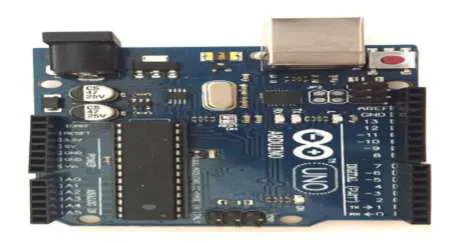
Fig1. Arduino Uno Board
4.2. 16x2 character LCD display:
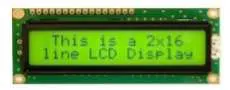
Fig 2: 16x2 character LCD display
- 5 x 8 dots with cursor
- Built-in controller
- + 5V power supply (Also available for + 3V)
- 1/16 duty cycle
- N.V. optional for + 3V power supply
4.3. Heart Beat sensor:
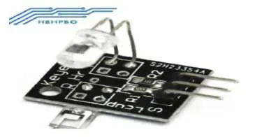
Fig3. Heart Beat sensor
- The patient’s heart beat rate is monitored using a photoelectric sensor which can Sense the patient’s pulse rate.
- This method of tracking the heart rate is more efficient than the traditional method which derives the same from the ECG graph.
- This project uses bright infrared (IR) Led and photo detector to detect the pulses of finger.
- The LED is the light side of finger and photo detector on the other side of the finger.
- Phototransistor used to obtain the flux emitted, when the blood pressure pulse by the finger when the resistance of the transistor will be slight changed.
4.4. LM35 sensor:
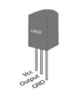
- Fig4. LM35 sensor
- Calibrated directly in ˚ Celsius (Centigrade).
- Linear + 10.0 mV/˚C scale factor.
- 0.5˚C accuracy guarantee able (at +25˚C).
- Rated for full −55˚ to +150˚C range.
- Suitable for remote applications.
- Low cost due to wafer-level trimming.
- Operates from 4 to 30 volts.
- Less than 60 μA current drain.
- Low self-heating, 0.08˚C in still air.
- Nonlinearity only ±1⁄4˚C typical.
- Low impedance output, 0.1 Ω for 1 mA load.
- The LM35 series are precision integrated-circuit temperature sensors, whose output voltage is linearly proportional to the Celsius (Centigrade) temperature.
5. Power Supply
- The power supply circuits built using filters, rectifiers, and then voltage regulators. Starting with an ac voltage, a steady dc voltage is obtained by rectifying the ac voltage, then filtering to a dc level, and finally, regulating to obtain a desired fixed dc voltage.
- The regulation is usually obtained from an IC voltage regulator unit, which takes a dc voltage and provides a somewhat lower dc voltage, which remains the same even if the input dc voltage varies, or the output load connected to the dc voltage changes.
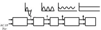
- Transformer Rectifier Filter Regulator Load
- Fig4. Block diagram of power supply.
5.1. Transformer:
- The potential transformer will step down the power supply voltage (0-230V) to (0-6V) level.
- Then the secondary of the potential transformer will be connected to the precision rectifier, which is constructed with the help of op–amp.
- The advantages of using precision rectifier are it will give peak voltage output as DC, rest of the circuits will give only RMS output.
5.2. Bridge rectifier:
- Bridge rectifier is used to maintain the proper DC polarity at the input to the circuit, irrespective of telephone line polarity.
- It comprises of four diodes connected to form a bridge. It uses the entire AC wave (both positive and negative sections).
- 1.4V is used up in the bridge rectifier because each diode uses 0.7V when conducting and there are always two diodes conducting, as shown in fig below.
- Fig 5. Bridge Rectifier
5.3. IC Voltage Regulators:
- Voltage regulators comprise a class of widely used ICs.
- Regulator IC units contain the circuitry for reference source, comparator amplifier, control device, and overload protection all in a single IC.
- Although the internal construction of the IC is somewhat different from that described for discrete voltage regulator circuits, the external operation is much the same.
- IC units provide regulation of either a fixed positive voltage, a fixed negative voltage, or an adjustably set voltage.
5.4.Three terminal Voltage Regulators:
- Fig shows the basic connection of a three-terminal voltage regulator IC to a load.
- The fixed voltage regulator has an unregulated dc input voltage, Vin, applied to one input terminal, a regulated output dc voltage, Vout, from a second terminal, with the third terminal connected to ground.
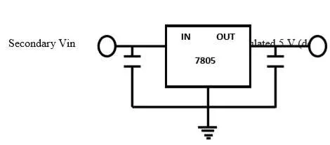
- Fig 6. Fixed Voltage Regulator.
5.5.Circuit Diagram Power Supply:
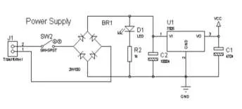
Fig7. Circuit Diagram of Power Supply.
6. APPLICATIONS:
- 1. NIR(Near Infrared) in present day study is evaluated the long term accuracy and stability of the method.
- 2. In clinical brain sciences drug delivery and in vivo Bio-MEMS based biosensors may assist with preventing and early treatment of mental disorders
- 3. Provide accurate statistics.
- 4. It also stores the data in the system memory for the future use.
- 5. Less sensitive towards temperature.
Even less sensitive to water.
7. CONCLUSION
- In the proposed system the low-cost biomedical measurement system with the ability of storage in digital format as well as sending the data to the remote area has been presented.
- This indeed is an easy, practical, inexpensive and yet very effective way for transmitting vital information to the healthcare staff and healthcare provider.
- The system monitors patient's health status, such as heart rate, and temperature.
- All the information obtained from the human body from sensors is then transmitted to the Arduino microcontroller system as digital values.
- The values obtained from like heart rate and temperature is also displayed on to the attached LCD in alphanumerical form.
- In the conclusion we consider how this system can be further improved in future, may be by adding new type of sensors as well as using new approaches for the security and triggering alarm
Block diagram level design of the multi-parameter monitor
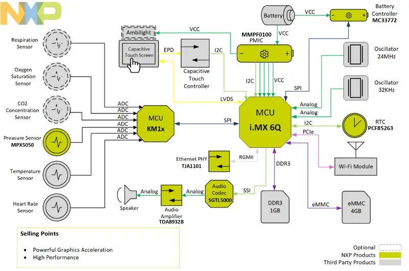
-
In today's world of automation, the field of biomedical is no longer aloof.
-
The application of engineering and technology has proved its significance in the field of biomedical.
-
It not only made doctors more efficient but also helped them in improving the total process of medication.
-
in multispecialty hospitals, where there are a huge number of wards and in each ward there is a spate ofpatients, doctors cannot supervise the patient each and every moment.
-
For these doctors form the time slots and each ward is visited after a specific time difference.
-
But patients may have some problems in between these timeslots.
- The present patient monitoring systems in hospitals allow continuous monitoring of patient vital signs, which require the sensors to be hardwired to nearby, bedside monitors or PCs, and essentially confine the patient to his hospital bed.
- Even after connecting these systems to a particular patient, a paramedical assistant needs to continuously monitor and note down all the vital parameters of a given patient by keeping track of all of his/her records manually.
- Adopting such a method is error-prone and may lead to disaster in the case of a human error.
- In the currently proposed system, patient health is continuously monitored by the Mobile multi-patient monitoring system, and the acquired data is transmitted to a centralized microcontroller using Wireless sensor networks.
- A Bluetooth transceiver is connected to every patient monitor system that consumes very low power and is extremely small in size.
- These are specifically designed for low power consumption, with minimal circuit components intended for a small packet, and long-distance range applications, and typically consist of a low power controller with minimal resources and interface capabilities.
- This Bluetooth is having a data transfer rate of about 10 m.
- So the Wireless Sensor Networks seems to be a perfect fit for remote patient monitoring.
- To improve the accuracy and to increase the efficiency of the above processes a real-time patient monitoring system based on Wireless Sensor Networks and a centralized microcontroller is integrated with a Bluetooth module is designed.
- This paper describes an independent system that automatically logs vital parameters of patients for easy access.
- The data is accessible to doctors through mobile devices for convenience if needed.
- The following figure shows the block diagram of a Bluetooth-based personal medical kit.
- Slight fluctuation in the normal heart rate and body temperature can be measured to the help of this medical kit. It will forward data to the microcontroller where it will be compared with the normal value of body temperature and heart rate.
- Depending on the parameters considered by the monitor, if it finds any parameter disturbed the result is sent to the doctor he may immediately take the necessary section.
- Thus without wasting time patient can be treated whereas sending the report can be done using Bluetooth. Bluetooth-based heart rate monitoring and the display system is portable and the best replacement for the old model stethoscope, which is less efficient.
- It is a combination of a HIGH POWER LED-based heart rate monitor interface with a Bluetooth module to transmit the heart rate of a patient to a remote location.
- The functioning of this device is based on the truth that the blood circulates for every heartbeat that can be sensed by LED. Depending upon the rate of circulation of the blood the heartbeat per minute is calculated.
- This calculated value is communicated to the person through Bluetooth to the receiver which is interfaced with it.
- Bluetooth-based heart rate monitoring and the display system is portable and the best replacement for the old model stethoscope, which is less efficient.
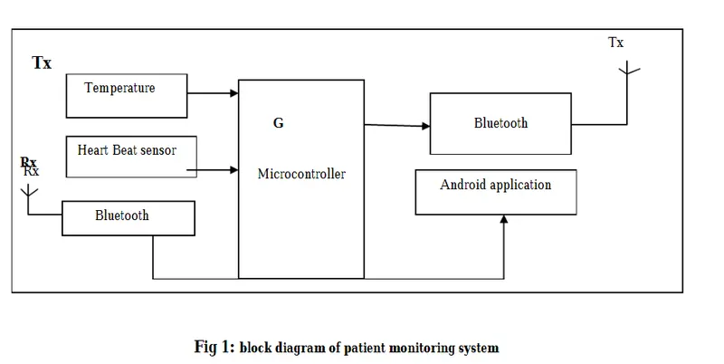
- It is a combination of a HIGH POWER LED-based heart rate monitor interface with a Bluetooth module to transmit the heart rate of a patient to a remote location.
- The functioning of this device is based on the truth that the blood circulates for every heartbeat that can be sensed by LED.
- Depending upon the rate of circulation of the blood the heartbeat per minute is calculated.
- This calculated value is communicated to the person through Bluetooth to the receiver which is interfaced with it.
- The temperature sensor we are using here in LM35 senses the body temperature of the patient and it is connected to the microcontroller.
- The heartbeat of the patient can be sensed by #1157 produced by SUN ROM technology, this is the advanced version of the sensor which is put into the finger of the patient and the output of this sensor is connected to the microcontroller.
- All this data is in analog form so we have to convert it into digital form, so we are using the ADC pins of the microcontroller for converting analog to digital form, all the digital data is transmitted to the receiver section where the current state of the patient can be seen.
- In the transmitter section, The received data is then connected to the microcontroller ATmega328, then this data is given to a computer or mobile port.
- The heartbeat sensor is based on the principle of photoplethysmography.
- It measures the change in volume of blood through any organ of the body which causes a change in the light intensity through that organ (a vascular region).
- In the case of applications where heart pulse rate is to be monitored, the timing of the pulses is more important. The flow of blood volume is decided by the rate of heart pulses and since light is absorbed by the blood, the signal pulses are equivalent to the heartbeat pulses.
- There are two types of photoplethysmography:Transmission: Light emitted from the light emitting device is transmitted through any vascular region of the body like the earlobe and received by the detector.
- Reflection: Light emitted from the light emitting device is reflected by the regions.
- The LM35 series are precision integrated-circuit temperature devices with an output voltage linearly proportional to the Centigrade temperature.
- The LM35 device has an advantage over linear temperature sensors calibrated in Kelvin, as the user is not required to subtract a large constant voltage from the output to obtain convenient Centigrade scaling.
- The LM35 device does not require any external calibration or trimming to provide typical accuracies of ±¼°C at room temperature and ±¾°C over a full −55°C to 150°C temperature range.
- Lower cost is assured by trimming and calibration at the wafer level.
- The low-output impedance, linear output, and precise inherent calibration of the LM35 device make interfacing to readout or control circuitry especially easy.
- The device is used with single power supplies, or with plus and minus supplies. As the LM35device draws only 60 µA from the supply, it has very low self-heating of less than 0.1°C in still air.
- The LM35 device is rated to operate over a −55°C to 150°C temperature range, while the LM35C device is rated for a−40°C to 110°C range (−10° with improved accuracy).
- The LM35-series devices are available packaged in hermetic TO transistor packages, while the LM35C, LM35CA, and LM35D devices are available in the plastic TO-92 transistor package.
- The LM35D device is available in an 8-lead surface-mount small-outline package and a plastic TO-220 package.
- HC-05module is an easy-to-use Bluetooth Serial Port Protocol module, designed for transparent wireless serial connection setup.
- the HC-05 Bluetooth Module can be used in a Master or Slave configuration, making it a great solution for wireless communication.
- This serial port Bluetooth module is fully qualified Bluetooth V2.0+EDR(Enhanced Data Rate) 3Mbps Modulation with a complete 2.4GHz radio transceiver and baseband.
- The Bluetooth module HC-05 is a master/slave module. By default, the factory setting is a slave.
- The Role of the module (Master or Slave) can be configured only by commands.
- The slave modules cannot initiate a connection to another Bluetooth device but can accept connections.
- The master module can initiate a connection to other devices.
- The user can use it simply for a serial to port replacement establish a connection between MCU and, PC to your embedded project, etc.
- The Arduino Uno is a microcontroller board based on the ATmega328 (datasheet).
- It has 14 digital input/output pins (of which 6 can be used as PWM outputs), 6 analog inputs, a 16 MHz crystal oscillator, a USB connection,a power jack, an ICSP header, and a reset button.
- It contains everything needed to support the microcontroller; simply connect it to a computer with a USB cable or power it with an AC-to-DC adapter or battery to get started.
- The Uno differs from all preceding boards in that it does not use the FTDI USB-to-serial driver chip.
- Instead, it features the Atmega8U2 programmed as a USB-to-serial converter.
- The Arduino Uno can be powered via a USB connection or with an external power supply.
- The power source is selected automatically.
- External (non-USB) power can come either from an AC-to-DC adapter (wall-wart) or a battery.
- The adapter can be connected by plugging a 2.1mm center-positive plug into the board's power jack.
- Leads from a battery can be inserted in the gnd and vin pin headers of the POWER connector.
- The board can operate on an external supply of 6 to 20volts.
- If supplied with less than 7V, however, the 5Vpin may supply less than five volts and the board may be unstable.
- If using more than 12V, the voltage regulator may overheat and damage the board.
- The recommended range is 7 to 12 volt.
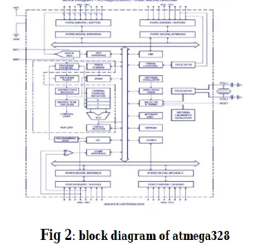
- The Atmel®AVR®AVR core combines a rich instruction set with 32 general-purpose working registers.
- All the32 registers are directly connected to the Arithmetic Logic Unit (ALU), allowing two independent registers to be accessed in one single instruction executed in one clock cycle.
- The resulting architecture is more code efficient while achieving throughputs up to ten times faster than conventional CISC microcontrollers The ATmega32provides the following features: 32Kbytes of In-System Programmable Flash.
- Program memory with Read-While-Write capabilities, 1024bytes EEPROM, 2Kbyte SRAM, 32general purpose I/O lines, 32 general purpose working registers, a JTAG interface for Boundary-scan, On-chip Debugging support and programming, three flexible Timer/Counters with compare modes, Internal and External Interrupts, a serial programmable USART, a byte-oriented Two-wire Serial Interface, an 8-channel, 10-bit ADC with optional differential input stage with programmable gain (TQFP package only), a programmable Watchdog Timer with Internal Oscillator, a SPIserial port, and six software selectable power saving modes.
- The Idle mode stops the CPU while allowing the UART, Two-wire interface, A/DConverter, SRAM, Timer/Counters, SPI port, and interrupt system to continue functioning.
- The Power-down mode saves the register contents but freezes the Oscillator, disabling all other chip functions until the next External Interrupt or Hardware Reset.
- In Power-save mode, the Asynchronous Timer continues to run, allowing the user to maintain a timer base while the rest of the device is sleeping.
- This allows very fast start-up combined with low-power consumption.
- In Extended Standby mode, both the main Oscillator and the Asynchronous Timer continue to run.
- The device is manufactured using Atmel’s high-density non volatile memory technology.
- The On-chip ISP Flash allows the program memory to be reprogrammed in-system through SPI serial interface, by a conventional nonvolatile memory programmer, or by an On-chip Boot program running on the AVR core.
- The boot program can use any interface to download the application program in the Application Flash memory.
- Software in the Boot Flash section will continue to run while the Application Flash section is updated, providing true Read-While-Write operation.
- By combining an 8-bit RISC CPU with In-System Self Programmable Flash on a monolithic chip, the AtmelATmega32 is a powerful microcontroller that provides a highly-flexible and cost-effective solution to many embedded control applications.
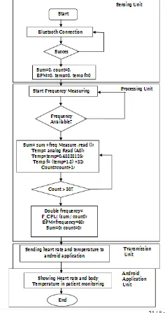
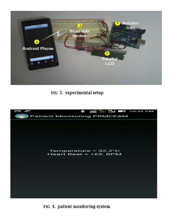
- The patient monitoring system using Bluetooth demonstrates significant improvements in outcomes by reducing hospital readmissions, and improving the quality of care for patients with severe parameters when combining aggressive remote Telemonitoring supported by a health care provider.
- The data provides evidence that the introduction of current state-of-the-art this technology allows rapid and accurate monitoring of patients with severe parameters.
- The combination of these technologies and that parameter management by a health care provider is cost-effective and leads to outcomes and care.
As the healthcare ecosystem has evolved over the years, one of the major feats has been the acceptance and evolution of patient monitoring systems to augment patient care and improve clinical performance. Assessing the condition or other medical parameters of a patient is essential in making informed clinical decisions. Patient monitoring inside and outside the hospital plays an important role in this process, as the infrastructure connects different modules of the healthcare ecosystem for effective patient care.
This addresses the advantages of remote patient monitoring, the shift toward wearables and other wireless-based systems for patient monitoring with examples of current systems, and the factors driving innovation and investment in the healthcare segment.
- “Monitoring” in medical terms attributes to observation of a condition, disease or several medical parameters over time.
- Assessing the patient’s condition, or any other medical parameters, to make informed clinical decisions supports the need for better patient monitoring.
- The healthcare ecosystem has evolved over years. One major feat has been the acceptance and evolution of patient monitoring systems to augment patient care and improve clinical performance.
- Physiological signals hold information that can be extracted to identify the functioning state of various bodily systems.
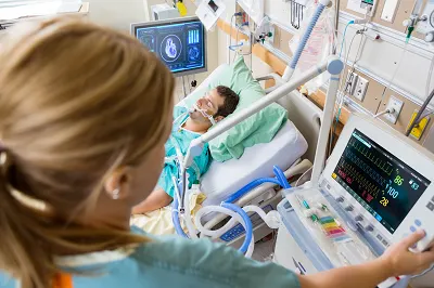
- Throughout treatment, it is extremely important to monitor the vital physiological signs of a patient on a continuous basis.
- Consequently, patient monitoring systems have always occupied a very significant position in the arena of medical devices.
- The increasing medical needs and growing demand for medical facilities are likely to drive patient monitoring systems future market growth.
Monitoring Classifications
- Patient monitoring systems can be classified in several ways. Common types are measured parameters and medical use cases.
- The classification based on measured parameters categorizes the monitoring systems into two broad categories – single parameter monitoring and multi-parameter monitoring.
- Single parameter systems can measure only single physiological signals like blood pressure, ECG, SPO2, and CO2. Multi-parameter monitoring systems can measure a combination of vital signals like ECG, respiration rate, blood pressure, SPO2, brain waves, gas anesthetic concentration, and body temperature.
- Alternatively, the classification based on medical use cases categorizes the patient monitors as follows:
- Blood glucose monitoring
- Hemodynamic monitoring – continuous cardiac output (CCO), stroke volume (SV), stroke volume variation (SVV), pulse pressure variation (PPV), mean arterial pressure (MAP), and systemic vascular resistance (SVR)
- Neurological monitoring – brain Waves (EEG and EMG), gas anesthetic concentration, bispectral index
- Cardiac monitoring – ECG
- Body temperature monitoring – thermometry
- Respiratory monitoring – pulse oximetry & capnography (CO2)
- Fetal and maternal monitoring – maternal and fetal heart rate
Infrastructure
- A typical patient monitoring system consists of one or more sensors, processing components, display devices and communication links for displaying or recording the results elsewhere through a monitoring network.
- Sensors used in the setup can be optical, electrochemical, electrodermal, or pressure and strain gauge.
- The processing and display components comprise microcontroller units (MCU) that can handle analog signal conditioning, digital signal processing, motor control, LCD control, touch sensing, wired and wireless connectivity.
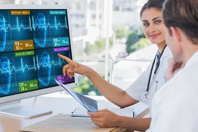
- On the communication front, several patient monitoring units are networkable, i.e., they can send their output to a central nursing station where an individual staff member can monitor and respond to multiple bedside monitors simultaneously.
- Different healthcare standards have been developed by organizations like IEEE and HL7 to facilitate integration of physiological data from patient-monitoring networks into hospital electronics’ health record and digital charting systems.
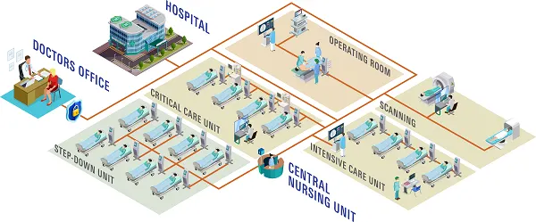
Patient Monitoring System Infrastructure
Wired to Wireless
- Healthcare providers are continuing to seek innovative products that can provide greater level of patient care to growing patient population.
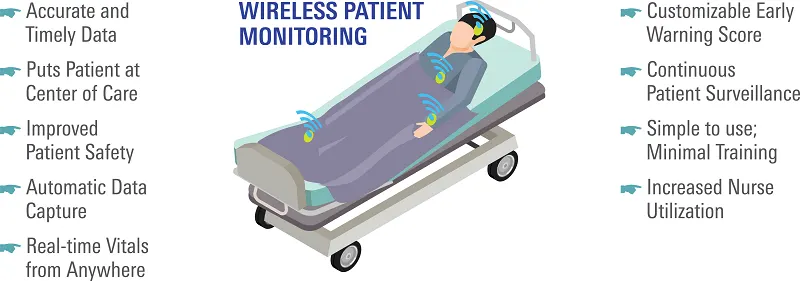
- The move from wired to wireless technology facilitates paramedics, clinicians and medical personnel with a hands-free, wireless device to monitor a patient’s vital signs, creating a safer environment for both EMS personnel and patients.
- The technology also allows real-time connectivity between the emergency room and medical technician in the field.
- Patient data can be transferred through a remote system from the ambulance to hospital to give doctors, nurses and other staff better situational knowledge prior to a patient’s arrival.
- A major advantage of Wi-Fi based monitoring is that it allows hospitals to leverage existing IEEE 802.11 (Wi-Fi) wireless network infrastructure and investment.
- Sixteen percent of U.S. Hospital and Health Systems have already implemented Wi-Fi based wireless patient monitoring and the coverage is expected to jump to 34 percent in the next few years.
- A major driving factor is the FDA 510K approval for recently introduced wireless monitoring devices.
- Low-acuity wearable patient monitoring devices with wireless capability are gaining recognition.
- They are compact, rechargeable and provide secure wireless connectivity.
- Similarly, implantable cardiac monitors that are used for long term monitoring of a patient’s heart electrical activity have secured FDA approval.
- These implantable monitors can be connected to smartphone via Bluetooth.
- The technology provides a new way to monitor for atypical heart rhythms, while staying connected to the doctor remotely.
Remote Patient Monitoring (RPM)
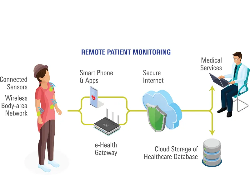
- RPM uses digital technologies to collect medical and other forms of health data from individuals in one location and electronically transmit that information securely to health care providers in a different location for assessment and recommendations.
- The RPM concept aligns well with point of care shifting from hospital to a patient’s home.
- A typical remote patient monitoring system will include a health hub used as the gateway device to collect and transmit vital sign measurements to the healthcare provider.
- The market for RPM solutions remain in a state of development. While growth is appreciable, the overall adoption is still low in comparison to the total number of chronic patients.
- The acceptance of RPM has been mainly in North American and Western European regions where the healthcare providers are incentivized to keep the readmission rates low.
Future Trends
- A patient monitor is considered the “data powerhouse”, capable of accumulating a significant amount of patient data.
- As the healthcare industry changes to improve competence in providing high quality patient care, there is growing demand for data integration and analytics to improve patient diagnosis and workflow.
- The future rests in understanding how patient data can fuel new product innovations, and be utilized in artificial intelligence algorithms within healthcare.
- Rather than making decisions on cloud-based data, emergency protocols could be implemented through decisions made on edge-node data.
- Moving forward, edge node computing combined with wireless patient monitoring will be considered an advantage for life critical applications.
Medical Device Equipment Calibration
- Medical Device Equipment Calibration is a procedure for detecting and fixing the uncertainties in measurements and bringing them to an acceptable level.
- The accuracy of the device has a great deal of importance, as it can seriously affect the diagnostic procedure and endanger patients’ life.
- The deviation in output is not necessarily the fault of the device itself, nor can it represent the manufacturer’s incompetence.
- Most commonly, it results from continuous device usage, which over time affects its internal components, resulting in a deviation from the device standard measurement output.
What is Medical Device Test Equipment Calibration?
- There is a difference between medical device test equipment calibration and maintenance activities.
- Maintenance removes fault in a device, whereas calibration helps to detect the defective instrument that has failed to output values according to the manufacturer’s standard baseline readings.
- The calibration activity varies from instrument to instrument.
- The manufacturer, in most cases, provides a built-in system for performing calibration processes.
- The manufacturer also recommends calibration frequency, although the user itself can devise its frequency, depending on the instrument application and frequency of use.
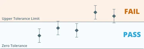
- Every device has its tolerance limit, as defined by the manufacturer.
- If the testing device’s calibration results fall within the tolerance limit, the device can be used further.
- On the other hand, if the calibration fails the device cannot be used for any process.
- Corrective actions are required for devices that fail the calibration.
- Such actions can either be performed in-house or the device can be sent to device specialists. Upon successful corrective actions, the device is again calibrated for verification. If results are satisfactory, the device is allowed to be used again. However, if the defect cannot be fixed, the instrument is permanently damaged and cannot be used further.
- The calibration cycle is followed by complete documentation.
- Common documentation includes:
- Calibration Plan, divided into monthly, quarterly, and yearly basis
- Calibration steps or a Standard Operating Procedure (SOP)
- Calibration Results Sheet indicating the actual results and tolerance limits
- Calibration status is also indicated on the device, showing the calibration status, date of calibration, and next due date for calibration.
- For example, MRI scanners are regularly calibrated for different sections, including the Imaging Gradients. It allows to keep the imaging output according to standard measurements and helps to accurately detect any changes in tissue structure up to the microscopic level.
Equipment Calibration Related Regulations and Standards
- The Medical Device Industry is strictly regulated by regulatory bodies such as Food and Drug Administration (FDA) in the United States to make them compliant with the regulations.
- Additionally, standardization bodies such as the International Organization for Standardization (ISO) also provide standards for performing medical device calibration.
- Some common regulations and standards for medical device companies include the following.
FDA 21 CFR Parts 11
- The FDA 21 CFR Part 11 is the FDA’s guidance regarding electronically saved records.
- Electronic records act as an alternative to manual records.
- Compliance with this act makes the manufacturer’s electronic records trustworthy.
- This regulation also guides electronic signatures, an alternative to manual signatures.
- However, this regulation does not apply to the electronic records transmitted by electronic means.
FDA 21 CFR Parts 820
- The FDA 21 CFR Part 820 is the FDA’s Quality System Regulation for production and process control of medical devices.
- This regulation allows manufacturers to provide valid inspection and test equipment results by establishing routine calibration procedures.
- This regulation also bounds manufacturers to handle all medical devices so as not to harm their accuracy.
Quality System Regulation (QSR)
- Quality System Regulation (QSR) is the FDA’s quality policy for medical device manufacturers, similar to the Current Good Manufacturing Practices (cGMP) for Food, Drugs, and Biologics.
- The QSR only applies to finished medical devices that are ready to be used.
- The QSR does not target any particular medical device but provides flexible guidelines to cover all medical devices.
- QSR puts the responsibility on the manufacturer to develop and establish their quality policies in light of QSR guidelines.
ISO 13485:2016
- ISO 13485 is a standard for medical device manufacturers developed by the International Organization for Standardization (ISO).
- It was reviewed and updated in the year 2016. The number 2016 comes with its terminology.
- ISO 13485:2016 is the set of requirements for a Quality Management System (QMS) in the medical device industry.
- This standard covers all life cycle stages of the medical device, including manufacturing, storage, distribution, installation, and servicing.
- Recommended Reading: Medical Device Quality Management System (QMS)
Why is Medical Device Test Equipment Calibration Important?
- Medical devices can drift away from the measurements due to their internal components.
- This phenomenon is common among all manufacturers, even with the most advanced ones.
- Over time, the internal components – both mechanical and electronics wear and tear.
- As a result, their performance starts to decrease and results in erratic readings.
- These erratic readings can directly affect the patient life, such as in the case of ultrasound units.
- Secondly, as mentioned above, regulatory bodies such as the FDA in the US have made it mandatory for the manufacturers to perform routine calibration and maintain calibration records.
- Otherwise, the manufacturer is not granted accreditation and is not allowed to start or continue its operations.
- The calibration process is necessary to detect these erratic readings and comply with the regional regulatory bodies.
- The calibration must be regularly performed, and documentation maintained for presenting during a routine inspection.
Medical Equipment Calibration Requirements
- The calibration is critical in the medical device industry and must be performed according to regulatory requirements.
- Considering its importance and significance, manufacturers prefer to dedicate separate resources for calibration activity such as:
- Establishing a department
- Hiring skilled personnel
- Implementing an Equipment Calibration Management Software
- Maintaining instruments to perform calibration
- Common requirements for calibration, as per the FDA 21 CFR Parts 820, section – 820.72 “Inspection, measuring, and test equipment”, are summarized below.
Procedures and Instructions
- It is the manufacturer’s responsibility to develop procedures for regular calibration of each device.
- The procedure should contain tolerance limits that help in qualifying instruments (fail or pass).
- In case the device calibration fails, the procedures should guide how to meet the acceptable limit.
- Secondly, procedures should document what to do if the device completely fails the calibration.
- For example, if a department replaces a device due to fault, it cannot use the new device without calibration and verification from relevant departments such as the Quality Assurance Department.
- In this scenario, the procedure for a new device calibration is required and should be developed by the device user department.
- The procedure should also contain steps to replace and update the relevant documents.
Regulatory Standards
- This includes the applicable standards for performing routine equipment calibration.
- The calibration standards should be traceable to national and international standards such as the ISO 13485:2016.
- In the absence of available standards, the manufacturer shall establish its own, in-house standards.
Documentation and Records
- The calibration activities must be documented and calibrations record well-maintained. A proper calibration plan should be in place, indicating each device’s calibration frequency, i.e., weekly, monthly, quarterly, or yearly. All the calibration records should be readily available during an inspection.
- The calibration records should contain the following information:
- Equipment identification number
- The date when calibration was performed
- Name of the person performing the calibration
- The due date for the next calibration
- In addition to records, the calibration status or tag should be indicated on the individual device, including the calibration date, the next calibration due date, and the name of the person performing the calibration.
- A software solution such as SimplerQMS can help you schedule each device’s calibration frequency and remind you when it’s time for the next calibration.
- The software is 21 CFR Part 11 compliant and allows you to sign calibration records with 21 CFR Part 11 compliant electronic signatures.
Training of Personnel
- These include training methods for the personnel involved in calibration activities, including the instructions on how to handle and store the calibrated devices.
- The manufacturer is responsible for conducting training for the personnel involved in calibration activities. Therefore, a proper training calendar and a training plan must be in place.
- All training activities must be recorded and maintained, including the topic details, attendance sheet, and trainer details.
- For example, the training plan for the instrument calibration can include the following:
- The personnel must be trained to use the master instrument according to standard specifications.
- The personnel must understand the instrument’s operational requirements, such as power or utility for normal operation.
- The personnel must be trained to establish a safe and secure connection between the master and calibrating instrument.
- The personnel must be trained about the device being calibrated. It includes operational training, available functions and features of the device, and how it connects to the master instrument.
- The personnel must be trained about the relevant applicable standards and their requirements.
- The personnel must be trained to complete all the documentation correctly. It includes recording the observed values against the standard values, identifying tolerance levels. The personnel must indicate calibration status on the device according to approved formats such as tags.
How to Manage Medical Equipment Calibration Effectively
- As mentioned above, calibration is of utmost importance for manufacturers when meeting compliance requirements.
- Regulatory bodies rely on the documentation developed by the manufacturer to decide whether or not the manufacturer has effectively established and followed calibration procedures.
- First of all, all the documents should be readily available for inspection by the regulatory bodies.
- If not produced timely, the regulatory body can assume that the calibration was not performed.
- The documents should be up-to-date and follow the approved calibration plan.
- The records for defective devices should not be included, and they must be separated from the supported devices.
- An effective way of managing calibration activities is by using digital solutions such as Equipment Calibration Management Software by SimplerQMS.
- SimplerQMS software solution is compliant with FDA 21 CFR Parts 11, FDA 21 CFR Parts 820, Quality System Regulation (QSR), ISO 13485:2016.
- It allows users to store all the calibration documents in a centralized system, in secured cloud-based storage.
- The system tracks the documentation status and automatically prompts email alerts to the responsible person to complete assignments.
- Equipment Calibration Software by SimplerQMS allows you to create a calibration plan of the device, so you don’t need to inspect device by device ‘on the floor’ to plan the device calibration.
- The software automatically creates a complete equipment calibration log that can easily be presented during audits or inspections.
- The system follows the calibration schedule and automatically alerts the responsible person when calibration is due for a particular device.
- This prevents you from missing the calibration for a particular device, as missed calibration can seriously cause regulatory non-conformance.
- Calibration is becoming an important tool for the regulatory bodies to gauge medical device manufacturer compliance adherence.
- It sometimes becomes difficult to track calibration of all devices, for example, due to human negligence error.
- In large manufacturing facilities, some devices are left without their documentation, although they have undergone proper calibration procedures.
- Sometimes due to equipment or area shifts, device identification is not updated.
- All the problems mentioned above can be effectively solved by using digital tools like the SimplerQMS Calibration Management Software.
- It helps the manufacturer to adhere to regulatory body requirements streamline various calibration process
- Design ICU with 8 beds using the multi-parameter monitor and how is it proposed to be networked. How is the Central Nursing station proposed to be designed and explain in detail?
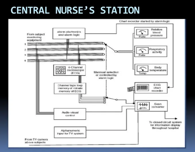
- the monitoring mode, there are no monitors at the bedside, and patients are managed from the nurse's station using central monitors.
- Telemetry monitoring is commonly used to manage less-critical patients who are able to (and sometimes required to) ambulate.
- Current telemetry systems are capable of monitoring in real time, 5-lead ECG, noninvasive blood pressure and oxygen saturation.
- One example of the state of the art in telemetry monitoring (Figure 98-4) is the Apex Pro Telemetry System product from GE Medical Systems.
- A telemetry monitoring system consists of transmitters, antenna subsystems, receiver cabinets and central station monitors.
- The transmitter, attached to the patient, sends real-time digitized vital signs data over a radio-frequency (RF) carrier signal.
- A network of antenna subsystems, installed at strategic locations in the care unit for maximum coverage, communicates the digitized data to a set of receivers for processing.
- The central monitor located in the nurse station displays and trends the real time information.
- All patient transactions, such as admitting, transferring, discharging, alarm configuration, and printing are performed and managed at the central station.
- The patient also can initiate a print transaction from the transmitter.
- The central monitors store alarm events and parameter trends.
- They can be complemented by dynamic ticker displays and slave monitors placed strategically around the unit.
- These complementary displays can provide up-to-date patient status information to the clinicians when they are away from the nurse station.
- The slave monitors display the same information as the central station.
- The telemetry configuration requires dedicated staff at the nurse station to monitor the patients and to alert the clinicians when there is a change in patient status.
- Clinical staff in the telemetry unit will use the hospital paging system to alert the clinicians.
- Step-down care units typically have a high patient-to-nurse ratio and also a high patient turnover every day.
- The acuity of the patients in these units, in many cases, calls only for telemetry monitoring.
- It is also likely that patients from multiple-care units located on different floors are monitored centrally from one nurse station.
- The total number of patients managed at these nurse stations can be upwards of 40.
- The biggest challenge in this environment of high patient turnover and inadequate staffing is to keep the clinicians informed of the changing status of their patients.
- Fast and reliable methods to communicate alerts to the caregivers are available in the market today that communicate alarms from a monitoring station to the alphanumeric pagers carried by clinicians.
- These systems reside on the same network as the telemetry receivers and the central monitors, receiving patient alarm information in real time, just like the central monitors.
- They are programmable to send alarm messages to one or more pagers, depending on the severity of the alarm and the responsibilities of the clinician.
- The alarm messages include the patient's name and room, as well as the date, time, and type of the alarm.
- The system can be programmed to send alarm messages from selected patients to a designated pager and a backup pager.
- Management reports generated by these systems can provide data on frequency and response times for different alarm categories that can be utilized by the institution for implementing changes in clinical practice and adjusting staffing levels in the unit.
- Note that these alarm communication systems were designed and sold only as adjuncts to the traditional monitoring systems and that they cannot replace the role of dedicated and knowledgeable clinical staff.
- The telemetry receiver cabinets and central monitors can reside on the same network with hardwire monitors.
- Because bedsides can be configured for both telemetry and hardwire monitoring, the clinician can tailor monitoring system capability to the acuity of the patient.
- Patients on telemetry and hardwire monitoring systems can be viewed on the same central station monitor, and managed from one nurse station.
- This flexibility significantly reduces the need for moving the patient within the unit and greatly benefits bed-management operation.
- Because both the telemetry and hardwire systems use the same patient interfaces (e.g., cables and leads), patient discomfort is also greatly reduced when switching between the two systems.
- Intermediate care units, with varying patient acuities, are usually equipped with hardwire monitors and an antenna network for the telemetry system for this very reason.
Patient monitoring alarms in the ICU and in the operating room
Introduction
- Historically, the word 'alarm' originates from the Latin, 'ad arma', or the French, 'à l'arme', which can be translated into 'to your weapons'.
- Hence, the word indicates a call for immediate action, for attack or for defense.
- Alarms have existed ever since humans have lived in groups. Some of the first documented alarms are watchmen on towers in the Middle Ages, who warned of fires or enemies by ringing bells.
- Warning fires provided a visual alert to enemy attacks, visible across long ranges and enabling an early reaction of armed forces.
- Today, comparable systems are available that send warning-SMSs (Short Message Service) of nearing tsunamis to mobile phones .
- In complex fields of work like aviation, mining, anesthesiology, and intensive care medicine - and here particularly with regard to monitoring of vital functions - alarms are ubiquitous and have been the subject of medical, technical, and psychological research for decades .
- Monitoring of vital functions and function of life-support devices is essential for critically ill patients, although real evidence based data are missing.
- However, modern patient monitors and implemented risk management (including alarms) must be constructed in accordance to approved and current international standards IEC 60601-1-11 and IEC 80001-1 . Tinker et al. surveyed 1,175 anesthetic-related closed malpractice claims from 17 professional liability insurance companies.
- It was determined that 31.5% of the negative outcomes could have been prevented by use of additional monitors.
- The authors concluded that monitoring with adequate thresholds appeared able to improve patient outcomes ].
- Cooper e t al. showed in the 1980s that 70% of all anesthesia-related critical incidents were caused by human error .
- Similar data are available from the aviation industry .
- Inevitable mistakes may be corrected in time if detected by a monitoring system (including alarms) before physiological variables run out of range.
- An alarm is an automatic warning that results from a measurement, or any other acquisition of descriptors of a state, and indicates a relevant deviation from a normal state .
- Loeb surveyed the reaction of anesthesiologists to relevant changes in monitoring parameters, and showed that anesthesiologists needed a mean time of 61 seconds to recognize a change in the parameters; 16% of the changes were unrecognized for over 5 minutes .
- In contrast, Morris and Montano studied the reaction of anesthesiologists to optical and acoustic warnings during maintenance of general anesthesia.
- The anesthesiologists showed a reaction time of 6 seconds to optical warnings and 1 second to acoustic warnings .
- An ideal alarm should only detect immediate or threatening danger that requires prompt attention.
- The alarm design should adequately represent the underlying situation. The announcement of the alarm should be instantly perceptible in critical situations.
- Additionally, the user should be informed of circumstances that impair the reliability of the alarming system.
- In addition to these general properties, device alarms have various goals, which follow a certain hierarchy :
-
Detection of life-threatening situations: The detection of life-threatening situations was the original purpose of monitor alarms. False negative alarms are not acceptable in such situations because of the danger of severe patient harm or death.
-
Detection of imminent danger: The early detection of gradual change that might indicate imminent danger.
-
Diagnostic alarms: These alarms indicate a pathophysiological condition (e. g., shock) rather than warning of 'out-of-range' variables.
-
Detection of life-threatening device malfunction: This ability is essential for all life-support devices, which must recognize malfunctions, such as disconnection from the patient, occlusion of the connection to the patient, disconnection from power, gas, or water supply, and internal malfunction.
-
Detection of imminent device malfunction: The early detection of device-related problems that could result in malfunction is an integral part of many therapeutic devices. These warning mechanisms range from simple aspects (e. g., low-battery warnings) to complex algorithms and sensors that track the wear of respiratory valves.
Alarm design
Alarms are typically displayed in two ways or as a combination of both:
1. Acoustic
- The alarm is given as a warning sound. Most manufacturers distinguish the priority of an alarm with different signals.
- Intuitive alarms with different tone sequences (e. g. 'short-long-short' for 'ven-ti-late') have been the object of research but have not found their way into routine clinical practice.
- Alarms directly mentioning organ systems, device hardware, or parts of it (e. g., ventilation or circulation) or alarms with direct labeling of the physiological problem ('blood pressure' or 'oxygen') have also not been introduced into practice .
2. Visual
- Visual alarms involve mostly flashing or coloring of the related parameter in an eye-catching manner.
- Some systems provide integrated displays of several parameters.
- One example is a spider-display, which shows the relationship of different parameters in a stylized spider web.
- Such applications can be useful to display different parameters in context.
- Compared to other professions in industry and aviation, adoption of such new displays in healthcare has been slow.
Alarm-related problems
- Alarms help to prevent patient harm by providing rapid recognition of and reaction to critical situations, but only if they are not 'false alarms'.
- Medical progress leads to an increasing number of 'monitorable' parameters and thus an increasing number of possible alarms.
False alarms
- In medicine, false alarms are conventionally defined as alarms without clinical or therapeutic consequence.
- Today's monitoring systems are still designed using a 'better-safe-than-sorry'-logic:
- A large number of false alarms are accepted rather than risking missing one valid alarm .
- Alarms can be differentiated into technically correct/technically false and clinically relevant/clinically not relevant.
- Alarms can be classified as technically correct, if they are based upon a technically correct measurement.
- Technically false alarms are not based on a technically correct measurement (e. g., interference with pulse oximetry caused by ambient light).
- Because not all technically correct alarms are clinically relevant, they can be further differentiated into clinically relevant or not relevant (e. g., inadequate thresholds).
False alarm rates
- There are several studies in the medical literature about monitoring alarms in anesthesiology and intensive care medicine.
- Lawless suggested that 94% of all alarms in a pediatric intensive care unit (PICU) were clinically irrelevant.
- Tsien and Fackler also found that 92% of alarms were false alarms in their observation in a PICU.
- In both studies, all alarms were recorded by the nursing staff, who also assessed their relevance and validity. O'Carroll reported that only 8 of 1,455 alarms were caused by potentially life-threatening situations. An observation by Liebig and co-workers showed that these results are not limited to the PICU.
- These authors digitally recorded all the alarms for 38 patients on a 12-bed medical ICU and retrospectively assessed their relevance and validity:
- Only 17% of the alarms were relevant, with 44% being technically false.
- Chambrin et al. conducted a multicenter study in 1999, including 131 medical ICU patients. The medical staff recorded all alarms, which were assessed according to their relevance and the reaction of the medical staff.
- Twenty-six percent of the alarms had marginal consequences, for example leading to the re-positioning of sensors.
- In only 6% did the alarm lead to a call for a doctor. Seventeen percent were the result of technical problems and 24% were caused by staff manipulation
- In contrast to ICU observations, there are only a few studies about false alarms in perioperative settings.
- Comparison between the ICU and the operating room (OR) is limited in part because ICU patients are only sedated and not anesthetized, causing higher rates of patient movement artifacts.
- Furthermore, in the OR, changes in patients' conditions often occur much more rapidly than in the ICU because of changes in the depth of anesthesia and surgical manipulation (e. g., extensive blood loss).
- Schmid et al. studied perioperative alarms in a highly complex surgical setting and included 25 patients undergoing elective cardiac surgery with extracorporeal circulation.
- All patient monitor and anesthesia workstation alarms were digitally recorded.
- Additionally, the anesthesiology workplace was videotaped from two angles to allow better assessment of external influences, retrospectively. During 124 hours of monitoring, 8,975 alarms were recorded: 7,556 alarms were hemodynamic alarms, 1,419 alarms were ventilation-related.
- This corresponded to 359 ± 158 alarms per procedure (1.2 alarms/minute). The reaction time to the alarms was on average 4 seconds. Of all the alarms, 96% were caused by threshold violations.
- Of the 8,975 alarms, 6,386 were classified as serious and life-threatening and analyzed further: 4,438 (70%) of these were labeled as valid, 1,948 (30%) were caused by artifacts; 1,735 (39%) of the valid alarms were classified as relevant, 2,703 (61%) were not relevant.
- These results supported earlier studies in less complex settings.
- Seagull and Sanderson surveyed perioperative alarms in different surgical disciplines (arthroscopic, cardiac surgery, abdominal surgery, and neurosurgery) with 6 cases in each discipline.
- The authors found 72% of alarms had no clinical consequences .
- A study by Kestin et al. included 50 pediatric patients (1 month to 10 years old) in the OR of a pediatric hospital (pediatric surgery, eye surgery, dental surgery, orthopedic surgery) and also found that 75% alarms had no therapeutic consequences (1 alarm per 4.5 minutes on average).Only 3% of all alarms indicated critical situations ].
- However, the studies by Kestin ] and Seagull were limited by the fact that 5 and 6 different monitors, respectively, were used in these observational studies.
- Artifacts: a common source of false alarm
- Many false alarms are caused by artifacts. The main sources of artifact are well known and are of physiological and non-physiological origin. Most of these artifacts directly influence the measured signals [ leading to incorrect measurements and this, in turn, triggers the alarm. The most common artifacts and their sources.
- The story of "the shepherd who cried wolf" appears in Aesop's Fables and, with some minor variations, can be found in the folklore of many different cultures.
- "One day, just to stir up excitement, the shepherd boy rushed down from the pasture, crying 'Wolf! Wolf!' The villagers heard the alarm and came running to help chase the marauder away, only to find the sheep peaceful and no wolf in sight. But there came a day when a wolf really came.
- The boy screamed and called for help. But all in vain! The neighbors, supposing him to be up to his old tricks, paid no heed to his cries, and the wolf devoured the sheep!" .
- Based on this Fable, Breznitz formed the phrase "Crying-Wolf-Phenomenon" for the desensitization caused by high false alarm rates, with the possible consequence of ignoring relevant alarms .
- The high incidence of false alarms in anesthesiology and intensive care medicine is not only a disturbance but a risk factor when relevant alarms in critical situations are ignored.
- That this phenomenon is not limited to monitoring alarms was impressively demonstrated by the attack of the Japanese air force on the United States Navy at Pearl Harbor, Hawaii on December 7, 1941.
- Despite a valid advance warning by the new radar technology, no appropriate reaction followed.
- The simultaneous report of a contact and the destruction of an enemy submarine also did not lead to a reaction because the commanding admiral wanted to wait for confirmation due to frequent false alarms
- Various studies have shown that anesthesiologists' reaction times to alarms increases in situations where there is low alarm validity . The annoyance from false alarms may also lead to complete inactivation of alarms or to inappropriately wide threshold settings by the clinical user to limit alarms as much as possible. Thereby, the 'mesh of the alarm-net' gets wider and the risk of missed relevant alarm increases
Medical staff and alarms
- Alarms in the ICU and in the OR frequently lead to sound levels up to 70 dB(A).
- This level corresponds to heavy traffic. Sound levels up to 90 dB are not rare . In a study by Hagerman et al., 94 patients with chest pain were retrospectively distributed into a good and poor acoustic group.
- Acoustics were altered during the study period by changing the ceiling tiles throughout the ICU from sound-reflecting (poor acoustics) to sound-absorbing tiles (good acoustics) of similar appearance. The patients were asked to complete a questionnaire about the quality of care.
- The patients considered the staff attitude was much better in the good acoustics period . Increased sound levels caused by alarms can impact on the health of the medical personnel.
- In 1988, Topf and Dillon demonstrated the relationship between in creased sound levels in ICUs and burn-out syndromes in ICU nurses .
Patients and alarms
- For undisturbed night-sleep, sound levels below 40 dB(A) are recommended. As sound levels on the ICU are frequently above this level, sleep deprivation in ICU patients is well recognized .
- Sleep deprivation in ICU patients leads to an impairment of the immune response and increased sympathetic nervous system activity: Catecholamine secretion increases heart rate, metabolism, and oxygen consumption .
- Frequent arousal from sleep may lead to cardiac arrhythmias in patients with pre-existing heart disease but also in healthy patients .
- Minckley reported significantly increased opioid needs when noise levels were high in her observation of 644 postoperative patients .
- In the study by Hagerman et al., patients during the good acoustic (sound-absorbing ceiling tiles) period had lower pulse amplitude values than those in the bad acoustic group; patients in the bad acoustics group also had higher rates of re-hospitalization after 1 (18% vs. 10%) and 3 months (48% vs. 21%) . In a recent study, Van Rompaey et al. found reduced rates and later onset of delirium in patients who slept with earplugs at night .
Technical approaches for false alarm reduction
- Essentially, there are three technical approaches to help reduce false alarms:
- (1) Improving signal extraction(prevention or detection of artifacts);
- (2) improving algorithms for alarm generation;
- (3) improving alarm validation. An algorithm for alarm generation can be based on a single parameter (e. g., heart-rate or mean arterial pressure) or on several parameters simulta neously (e. g., heart rate detection from elecrocardiogram [EKG], pulseoximetry oxygen saturation [SpO2] and arterial line).
- Most devices are equipped with alarms based on a single parameter. In recent years, different approaches for false alarm reduction have been developed.
Phase specific settings
- Observations, especially in surgical settings, have shown that different phases of the surgical procedure are characterized by different numbers and types of false alarms and also different patterns of alarms and specific reactions by the medical staff (e. g., during induction and emergence of anesthesia, during extracorporeal circulation, or single lung ventilation, or suctioning of patients).
- Schmid et al. found different characteristic patterns and density of alarms in 4 different intraoperative phases (beginning of surgery, start of extracorporeal circulation [ECC], end of ECC, end of surgery) .
- Seagull and Sanderson also differentiated three different phases in anesthesia procedures (introduction, maintenance and emergence) and found characteristic patterns of alarms and alarm reactions in each phase .
- This knowledge could be used for the development of phase specific settings to reduce false alarms (e. g., for specific settings for surgery or on the ICU).
Integrated validation of alarms (cross checking)
- Matching of different parameters can be used for the reduction of false alarms: e. g., a 'ventricular fibrillation' alarm can be assumed to be false in the presence of undisturbed pulse oximetry and arterial blood pressure waveforms. Aboukhalil et al. were able to reduce the incidence of false arrhythmia-related alarms from 42.7% to 1 7.2% in an offline application on a database of 447 patients (5,386 arrhythmia alarms).
- However, 9.7% of the true cases of ventricular fibrillation were not detected; this situation is in admissible for a lethal arrhythmia.
- Removal of ventricular arrhythmias from the algorithm still resulted in reduction in the incidence of false alarms to 22.7% .
Implementation of time delays
- Görges et al. in an ICU setting and Schmid et al. in an intraoperative setting that a great number of false alarms are caused by only mild threshold violations of short duration.
- In an offline validation, Görges et al. showed that the implementation of a 14-second delay reduced false alarms by 50%; a 19-second delay reduced false alarms by 67%. However, a simple delay carries the risk of unrecognized critical situations of short duration (e. g., short self-limiting tachycardia).
- The implementation of a graduated delay brings additional safety and flexibility to that approach.
- First, severe deviations are alarmed faster; this results in improved patient safety.
- Second, the graduation offers the possibility of a prolonged delay (more than 14 seconds) in cases of only moderate and clinically not-relevant deviations. However, studies using such an approach are still missing.
Statistical approaches for false alarm reduction
- Improved signal extraction is an essential approach for reduction of false alarms caused by artifacts. Several approaches have been developed over the last decades.
Autoregressive models and self-adjusting thresholds
- An autoregressive model describes measurement values as a linear transformation and integrates previous values plus a random error.
- An autoregressive model is appropriate for the observation of values in steady-state and for alarm generation caused by deviation from steady-state.
- It is also used for the generation of self-adjusting thresholds, because of the integration of the individual patient's condition.
- However, self-adjusting thresholds always have to be elaborated on and confirmed by the user .
Statistical process control
- Statistical approaches are commonly used in alarm systems to detect 'out-of-control' states in a process.
- The original applications were in industrial production processes but they are also used for alarm generation.
- Kennedy used a process control approach to detect the onset of changes in systolic blood pressure .
- The algorithm was tested on an existing database and detected 94% of changes correctly, whereas anesthesiologists only detected 85%.
Median filters
- The median filter is a non-linear, signal processing method used for removal of short-term noise in measurement signals without influencing the baseline signal.
- For this purpose, the median is calculated for a defined interval. Thus, the signal is 'smoothed' and short noises, such as movement artifacts or interference from electro surgery, are eliminated.
- This method is limited in long-lasting interferences that exceed the adjusted duration of the filter.
- Mäkivirta et al. evaluated the effectiveness of a combination of a "short" (15 seconds) and a "long" (2.5 min utes) filter in a database of 10 cardiac surgery patients.
- The use of the filter increased the alarms that had therapeutic consequences from 12 to 49%; the authors declared that no relevant alarms were missed.
Artificial intelligence
- Although statistical approaches are predominantly used for the reduction of artifacts, artificial intelligence offers the possibility of integrating more complex contexts.
- This approach tries to validate alarms by imitating human thinking. Artificial intelligence can be embedded into decision-making systems.
- A paper by Imhoff and Kuhls provides an overview of artificial intelligence use in intensive care monitoring .
Rule-based expert systems
- Rule-based expert systems are based on an integrated expert knowledge database. Some early rule-based expert systems were developed for medical use in the 1970s (MYCIN-System, ONCOCIN-System) .
- These early systems applied expert knowledge from a database into a new context and simulated expert decisions in oncology and therapy of infectious diseases.
- In 1993, Sukuvaara et al. developed an alarm system for the detection of hypovolemia, hyperdynamic circulation, left-heart failure, and hypoventilation.
- Although results showed that rule-based expert systems work well in the context of pathologic conditions, they have not been introduced into the clinical arena.
- The expansion of rule-based expert systems by so-called machine-learning is possible, whereby the pre-existing database is updated by actual patient data.
Neural networks
- Neural networks were developed to imitate the neuronal process of human thinking. They are able to anticipate the presence of diseases on the basis of advance information (e. g., hemodynamic data from a myocardial infarction study group).
- Baxt and Skora developed a neural network for early detection of myocardial infarction in patients admitted to a hospital with chest pain.
- The system was "trained" to detect specific changes in patients with myocardial infarction by implementation of a database (350 patients, 120 of whom had myocardial infarction).
- The results showed that the neural network was able to detect or to exclude infarction with a sensitivity and specificity of 96%.
- The doctors at the respective emergency department achieved an average sensitivity of 73.3% and specificity of 81.1%. Neural networks have also been used for alarm generation in anesthesia ventilators .
Fuzzy logic
- Fuzzy logic was introduced by Zadeh in the 1960s . A common problem in clinical routine is the aim for objectivity and precision when the information does not allow an explicit conclusion.
- Fuzzy logic allows diffuse processing of exact data. Fuzzy logic is widely used in industry (e. g., for picture stabilization in cameras).
- Gold-man and Cordova demonstrated a patient monitor that was able to diagnose a simulated cardiac arrest by evaluation of EKG, capnography, and arterial blood pressure using fuzzy logic.
Bayesian networks
- Bayesian networks have been used for estimation of event occurrence. In patient monitoring, they can be used for decision support.
- Laursen developed software for cardiac event detection .
- The software continuously compared different physiologic parameters and their changes; thus, it was possible to check values against each other for plausibility and to anticipate cardiac events.
Conclusion
- Medical progress has led to obvious improvements in ICU and perioperative monitoring over recent decades.
- With the increase in 'monitorable' parameters, rates of alarms have also increased. But technical progress has rarely affected the rates of false alarms.
- In addition to noise-related increase in burn-out rates, false alarms lead to desensitization of medical staff to alarms with the risk of critical situations potentially being ignored despite correct alarming.
- Patients are also directly affected by alarm-related sleep disorders with subsequent development of delirium and increased sympathetic nervous system activity and catecholamine secretion.
- In recent years, many promising approaches using statistical methods and artificial intelligence have been developed for the reduction of false alarms without obvious changes in false alarm rates in our clinical reality
Health Data Management: Benefits, Challenges and Storage – Cloudian
- Healthcare data is increasingly digitized and, like in most other industries, data is growing in Velocity, Volume and Value.
- Health Data Management is the practice of making sense of this data and managing it to the benefit of healthcare organizations, practitioners, and ultimately patient well being and health.
- Read on to learn what types of data are managed in Health Data Management, the benefits and unique challenges of the field, including data protection considerations and measures to avoid data breaches when storing healthcare data.
What is Health Data Management?
- Health Data Management (HDM), also known as Health Information Management (HIM) is the systematic organization of health data in digital form. This can be anything from Electronic Medical Records (EMR) generated as a result of doctor visits, to Electronic Health Records (EHR), to handwritten medical notes scanned to a digital repository.
- Health Data Management is tasked not only with organizing medical data but also integrating it and enabling its analysis to make patient care more efficient, and derive insights that can improve medical outcomes, while protecting the privacy and security of the data.
- According to a Deloitte survey:
- Most healthcare organizations collect EMR abstracts, claims data, and data about enrollment and medical programs.
- Only leading organizations additionally use electronic EMR feeds and disease management program data.
- Very few organizations use non-health data sources that can be used to augment formal medical data, such as patient lifestyle information, remote monitoring and wearable devices, and survey data about patient experience.
- As Health Data Management progresses, more organizations will collect a larger variety of health-related data, and integrate it to generate new insights that can enhance patient health.
- Five Health Data Management Benefits
- Health Data Management can have significant benefits for healthcare organizations, medical staff and patients:
- Create a comprehensive view of patients, households, and patient groups—composite profiles that provide status and enable predictions.
- Improve patient engagement—target patients with reminders and care suggestions that can be relevant for them, based on predictive modeling.
- Improve health outcomes—track health trends in certain areas or among specific populations, predict new trends and suggest proactive measures to counter rising health issues.
- Business decision making—help healthcare providers make better data-driven decisions, such as which types of medical professionals to recruit, what equipment to invest in, or which types of patients to focus on in marketing efforts.
- Analyze physician activity—analyze data on medical practitioners such as success rates, time invested in different treatments and medical decisions, and aligning physicians with the goals of the healthcare organization.
- Three Health Data Management Challenges
- In the past forty years, medical data began a transition from purely paper-based tracking to digitized information. Even today, many types of medical data have yet to be digitized, or have not yet been integrated into Health Data Management systems.
- Here are a few important challenges facing health data professionals today:
- Fragmented data—medical data can be structured data in spreadsheets or databases, images or video files, digital documents, scanned paper documents, or may be stored in specialized formats such as the DICOM format used for MRI scans. Data is widely duplicated, collected multiple times and stored in different versions by healthcare providers, public health organizations, insurance bodies, pharmacies, and patients themselves. There is no one source of truth for information on patient well being.
- Changes to data—medical data constantly changes as do the names, professions, locations and conditions of patients and physicians. Patients undergo numerous tests and are administered many types of treatment over the years, and the treatments and medications themselves evolve over time. New types of medical treatment, such as telehealth models, create new types of data.
- Regulations and compliance—medical data is sensitive and must adhere to government regulations, such as the USA Health Insurance Portability and Accountability Act (HIPAA). Data discovery challenges and poor data quality make it much more difficult to perform the required audits and meet regulatory requirements and limits the diversity of data healthcare providers can use for the benefit of patients.
- Storage Considerations for Healthcare Data
- According to research from IDC, the amount of data generated by healthcare information and imaging systems is expected to grow from 153 Exabytes in 2013 to 2,300 Exabytes in 2020.
- Beyond the above challenges, healthcare providers need to consider how they will store all that data:
- Scalability—the fastest-growing segment of healthcare data is unstructured data such as MRIs, CT scans, X-ray and PET scans. As this data grows to Petabytes, healthcare organizations need a highly scalable, low-cost storage solution.
- Compliance—storage must be compliant with regulations. HIPAA and other healthcare industry regulations require that data be protected with Role-Based Access Controls (RBAC), audit trail logging and data encryption at rest and SSL for data in transit, with properly-audited key management procedures.
- Vendor-neutral archive (VNA)—a VNA provides one interface for multiple healthcare information platforms. It makes it easier to consolidate many types of healthcare information into a central repository that provides a central view of patient records. Storage platforms should provide VNA integration.
- Data resiliency and protection—health data is a frequent target for cyberattacks and represents a major risk for providers if it is accidentally lost or deleted. Storage systems should provide redundancy, replication, data backup, and erasure coding that can distribute fragments of data across multiple nodes.
- Secure, Low-Cost Health Data Storage with Cloudian
- Cloudian provides HyperStore, an Exabyte-scale, on-premise, cost-effective storage platform for healthcare environments. It allows healthcare organizations to store, move and protect files across locations at up to 70% lower cost than disk-based storage.
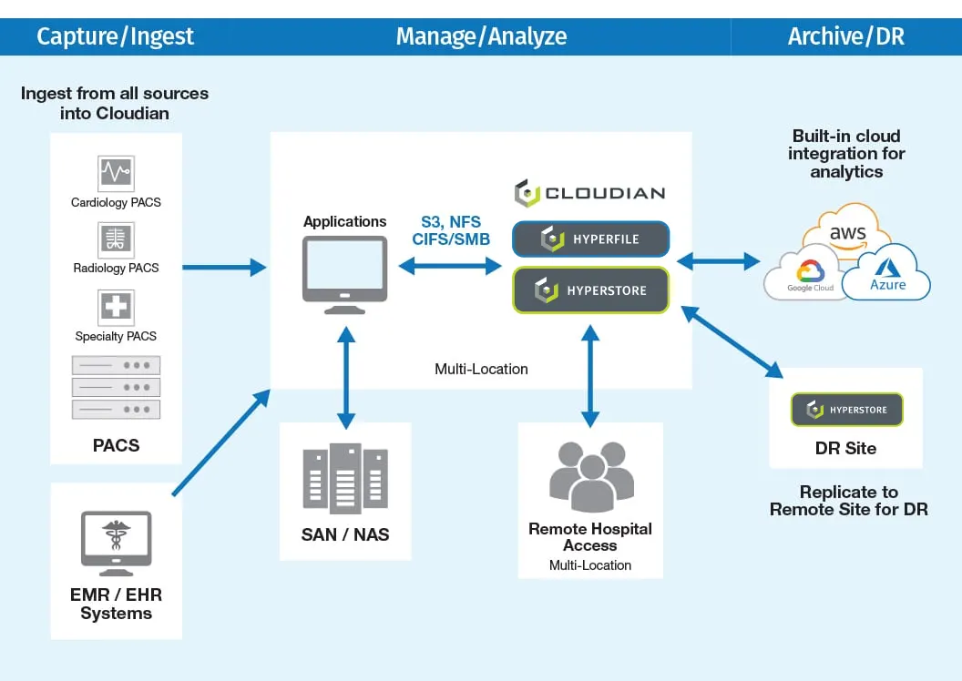
- Hyperstore provides the following benefits for healthcare organizations:
- Cloud flexibility—deployments can start small and grow as needed, from a 3-node configuration to thousands of nodes as needed. Each node can be a physical appliance or virtual, running on commodity hardware.
- Broad integrations—Hyperstore integrates with numerous healthcare applications and archive solutions, allowing it to be used as a central repository, with access to a complete view of patient information.
- Security and compliance—Hyperstore is HIPAA-compliant, with security features including data encryption and transparent key management, AES-256 server-side encryption for data at rest, SSL for data in transit, RBAC with specified levels of access, audit trail logging, WORM (Write Once Read Multiple) for storage of immutable data.
- Rich metadata—HyperStore has rich metadata tagging features built-in, which lets data scientists to discover new patterns and insights in healthcare data.
- Data protection—HyperStore provides data durability of 99.999999999999% (14 nines), by distributing data using replication or erasure coding. The number of replicas and erasure code scheme can be customized to meet the SLA.
- Learn More About Health Data Management
- Health Data Management (HDM) is the methodical organization of health data in digital form. This can be anything from Electronic Health Records (EHR), Electronic Medical Records (EMR) generated as a result of doctor visits, to handwritten medical notes.
- Health Data Management enables you to integrate and analyze medical data to make patient care more efficient, and extract insights that can improve medical outcomes, while protecting the security and privacy of the data.
- There’s a lot more to learn about health data management. To continue your research, take a look at the rest of our blogs on this topic:
- Medical Records Retention: Understanding the Problem
- Every person in the developed world has a medical record. Medical records collect valuable and sensitive information about the health of an individual. This includes their well being, medical conditions they suffer from and required medical treatments.
- The need to retain medical records for long periods of time, makes them complex to manage. This article reviews the concept of medical records, how long you need to retain them, and how to manage organizational and storage aspects of medical records.
- HIPAA Compliant Cloud Storage and On-Premises Alternatives
- HIPAA regulations apply to healthcare organizations, healthcare professionals, healthcare insurance companies, and business services providers. Protected Health Information (PHI) is increasingly digitized, and the exploding volume of protected data raises the question of how to store the data in a way that is HIPAA compliant and conducive to protecting data privacy and security.
- This article reviews the basics of HIPAA compliance, how it applies to storage both in the cloud and on-premises, HIPAA compliance offered by the major public cloud vendors, and how you can achieve cloud-like benefits with HIPAA compliance on-premises.
- Vendor Neutral Archive: From Silos to Unified Medical Image Data
- A Vendor Neutral Archive (VNA) stores images generated by medical devices and scanners using a standard format. It enables interoperability between the systems that store the images and Communication Systems (PACS). Medical staff can view images from across the organization using multiple screens or devices.
- it explains how VNA systems can standardize and connect imaging silos across a medical organization, and allow medical staff and patients to access and view medical images from anywhere.