Project 1
Part A - Detailed study of anatomical basis of surgical procedures or Clinical management in association with any of the organ system (select 3 system from Module 1 to module 6))
Description:
1. Describe one common clinical condition or disorder associated with the system selected ( Minimum 3 clinical condition / disorder with respect to each system ) example : compound Fracture of tibia bone (system involved musculoskeletal system)
Factors to be remembered
- Describe the gross anatomy of the organ involved
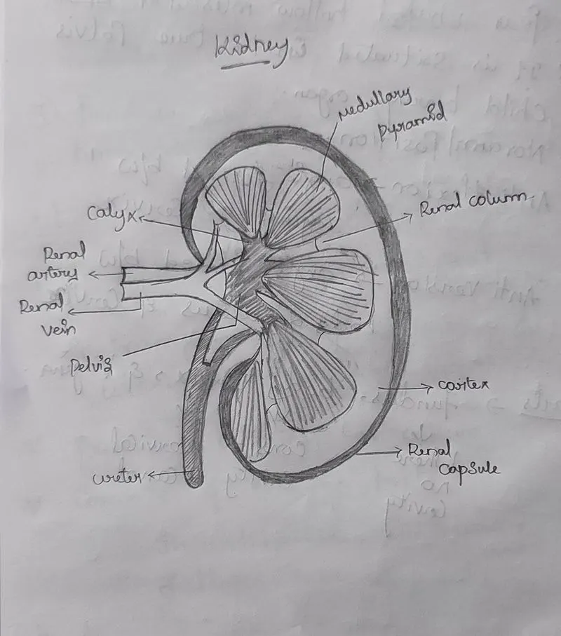
- Describe the microscopic and macroscopic structure of the organ involved with relevant labelled diagram
- Describe the blood supply and nerve supply of the organ involved with suitable diagram
Disorders of the Urinary System
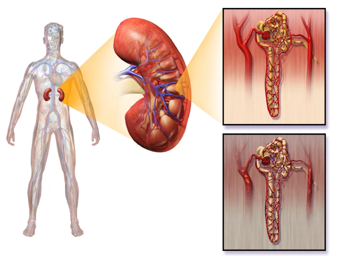
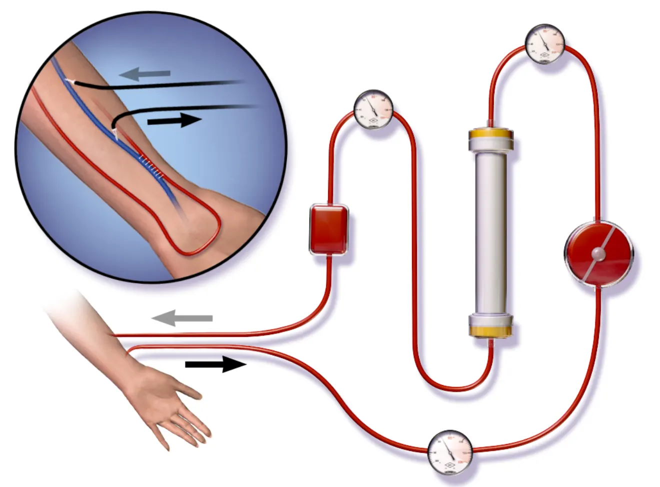
The kidneys play such vital roles in eliminating wastes and toxins and maintaining body-wide homeostasis that disorders of the kidneys may be life-threatening. The gradual loss of normal kidney function commonly occurs with a number of disorders, including diabetes mellitus and high blood pressure. Other disorders of the kidneys are caused by faulty genes that are inherited. Loss of kidney function may eventually progress to kidney failure.
Proteins are large molecules that usually are not filtered out of blood in the glomeruli. When the glomerular capillaries are damaged, it allows proteins such as albumin to leak into the filtrate from the blood. As a result, albumin ends up being excreted in the urine. Finding a high level of albumin in the urine is one indicator of diabetic nephropathy and helps to diagnose the disorder. Drugs may be prescribed to reduce protein levels in the urine. Controlling high blood sugar levels and hypertension (high blood pressure) is also important to help slow kidney damage, as is a reduction of sodium intake.
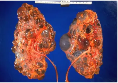
Polycystic Kidney Disease
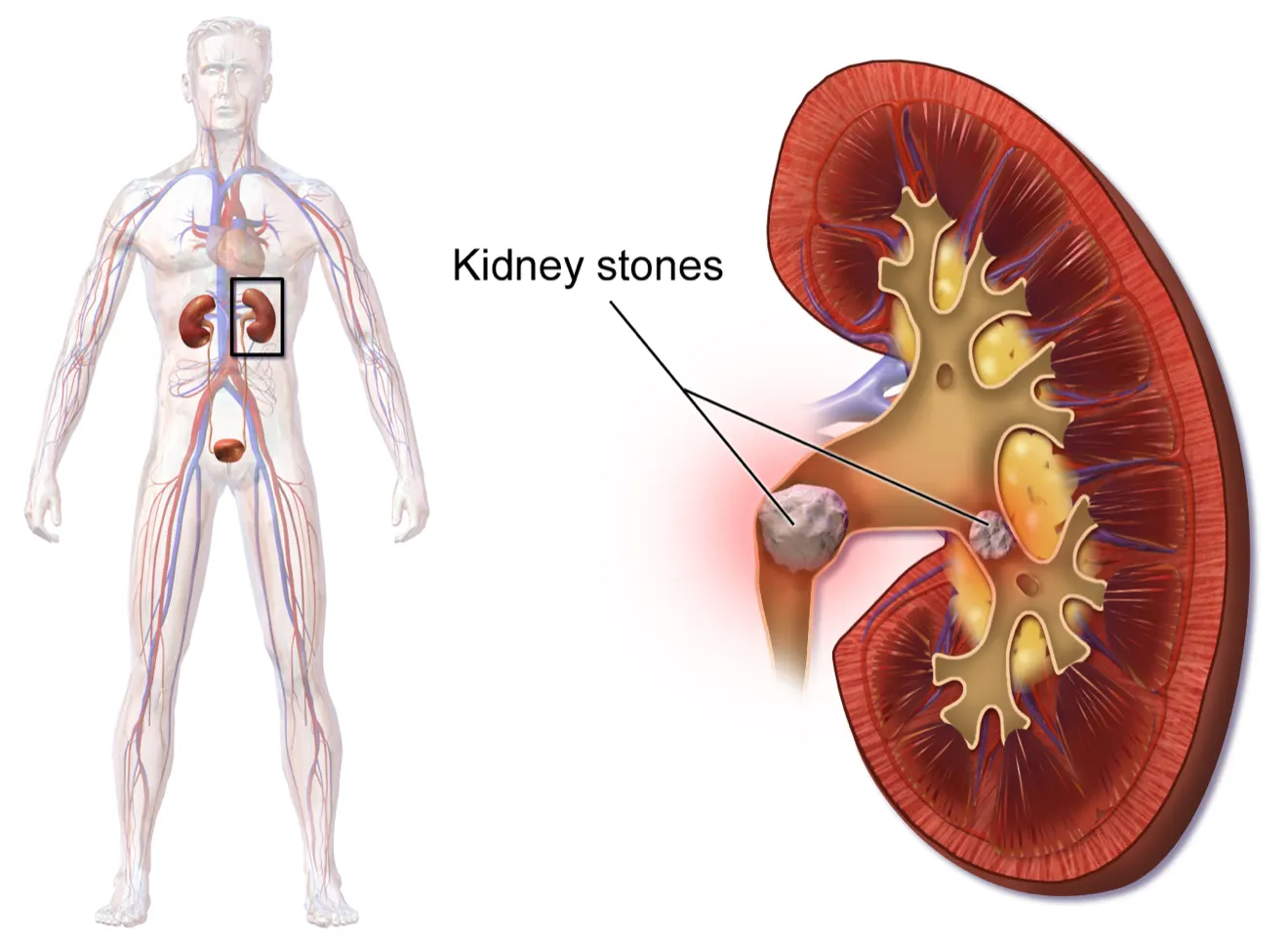
A kidney stone, also known as a renal calculus, is a solid crystal that forms in a kidney from minerals in urine. The majority of kidney stones consist of crystals of calcium salts. Kidney stones typically leave the body in the urine stream. A small stone may pass through the ureters and other urinary tract organs without causing symptoms and go undetected. A larger stone may cause pain when it passes through the urinary tract. If a kidney stone grows large enough, it may block the ureter. Blockage of a ureter may cause a decrease in kidney function and damage to the kidney.A kidney stone that causes pain is generally treated with pain medication until it passes through the urinary tract. A stone that causes a blockage may be treated with lithotripsy. This is a medical procedure in which high-intensity ultrasound pulses are applied externally to cause fragmentation of the stone into pieces small enough to pass easily through the urinary tract. Although lithotripsy is noninvasive, it can cause damage to the kidneys. An alternative treatment for a stone that blocks urine flow is to insert a stent into the ureter to expand it and allow both urine and the stone to pass. In some cases, surgery may be required to physically remove a large stone from the ureter.A combination of lifestyle and genetic factors seem to predispose certain people to develop kidney stones. Risk factors include high consumption of cola soft drinks, eating a diet high in animal protein, being overweight, and not drinking enough fluids. Preventive measures are obvious. They include limiting cola consumption, eating less animal protein, losing weight, and increasing fluid intake.
Other Urinary System Disorders
Although disorders of the kidneys are generally the most serious urinary system disorders, problems that affect other organs of the urinary tract are generally more common. They include bladder infections and urinary incontinence.
A bladder infection, also called cystitis, is a very common type of urinary tract infection in which the urinary bladder becomes infected by bacteria (typically Escherichia coli), rarely by fungi. Symptoms of bladder infections may include pain with urination, frequent urination, and feeling the need to urinate despite having an empty bladder. In some cases, there may be blood in the urine. A much less common type of urinary tract infection is pyelonephritis, in which the kidney becomes infected. If a kidney infection occurs, it is generally because of an untreated bladder infection. Bladder infections are treated mainly with antibiotics.
Using a diaphragm like this one as a barrier method of birth control may increase the risk of a woman developing a bladder infection following sexual intercourse.
Risk factors for urinary bladder infections include sexual intercourse (especially when spermicide or a diaphragm. In fact, in women, they are the most common type of bacterial infection, and as many as 1 in 10 women has a bladder infection in any given year. Female anatomy explains the sex difference in the incidence of bladder infections. The urethra is much shorter and closer to the anus in females than in males, so contamination of the urethra and then the bladder with GI tract bacteria is more likely in females than in males. Once the bacteria reach the bladder, they can attach to the bladder wall and form a biofilm that resists the body’s immune response.
- Stress urinary incontinence is caused by loss of support of the urethra, usually due to stretching of pelvic floor muscles during childbirth. It is characterized by leakage of small amounts of urine with activities that increase abdominal pressure, such as coughing, sneezing, or lifting. Treatment of stress urinary incontinence may include Kegel exercises to strengthen the pelvic muscles (see Explore More below). More serious cases may call for surgery to improve support for the bladder.
- Urge urinary incontinence (commonly called “overactive bladder”) is caused by uncontrolled contractions of the detrusor muscle in the wall of the bladder. This causes the bladder to empty unexpectedly. Urge incontinence is characterized by leakage of large amounts of urine in association with an insufficient warning to get to the bathroom in time. Treatment of urge incontinence may include taking medication to relax the detrusor muscle.
Diagnostic Technique:
You probably have had to “donate” a urine specimen for analysis in conjunction with a medical visit. A thorough medical exam often includes clinical tests for urine. Understanding what your urine may reveal about your health may help you appreciate the need for such tests.
The most common urine test is called a urinalysis. In a routine urinalysis, a urine sample may be analyzed by sight and smell and with simple urine test strips. If a particular disorder is suspected, urinalysis may be more extensive. For example, the urine may be analyzed with specific tests or viewed under a microscope to identify abnormal substances in the urine. If a bacterial infection is suspected, a sample of urine may be cultured in the lab to see if it grows bacteria and which type of bacteria grows. Knowing the type of bacteria is important for deciding which class of antibiotics is likely to be most effective in treating the infection.
The color and clarity of urine may be obvious first indicators of disorders or other abnormalities. Normal urine is yellow to amber in color and looks clear. If urine is nearly colorless, it could be a sign of excessive fluid intake, or it might be a sign of diabetes. Very dark urine may indicate dehydration, but it could also be caused by taking certain medications or ingesting some other substances. If the
Normal urine may have virtually no odor if it is very dilute. It will have a stronger odor if it is concentrated. Brief changes in the normal odor of urine often occur due to the ingestion of certain foods or medications. For example, after eating asparagus, urine may have a peculiar and distinctive odor for several hours. More significant is urine that has a sweet smell, because this may indicate sugar in the urine, which is a sign of diabetes.
Besides the use of urine test strips, other simple urine tests that are often performed include Benedict’s test, which is a test for the presence and quantity of glucose in urine. If the level is high, it is likely to indicate diabetes. The test is so simple that it may even be done in the home by the patient to monitor how well sugar levels are being controlled. Testing for some other substances in urine requires the patient to collect urine over a 24-hour period. This is the case, for example, when testing for the adrenal hormone cortisol in urine. When urine cortisol levels are higher than normal it may indicate Cushing’s syndrome, and when the levels are lower than normal it may indicate Addison’s disease.
2. Describe the common disorders in association with the organ
Respiratory Disorders
Respiratory disease is a medical term that encompasses pathological conditions affecting the organs and tissues that make gas exchange possible in higher organisms and includes conditions of the upper respiratory tract, trachea, bronchi, bronchioles, alveoli, pleura, and pleural cavity, and the nerves and muscles of breathing. Respiratory diseases range from mild and self-limiting, such as the common cold, to life-threatening entities like bacterial pneumonia, pulmonary embolism, and lung cancer.
In humans, the anatomical features of the respiratory system include airways, lungs, and the respiratory muscles. Molecules of oxygen and carbon dioxide are passively exchanged, by diffusion, between the gaseous external environment and the blood. This exchange process occurs in the alveolar region of the lungs.
The respiratory system can be subdivided into an upper respiratory tract and a lower respiratory tract based on anatomical features. The upper respiratory tract includes the nasal passages, pharynx, and the larynx, while the lower respiratory tract comprises the trachea, the primary bronchi and lungs.
The primary function of the respiratory system is to supply the blood with oxygen in order for the blood to deliver oxygen to all parts of the body. The respiratory system does this through breathing. When we breathe, we inhale oxygen and exhale carbon dioxide. This exchange of gases is the respiratory system's means of getting oxygen to the blood.
The respiratory system lies dormant in the human fetus during pregnancy. At birth, the respiratory system becomes fully functional upon exposure to air, although some lung development and growth continues throughout childhood. Pre-term birth can lead to infants with under-developed lungs. Smoking and air pollution are two common causes of respiratory problems.
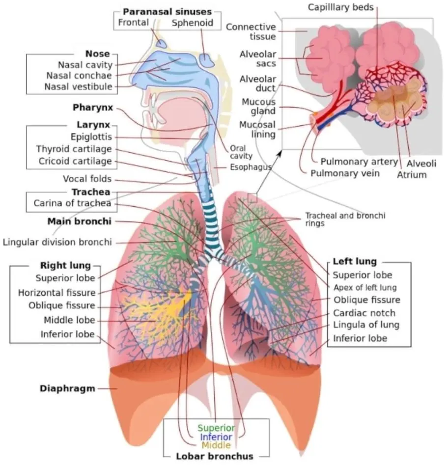 Labeled illustration of the human respiratory system which consists of the airways, the lungs, and the respiratory muscles that mediate the movement of air into and out of the body.
Disorders of the respiratory system can be classified into four general areas:
- Obstructive conditions (e.g., emphysema, bronchitis, asthma attacks)
- Restrictive conditions (e.g., fibrosis, sarcoidosis, alveolar damage, pleural effusion)
- Vascular diseases (e.g., pulmonary edema, pulmonary embolism, pulmonary hypertension)
- Infectious, environmental and other "diseases" (e.g., pneumonia, tuberculosis, asbestosis, particulate pollutants): Coughing is of major importance, as it is the body's main method to remove dust, mucus, saliva, and other debris from the lungs. Inability to cough can lead to infection. Deep breathing exercises may help keep finer structures of the lungs clear from particulate matter, etc.
The respiratory tract is constantly exposed to microbes due to the extensive surface area, which is why the respiratory system includes many mechanisms to defend itself and prevent pathogens from entering the body.
Disorder of Common Respiratory Disorders
- Chronic Obstructive Pulmonary Disease (COPD) - Irritation of the lungs can lead to asthma, emphysema, and chronic bronchitis and people can develop two or three of these together.
- Chronic Bronchitis - Any irritant reaching the bronchi and bronchioles will stimulate an increased secretion of mucus. In chronic bronchitis, the air passages become clogged with mucus, and this leads to a persistent cough.
- Emphysema - The delicate walls of the alveoli break down, reducing the gas exchange area of the lungs. The condition develops slowly and is seldom a direct cause of death.
- Asthma - Periodic constriction of the bronchi and bronchioles makes it more difficult to breathe.
- Pneumonia - An infection of the alveoli. It can be caused by many kinds of, both bacteria and viruses. Tissue fluids accumulate in the alveoli, reducing the surface area exposed to air. If enough alveoli are affected, the patient may need supplemental oxygen.
Disorders of the respiratory system are usually treated internally by a pulmonologist or respiratory physician.
Respiratory Disorder Facts and Statistics
- In the US, approximately 1 billion "common colds" occur each year.
- Respiratory disease is a common and significant cause of illness and death around the world.
- Respiratory diseases (including lung cancer) are responsible for over 10% of hospitalizations and over 16% of deaths in Canada.
- A study found that in 2010, there were approximately 6.8 million emergency department visits for respiratory disorders in the U.S. for patients under the age of 18.
- In the UK, approximately 1 in 7 individuals are affected by some form of chronic lung disease, most commonly chronic obstructive pulmonary disease, which includes asthma, chronic bronchitis and emphysema.
- According to the WHO Global Status Report on NCDs 2010, smoking is estimated to cause about 71% of all lung cancer deaths and 42% of chronic respiratory disease worldwide. Of the six WHO regions, the highest overall prevalence for smoking in 2008 was estimated to be the in the European Region, at nearly 29%.
- According to the Centers for Disease Control and Prevention (CDC), COPD is the fourth leading cause of death in the United States. Its prevalence increases with age. Men are more likely to have the disease, but the death rate for men and women is about the same.
- Diseases of the lung and airways are the most common cause of illness in children in developed countries and a leading cause of death in children in developing areas.
- In developed countries, the frequency of life-threatening acute respiratory infections has dropped over the last 50 years. This is probably due to improved living conditions and health care. Within Europe, there tends to be more asthma and allergy in the West and more infectious diseases in the East.
- Symptoms Patients with respiratory disease may report wheezing, which is suggestive of airways disease, particularly asthma. Hemoptysis can be a symptom of a variety of lung diseases, including infections of the respiratory tract, bronchogenic carcinoma, and pulmonary embolism. In addition, chest pain or discomfort is often thought to be respiratory in origin. As the lung parenchyma is not innervated with pain fibers, pain in the chest from respiratory disorders usually results from either diseases of the parietal pleura (e.g., pneumothorax) or pulmonary vascular diseases (e.g., pulmonary hypertension). As many diseases of the lung can result in strain on the right side of the heart, patients may also present with symptoms of cor pulmonale, including abdominal bloating or distention and pedal edema
Physical Examination The clinician’s suspicion of respiratory disease often begins with a patient’s vital signs. The respiratory rate is often informative, whether elevated (tachypnea) or depressed (hypopnea). In addition, pulse oximetry should be measured, as many patients with respiratory disease have hypoxemia, either at rest or with exertion. The classic structure of the respiratory examination proceeds through inspection, percussion, palpation, and auscultation as described below. Often, however, auscultatory findings will lead the clinician to perform further percussion or palpation in order to clarify these findings.
The first step of the physical examination is inspection. Patients with respiratory disease may be in distress, often using accessory muscles of respiration to breathe. Severe kyphoscoliosis can result in restrictive pathophysiology. Inability to complete a sentence in conversation is generally a sign of severe impairment and should result in an expedited evaluation of the patient.
Percussion of the chest is used to establish diaphragm excursion and lung size. In the setting of decreased breath sounds, percussion is used to distinguish between pleural effusions (dull to percussion) and pneumothorax (hyper-resonant note).
The role of palpation is limited in the respiratory examination. Palpation can demonstrate subcutaneous air in the setting of barotrauma. It can also be used as an adjunctive assessment to determine whether an area of decreased breath sounds is due to consolidation (increased tactile fremitus) or a pleural effusion (decreased tactile fremitus).
The majority of the manifestations of respiratory disease present as abnormalities of auscultation. Wheezes are a manifestation of airway obstruction. While most commonly a sign of asthma, peribronchial edema in the setting of congestive heart failure can also result in diffuse wheezes, as can any other process that causes narrowing of small airways. For this reason, clinicians must take care not to attribute all wheezing to asthma.
Rhonchi are a manifestation of obstruction of medium-sized airways, most often with secretions. In the acute setting, this manifestation may be a sign of viral or bacterial bronchitis. Chronic rhonchi suggest bronchiectasis or COPD. Stridor, a high-pitched, focal inspiratory wheeze, usually heard over the neck, is a manifestation of upper airway obstruction and should prompt expedited evaluation of the patient, as it can precede complete upper airway obstruction and respiratory failure.
Crackles, or rales, are commonly a sign of alveolar disease. A variety of processes that fill the alveoli with fluid may result in crackles. Pneumonia can cause focal crackles. Pulmonary edema is associated with crackles, generally more prominent at the bases. Interestingly, diseases that result in fibrosis of the interstitium (e.g., IPF) also result in crackles often sounding like Velcro being ripped apart. Although some clinicians make a distinction between “wet” and “dry” crackles, this distinction has not been shown to be a reliable way to differentiate among etiologies of respiratory disease.
One way to help distinguish between crackles associated with alveolar fluid and those associated with interstitial fibrosis is to assess for egophony. Egophony is the auscultation of the sound “AH” instead of “EEE” when a patient phonates “EEE.” This change in note is due to abnormal sound transmission through consolidated parenchyma and is present in pneumonia but not in IPF. Similarly, areas of alveolar filling have increased whispered pectoriloquy as well as transmission of larger-airway sounds (i.e., bronchial breath sounds in a lung zone where vesicular breath sounds are expected).
The lack or diminution of breath sounds can also help determine the etiology of respiratory disease. Patients with emphysema often have a quiet chest with diffusely decreased breath sounds. A pneumothorax or pleural effusion may present with an area of absent breath sounds.
Other Systems Pedal edema, if symmetric, may suggest cor pulmonale; if asymmetric, it may be due to deep venous thrombosis and associated pulmonary embolism. Jugular venous distention may also be a sign of volume overload associated with right heart failure. Pulsus paradoxus is an ominous sign in a patient with obstructive lung disease, as it is associated with significant negative intrathoracic (pleural) pressures required for ventilation and impending respiratory failure.
As stated earlier, rheumatologic disease may manifest primarily as lung disease. Owing to this association, particular attention should be paid to joint and skin examination. Clubbing can be found in many lung diseases, including cystic fibrosis, IPF, and lung cancer. Cyanosis is seen in hypoxemic respiratory disorders that result in >5 g of deoxygenated hemoglobin/dL.
DIAGNOSTIC EVALUATION
The sequence of studies is dictated by the clinician’s differential diagnosis, as determined by the history and physical examination. Acute respiratory symptoms are often evaluated with multiple tests performed at the same time in order to diagnose any life-threatening diseases rapidly (e.g., pulmonary embolism or multilobar pneumonia). In contrast, chronic dyspnea and cough can be evaluated in a more protracted, stepwise fashion.
Pulmonary Function Testing
The initial pulmonary function test obtained is spirometry. This study is an effort-dependent test used to assess for obstructive pathophysiology as seen in asthma, COPD, and bronchiectasis. A diminished-forced expiratory volume in 1 sec (FEV1)/forced vital capacity (FVC) (often defined as <70% of the predicted value) is diagnostic of obstruction. In addition to measuring FEV1 and FVC, the clinician should examine the flow-volume loop (which is effort-independent). A plateau of the inspiratory and expiratory curves suggests large-airway obstruction in extrathoracic and intrathoracic locations, respectively.
Spirometry with symmetric decreases in FEV1 and FVC warrants further testing, including measurement of lung volumes and the diffusion capacity of the lung for carbon monoxide (DLCO). A total lung capacity <80% of the predicted value for a patient’s age, race, sex, and height defines restrictive pathophysiology. Restriction can result from parenchymal disease, neuromuscular weakness, or chest wall or pleural diseases. Restriction with impaired gas exchange, as indicated by a decreased DLCO, suggests parenchymal lung disease. Additional testing, such as measurements of maximal expiratory pressure and maximal inspiratory pressure, can help diagnose neuromuscular weakness. Normal spirometry, normal lung volumes, and a low DLCO should prompt further evaluation for pulmonary vascular disease.
Arterial blood gas testing is often helpful in assessing respiratory disease. Hypoxemia, while usually apparent with pulse oximetry, can be further evaluated with the measurement of arterial PO2 and the calculation of an alveolar gas and arterial blood oxygen tension difference ([A–a]DO2). Patients with diseases that cause ventilation-perfusion mismatch or shunt physiology have an increased (A–a) DO2 at rest. Arterial blood gas testing also allows the measurement of arterial PCO2. Hypercarbia can accompany severe airway obstruction (e.g., COPD) or progressive restrictive physiology, as in patients with neuromuscular weakness.
Chest Imaging () Most patients with disease of the respiratory system undergo imaging of the chest as part of the initial evaluation. Clinicians should generally begin with a plain chest radiograph, preferably posterior-anterior and lateral films. Several findings, including opacities of the parenchyma, blunting of the costophrenic angles, mass lesions, and volume loss, can be very helpful in determining an etiology. However, many diseases of the respiratory system, particularly those of the airways and pulmonary vasculature, are associated with a normal chest radiograph.
CT of the chest is often performed subsequently and allows better delineation of parenchymal processes, pleural disease, masses or nodules, and large airways. If the test includes administration of contrast, the pulmonary vasculature can be assessed with particular utility for determination of pulmonary emboli. Intravenous contrast also allows lymph nodes to be delineated in greater detail.
3. Describe the management and treatment plan utilized in the disorder
Factors to be remembered
- Medical management
- Surgical procedure applicable for the disorder
Breast cancer:
Breast cancer is a disease that is caused by malignant tumors. The breast is made up of lobes and ducts. Each breast has 15 to 20 sections called lobes. Each lobe has many smaller sections called lobules. Lobules end in dozens of tiny bulbs that can make milk. The lobes, lobules, and bulbs are linked by thin tubes called ducts.
There are different types of treatment for patients with breast cancer.
Different types of treatment are available for patients with breast cancer. Some treatments are standard (the currently used treatment), and some are being tested in clinical trials. A treatment clinical trial is a research study meant to help improve current treatments or obtain information on new treatments for patients with cancer. When clinical trials show that a new treatment is better than the standard treatment, the new treatment may become the standard treatment. Patients may want to think about taking part in a clinical trial. Some clinical trials are open only to patients who have not started treatment.
Six types of standard treatment are used:
Surgery
Most patients with breast cancer have surgery to remove cancer.
Sentinel lymph node biopsy is the removal of the sentinel lymph node during surgery. The sentinel lymph node is the first lymph node in a group of lymph nodes to receive lymphatic drainage from the primary tumor. It is the first lymph node the cancer is likely to spread to from the primary tumor. A radioactive substance and/or blue dye is injected near the tumor. The substance or dye flows through the lymph ducts to the lymph nodes. The first lymph node to receive the substance or dye is removed. A pathologist views the tissue under a microscope to look for cancer cells. If cancer cells are not found, it may not be necessary to remove more lymph nodes. Sometimes, a sentinel lymph node is found in more than one group of nodes. After the sentinel lymph node biopsy, the surgeon removes the tumor using breast-conserving surgery or mastectomy. If cancer cells were found, more lymph nodes will be removed through a separate incision. This is called a lymph node dissection.
Types of surgery include the following:
- Breast-conserving surgery is an operation to remove the cancer and some normal tissue around it, but not the breast itself. Part of the chest wall lining may also be removed if the cancer is near it. This type of surgery may also be called lumpectomy, partial mastectomy, segmental mastectomy, quadrantectomy, or breast-sparing surgery.
- Total mastectomy: Surgery to remove the whole breast that has cancer. This procedure is also called a simple mastectomy. Some of the lymph nodes under the arm may be removed and checked for cancer. This may be done at the same time as the breast surgery or after. This is done through a separate incision.
- Modified radical mastectomy: Surgery to remove the whole breast that has cancer. This may include removal of the nipple, areola (the dark-colored skin around the nipple), and skin over the breast. Most of the lymph nodes under the arm are also removed.
Chemotherapy may be given before surgery to remove the tumor. When given before surgery, chemotherapy will shrink the tumor and reduce the amount of tissue that needs to be removed during surgery. Treatment given before surgery is called preoperative therapy or neoadjuvant therapy.
After the doctor removes all the cancer that can be seen at the time of the surgery, some patients may be given radiation therapy, chemotherapy, targeted therapy, or hormone therapy after surgery, to kill any cancer cells that are left. Treatment given after the surgery, to lower the risk that the cancer will come back, is called postoperative therapy or adjuvant therapy.
If a patient is going to have a mastectomy, breast reconstruction (surgery to rebuild a breast’s shape after a mastectomy) may be considered. Breast reconstruction may be done at the time of the mastectomy or at some time after. The reconstructed breast may be made with the patient’s own (nonbreast) tissue or by using implants filled with saline or silicone gel. Before the decision to get an implant is made, patients can call the Food and Drug Administration's (FDA) Center for Devices and Radiologic Health at 1-888-INFO-FDA (1-888-463-6332) or visit the FDA website for more information on breast implants.
Radiation therapy
Radiation therapy is a cancer treatment that uses high-energy x-rays or other types of radiation to kill cancer cells or keep them from growing. There are two types of radiation therapy:
- External radiation therapy uses a machine outside the body to send radiation toward the area of the body with cancer.
- Internal radiation therapy uses a radioactive substance sealed in needles, seeds, wires, or catheters that are placed directly into or near the cancer.
The way the radiation therapy is given depends on the type and stage of the cancer being treated. External radiation therapy is used to treat breast cancer. Internal radiation therapy with strontium-89 (a radionuclide) is used to relieve bone pain caused by breast cancer that has spread to the bones. Strontium-89 is injected into a vein and travels to the surface of the bones. Radiation is released and kills cancer cells in the bones.
Chemotherapy
Chemotherapy is a cancer treatment that uses drugs to stop the growth of cancer cells, either by killing the cells or by stopping them from dividing. When chemotherapy is taken by mouth or injected into a vein or muscle, the drugs enter the bloodstream and can reach cancer cells throughout the body (systemic chemotherapy).
Hormone therapy
Hormone therapy is a cancer treatment that removes hormones or blocks their action and stops cancer cells from growing. Hormones are substances made by glands in the body and circulated in the bloodstream. Some hormones can cause certain cancers to grow. If tests show that the cancer cells have places where hormones can attach (receptors), drugs, surgery, or radiation therapy is used to reduce the production of hormones or block them from working. The hormone estrogen, which makes some breast cancers grow, is made mainly by the ovaries. Treatment to stop the ovaries from making estrogen is called ovarian ablation.
Hormone therapy with tamoxifen is often given to patients with early localized breast cancer that can be removed by surgery and those with metastatic breast cancer (cancer that has spread to other parts of the body). Hormone therapy with tamoxifen or estrogens can act on cells all over the body and may increase the chance of developing endometrial cancer. Women taking tamoxifen should have a pelvic exam every year to look for any signs of cancer. Any vaginal bleeding, other than menstrual bleeding, should be reported to a doctor as soon as possible.
Hormone therapy with a luteinizing hormone-releasing hormone (LHRH) agonist is given to some premenopausal women who have just been diagnosed with hormone receptor positive breast cancer. LHRH agonists decrease the body's estrogen and progesterone.
Hormone therapy with an aromatase inhibitor is given to some postmenopausal women who have hormone receptor positive breast cancer. Aromatase inhibitors decrease the body's estrogen by blocking an enzyme called aromatase from turning androgen into estrogen. Anastrozole, letrozole, and exemestane are types of aromatase inhibitors.
For the treatment of early localized breast cancer that can be removed by surgery, certain aromatase inhibitors may be used as adjuvant therapy instead of tamoxifen or after 2 to 3 years of tamoxifen use. For the treatment of metastatic breast cancer, aromatase inhibitors are being tested in clinical trials to compare them to hormone therapy with tamoxifen.
In women with hormone receptor positive breast cancer, at least 5 years of adjuvant hormone therapy reduces the risk that the cancer will recur (come back).
Other types of hormone therapy include megestrol acetate or anti-estrogen therapy such as fulvestrant.
Targeted therapy
Targeted therapy is a type of treatment that uses drugs or other substances to identify and attack specific cancer cells. Targeted therapies usually cause less harm to normal cells than chemotherapy or radiation therapy do. Monoclonal antibodies, tyrosine kinase inhibitors, cyclin-dependent kinase inhibitors, mammalian target of rapamycin (mTOR) inhibitors, and PARP inhibitors are types of targeted therapies used in the treatment of breast cancer.
-
- Trastuzumab is a monoclonal antibody that blocks the effects of the growth factor protein HER2, which sends growth signals to breast cancer cells. It may be used with other therapies to treat HER2 positive breast cancer.
- Trastuzumab deruxtecan is an antibody-drug conjugate that links trastuzumab to a type of chemotherapy. When this linked therapy binds to HER2 positive breast cancer cells, the chemotherapy enters the cells and kills them. Trastuzumab deruxtecan may be used to treat certain patients with HER2 positive breast cancer that has already been treated and has metastasized (spread to other parts of the body).
- Pertuzumab is a monoclonal antibody that may be combined with trastuzumab and chemotherapy to treat breast cancer. It may be used to treat certain patients with HER2 positive breast cancer that has metastasized (spread to other parts of the body). It may also be used as neoadjuvant therapy in patients with locally advanced, inflammatory, or early-stage breast cancer. It may also be used as adjuvant therapy in certain patients with early-stage HER2 positive breast cancer.
- Ado-trastuzumab emtansine is a monoclonal antibody linked to an anticancer drug. This is called an antibody-drug conjugate. It is used to treat HER2 positive breast cancer that has spread to other parts of the body or recurred (come back). It is also used as adjuvant therapy to treat HER2 positive breast cancer in patients who have residual disease after surgery.
- Sacituzumab govitecan is a monoclonal antibody that carries an anticancer drug to the tumor. This is called an antibody-drug conjugate. It is used to treat metastatic triple-negative breast cancer in patients who have received at least two previous chemotherapy regimens. Monoclonal antibodies: Monoclonal antibodies are immune system proteins made in the laboratory to treat many diseases, including cancer. As a cancer treatment, these antibodies can attach to a specific target on cancer cells or other cells that may help cancer cells grow. The antibodies are able to then kill the cancer cells, block their growth, or keep them from spreading. Monoclonal antibodies are given by infusion. They may be used alone or to carry drugs, toxins, or radioactive material directly to cancer cells. Monoclonal antibodies may be used in combination with chemotherapy as adjuvant therapy.
Types of monoclonal antibody therapy include the following:
- Tyrosine kinase inhibitors: This treatment block signals needed for tumors to grow. Tyrosine kinase inhibitors may be used with other anticancer drugs as adjuvant therapy. Tyrosine kinase inhibitors include the following:
- Tucatinib is a tyrosine kinase inhibitor used with trastuzumab and capecitabine to treat HER2 positive breast cancer that is advanced and cannot be removed by surgery or has spread to other parts of the body, including the brain. It is used in patients whose cancer has already been treated with at least one other anti-HER2 therapy.
- Neratinib is a tyrosine kinase inhibitor that blocks the effects of the HER2 protein and other proteins inside tumor cells. It is used with capecitabine in adults whose disease is advanced or has spread to other parts of the body and has already been treated with at least two other anti-HER2 therapies. It may also be used to treat patients with early-stage HER2 positive breast cancer after treatment with trastuzumab.
- Lapatinib is a tyrosine kinase inhibitor that blocks the effects of the HER2 protein and other proteins inside tumor cells. It may be used with other drugs to treat patients with HER2 positive breast cancer that has progressed after treatment with trastuzumab.
- Cyclin-dependent kinase inhibitors: This treatment blocks proteins called cyclin-dependent kinases (CDKs), which cause the growth of cancer cells. Combining CDK4/6 inhibitors with hormone therapy may be effective in treating advanced hormone receptor–positive HER2 negative breast cancer. Cyclin-dependent kinase inhibitors include the following:
- Palbociclib is a cyclin-dependent kinase inhibitor used with the drug letrozole to treat breast cancer that is estrogen receptor positive and HER2 negative and has spread to other parts of the body. It is used in postmenopausal women whose cancer has not been treated with hormone therapy. Palbociclib may also be used with fulvestrant in women whose disease has gotten worse after treatment with hormone therapy.
- Ribociclib is a cyclin-dependent kinase inhibitor used with letrozole to treat breast cancer that is hormone receptor positive and HER2 negative and has come back or spread to other parts of the body. It is used in postmenopausal women whose cancer has not been treated with hormone therapy. It is also used with fulvestrant in postmenopausal women with hormone receptor positive and HER2 negative breast cancer that has spread to other parts of the body or has recurred. It is also used in premenopausal women with hormone receptor positive and HER2 negative breast cancer that has spread to other parts of the body or has recurred.
- Abemaciclib is a cyclin-dependent kinase inhibitor used to treat hormone receptor positive and HER2 negative breast cancer that is advanced or has spread to other parts of the body. It may be used alone or with other drugs.
- Alpelisib is a cylin-dependent kinase inhibitor used with the drug fulvestrant to treat hormone receptor positive and HER2 negative breast cancer that has a certain gene change and is advanced or has spread to other parts of the body. It is used in postmenopausal women whose breast cancer has gotten worse during or after treatment with hormone therapy.
- Mammalian target of rapamycin (mTOR) inhibitors: This treatment blocks a protein called mTOR, which may keep cancer cells from growing and prevent the growth of new blood vessels that tumors need to grow. mTOR inhibitors include the following:
- Everolimus is an mTOR inhibitor used in postmenopausal women with advanced hormone receptor positive breast cancer that is also HER2 negative and has not gotten better with other treatment.
- PARP inhibitors: This treatment blocks DNA repair and may cause cancer cells to die. PARP inhibitors include the following:
- Olaparib is a PARP inhibitor used to treat patients with mutations in the BRCA1 or BRCA2 gene and HER2 negative breast cancer that has spread to other parts of the body. PARP inhibitor therapy is being studied for the treatment of patients with triple-negative breast cancer and as adjuvant therapy in patients with HER2 negative early breast cancer and mutations in the BRCA1 or BRCA2 gene.
- Talazoparib is a PARP inhibitor used to treat patients with mutations in the BRCA1 or BRCA2 genes and HER2 negative breast cancer that is locally advanced or has spread to other parts of the body.
Immunotherapy
Immunotherapy is a treatment that uses the patient’s immune system to fight cancer. Substances made by the body or made in a laboratory are used to boost, direct, or restore the body’s natural defenses against cancer. This cancer treatment is a type of biological therapy.
There are different types of immunotherapy:
- PD-1 and PD-L1 inhibitor therapy: PD-1 is a protein on the surface of T cells that helps keep the body’s immune responses in check. PD-L1 is a protein found on some types of cancer cells. When PD-1 attaches to PD-L1, it stops the T cell from killing the cancer cell. PD-1 and PD-L1 inhibitors keep PD-1 and PD-L1 proteins from attaching to each other. This allows the T cells to kill cancer cells. Atezolizumab is a PD-L1 inhibitor used to treat breast cancer that has spread to other parts of the body
4. Describe the surgical management of the disorder
Factors to be remembered
- Anatomical basis of the surgery
- Organ structure and anatomical relation of the organ to be noted while doing the procedure
- Detailed description of the instruments and equipments used for surgery
- Write a brief note on the post operative care
- Elaborate on the equipments/ medical devices used to monitor in post operative care
Open Heart Surgery- Types, Procedure, And Surgical Instruments
During this surgery, the chest wall is opened to get a view of the heart. This kind of surgery is done to treat multiple cardiologic issues like arrhythmias, aneurysms, coronary artery disease, and heart failure, thoracic aortic aneurysm, etc.
This procedure is only recommended to those patients who can bear it. This surgery is performed to treat muscles, arteries, or valves of the heart. It is the most common type of heart surgery. It is also referred to as ‘traditional heart surgery’. It has been in practice for years.
The term ‘open heart surgery’ can be misleading. It does not involve opening the full chest. This surgery can easily be performed through a small incision between the ribs on the right side of the chest.
Types of Open Heart Surgery:
On Pump means that a bypass machine is attached to the patient that temporarily takes over and circulates the blood through the body.
So, the heart is stopped during the surgery and does not pump blood. The surgeon disconnects the machine after the surgery, to the heart starts beating again.
Off-Pump
Off-pump surgery does not involve the use of any bypass machine. During this surgery, the heart keeps beating and circulating the blood. However, the off-pump procedure can only be used for coronary artery bypass grafting (CABG). It is also known as ‘beating heart surgery.
Procedure:
- Before open heart surgery, a chest x-ray, electrocardiogram, and ECG are conducted to plan the procedure accordingly.
- The surgical area is sterilized before the surgery to make sure there are no bacteria left.
- Fluids and medications are provided through an intravenous line (IV).
- A 6 to 8 inch long is incision is made in the middle of the chest with the help of a surgical knife.
- The breastbone is cut, and a rib spreader is used to spread the ribcage in order to reach the heart.
- The heart is connected to the bypass machine, and the heart is stopped through an IV medication.
- In the case of a blocked artery, a new healthy vein is selected to create a new path for the blood to follow.
- The surgeon then uses surgical wires to close the breastbone. Sometimes, to further avoid any dangers, the surgeon uses titanium plates to rejoin the breastbone. This is usually done for old patients or those who have had multiple surgeries in the past.
- In the end, the cut is stitched up to increase the healing process.
Open Heart Instrument Set:
Multiple surgical instruments are used during open heart surgery. There are companies that offer complete Open Heart Instrument Sets to facilitate this procedure.
These sets are comprised of high-quality German stainless steel instruments that are strong, durable, and rustproof.
This instrument kit includes the following instruments:
- Richardson Eastman Retractors
- Backhaus Towel Clamp
- Foerster Sponge Forceps
- Mayo Hegar Needle Holder
- Allis Willauer Tissue Forceps
- Tube Occluding Clamps
- Knife Handle
- Gemini Mixter Delicate
- Metzenbaum Scissors
- Debakey Needle Holder
- Debakey Thoracic Tissue Forceps
- Debakey Multipurpose Clamps
- Glover Ducts Clamp
- Halstead Mosquito Forceps
- Mayo Scissors
- Adson Tissue Forceps
- Russian Tissue
- Cushing Forceps 7″ 1×2 Teeth
- Yankauer Suction Tube
- Crile Hemostatic Forceps
- Coller Crile Forceps
- Rochester Pean Forceps
- Rochester Ochsner Forceps
- Mixter Hemostatic Forceps
- Adson Hemostatic Forceps Curved
All these surgical instruments are necessary to perform any open heart surgery. They play a remarkable role in cutting, grasping, holding, retracting, and manipulating the tissues, organs, or blood vessels.
You can get these instruments from those who have been the manufacturer and suppliers of surgical instruments for decades. Some companies also offer free delivery services for the U.S. and Canada. You can also look for discounted prices.
However, it is better not to compromise on the quality of instruments just to buy them at a low price. Some instruments also come with enhanced features like tungsten carbide inserts, titanium, and color coating. Tungsten carbide increases the strength of the instrument and makes it durable.
Titanium is more biocompatible and does not cause any side effects when inserted inside the body. Color-coated instruments are easier to use and identify. The ones with dark coating reflect less light which guarantees better accuracy and precision.
Risks:
Open heart surgery is a risky procedure. It can lead to multiple problems that can even prove to be fatal.
A person getting open-heart surgery can get a chest wound infection which can cause pain and discomfort. It is common among obese patients. The patient can also experience irregular heartbeat, chest pain, blood clots, anemia, breathing difficulty, or pneumonia.
In the worst-case scenario, the patient can get a heart attack, lung failure, kidney failure, or stroke. The on-pump open heart surgery is considered more dangerous as it can even cause neurological problems.
Open heart surgery is a major operation that requires a hospital stay of a week or more. An individual will often spend time in the intensive care unit immediately after surgery.
In adults, a surgeon will operate on the heart to treat problems with the valves, arteries supplying the heart, and aneurysms in the main vessel leaving the heart.
While it is an intensive surgery, the risk of mortality is very low. One 2013 study showed an in-hospital mortality rate of 2.94 percent.
This article will focus on the preparation, procedure, and recovery for open heart surgery in adults.
Procedures:
Open heart surgery has an excellent survival rate.
Open heart surgery is an operation to repair a fault or damage in the heart. The operation requires the surgeon to open the chest to access the heart.
The most common type of open heart surgery is a coronary artery bypass.
The coronary arteries supply the heart with blood. If the arteries become blocked or narrowed due to heart disease, a person may be at risk of a heart attack.
The operation involves taking a healthy blood vessel from another part of the body and using it to bypass the blocked arteries.
Another open-heart procedure involves replacing a faulty aortic valve. The aortic heart valve prevents blood from flowing back into parts of the heart after the heart has pumped it out.
Surgeons also perform open-heart surgery to repair aneurysms, which are bulges in the main artery leaving the heart.
A person undergoing open heart surgery will need to stay in the hospital for 7 – 10 days. This includes at least a day in the intensive care unit immediately after the operation.
Preparing for the surgery
Preparation for open heart surgery starts the night before. A person should eat an evening meal as usual but must not consume any food or drink after midnight.
It is a good idea to wear loose, comfortable clothing to assist with restricted movement following surgery, but wear whatever is comfortable.
Be sure to have all personal medical information on hand. This might include a list of medications, recent illness, and insurance information.
It is normal to feel anxious before an anesthetic, and people should not hesitate to seek reassurance from the healthcare team.
The doctor may request that the person washes their upper body with antibacterial soap. A member of the healthcare team may need to shave the person’s chest area before they can have the anesthetic.
The doctors may also need to run tests before surgery, such as monitoring the heart or taking blood samples. A doctor or nurse might place a line into a vein to enable the delivery of fluids.
After the medical team has completed the preliminary tasks, the anesthesiologist will administer general anesthesia.
During the operation
The length of time it takes to carry out open heart surgery depends on the type of procedure and the needs of the individual. As a guide, the National Heart, Lung, and Blood Institute (NHLBI) state that a coronary artery bypass takes 3 to 6 hours.
To access the heart, the surgeon makes a 6-to-8-inch incisionTrusted Source along the middle of the chest. The cut will go through the breastbone.
The medical team might use a heart-lung bypass machine during the surgery. This involves stopping the heart from beating. The bypass machine takes over the heart’s pumping action and removes blood from the heart via tubes. The machine then removes carbon dioxide from the blood, adds oxygen, and returns the blood to the body. This surgery is called “on-pump” surgery.
Sometimes, a surgeon might work “off-pump.” When a bypass machine is not in use, the person’s heart keeps beating. A member of the surgical team uses a device to steady the heart while the surgeon performs the procedure.
There is insufficient evidence to confirm whether on-pump open heart surgery is safer than off-pump surgery. However, according to the National Institute for Health and Clinical Excellence (NICE), survival rates 1 year after either form of open-heart surgery are similar at about 96–97 percent.
A team of doctors and other health professionals work together in the operating theater during open heart surgery.
The team is likely to include:
- the lead surgeon who will direct others surgeons who will assist during the operation
- the anesthesiologist, who is in charge of giving and anesthesia and monitoring vital signs
- the pump team, also known as perfusionists, operate the heart-lung machine and other technical equipment that supports open heart surgery
- nurses and technicians, who assist the surgical team and prepare the operating theater for surgery
Open heart surgery is a major operation that requires close monitoring and immediate post-operative support.
It is normal for a person to remain in the intensive care unit (ICU) for a couple of days after the procedure to receive further care.
People will often have to stay in ICU after open heart surgery.
After the operation, a breathing tube will remain in place for a period to assist with breathing. A line also stays in the vein to administer pain relief. A person may find themselves attached to a variety of other monitoring equipment.
After leaving ICU, a person will probably stay in the hospital for about a week. Recovery at home after leaving the hospital usually takes between 4 to 6 weeks.
The healthcare team will guide rehabilitation and advise on medications and restrictions on physical activity.
During recovery:
- It is perfectly normal to experience tiredness and some pain.
- Follow the advice of the medical team on wound care, and look out for signs of infection around the chest wound, such as redness or discharge.
- Seek urgent care for any potentially serious symptoms of infection. Symptoms include difficulty breathing, fever, and excessive sweating.
Be patient and take your time. It takes many weeks or months to return to usual levels of activity. Some doctors might offer specialist support for daily activities and other aspects of recovery as part of a specific cardiac rehabilitation program.
Aftercare varies for each individual but might include blood tests, heart scans, and stress tests. A stress test involves monitoring the heart during a treadmill exercise.
Medication might include blood-thinning drugs.
There are now some alternatives to opening the chest to perform heart surgery, including the use of endoscope cameras and robots.
Sometimes, surgeons can treat narrowed coronary arteries by making minimally invasive cuts to the skin and using the da Vinci robot to perform the surgery. The Da Vinci is a multi-limbed, robotic surgical tool, which the surgeon controls remotely.

The procedure is called endoscopic coronary artery bypass surgery.
This procedure has no advantages in terms of effectiveness or safety over open-heart surgery. Both types of surgery require a similar length of stay and care plan in the hospital.
The main difference is that people who have undergone this less invasive surgery recover in less time than those who have had more traditional surgery.
Other alternatives to open heart surgery include:
- Angioplasty: During this procedure, a surgeon places a stent inside the narrowed artery to widen it.
- Transcatheter aortic valve replacement: This involves inserting a new valve via a catheter. This opens out once in place, expanding the blood vessel.
- Aortic valve balloon valvuloplasty: This procedure involves inserting a balloon to enlarge a valve.
All forms of heart surgery come with risks. These risks include:
- being under anesthesia
- infection
- organ damage
- stroke
The severity of these risks depends on the individual. People with a more advanced heart condition face a higher risk of complications during and after surgery.
5. Self-study on recent advances in the field
Factors to be remembered
- Describe the recent advances with respect to the management of the clinical condition /disorder involved.
- Describe the possible newer techniques (diagnostic or therapeutic) involved in managing the disorder
Metabolic dysfunction-associated fatty liver disease: from basic research to clinical application
The increasing burden of non-alcoholic fatty liver disease: Non-alcoholic fatty liver disease (NAFLD) is the most common cause of chronic liver disease in the world. NAFLD encompasses a spectrum of liver disease, ranging from simple hepatic steatosis to non-alcoholic steatohepatitis (NASH), fibrosis, cirrhosis, and hepatocellular carcinoma (HCC). With the pandemic of obesity and type 2 diabetes mellitus (T2DM), there has been an exponential growth in the prevalence of NAFLD over the past two decades. The prevalence of NAFLD in most Asian countries, including China, is above 25% in the general adult population.[1] Furthermore, there is a developing childhood obesity pandemic, and a meta-analysis of 20,595 children in Asia generated a pooled NAFLD prevalence of 5.53%, which had increased by approximately 1.6-fold since 2010. The pooled prevalence of NAFLD in Asian children increased from those with normal weight (1.5%) to those who were overweight (16.7%) or obese (50.1%).
A recent study suggested that NAFLD is not uncommon in lean Chinese adults with a normal waist circumstance. Metabolic risk factors, rather than genetic factors, may play an important role in the development of lean NAFLD,and the hepatic and extra-hepatic complications can also develop in lean patients, which reinforces the importance of considering metabolic phenotype in the assessment of NAFLD, rather than using body mass index-based approaches.
Renaming of NAFLD to MAFLD: A diagnosis of NAFLD is made on the basis of histological or imaging-derived evidence of steatosis, in the absence of a known etiology of fatty liver. With advances in knowledge of the pathogenesis of the condition, the “exclusive” term NAFLD no longer serves to precisely describe a highly heterogeneous disease. In 2020, the novel term of metabolic dysfunction-associated fatty liver disease (MAFLD) was proposed in an attempt to create an “inclusive” diagnosis. Zeng et al performed a cross-sectional study of Chinese adults which showed that the prevalence of MAFLD is higher than that of NAFLD, and therefore the newly-defined label of MAFLD may better reflect the metabolic pathogenesis. Furthermore, a pathologic analysis of patients with MAFLD showed that a single metabolic defect can have a significant role in the development of fibrosis and that insulin resistance plays a key role in the progression of steatohepatitis and the development of significant fibrosis
As Zheng et al discussed, by using the new terminology, “cryptogenic cirrhosis” and MAFLD can now be diagnosed in lean individuals using metabolic criteria, rather than being viewed as completely separate entities. The renaming of NAFLD to MAFLD may result in significant improvements in awareness, advocacy, research, and the clinical management of the condition.
Update on the pathogenesis of MAFLD: The pathogenesis of NAFLD/MAFLD is a multifactorial process, involving interactions among nutrition, metabolism, genetic predisposition, the gut microbiota, and environmental factors. Although a great deal of progress has been made in recent decades, the pathogenic mechanism of NAFLD/MAFLD has yet to be fully elucidated. In this issue of the Chinese Medical Journal (CMJ), Pan et al give an overview of the role of hepatocyte nuclear factor 4α (HNF4α) in the pathogenesis of NAFLD. HNF4α has been shown to regulate bile acid, lipid, and glucose metabolism; and hepatic HNF4α expression is much lower in patients with NAFLD and mouse models of NASH. Furthermore, there is evidence that hepatic HNF4α plays a key role in the initiation and progression of NAFLD and may represent a therapeutic target for NAFLD.
Huang et al presented a systematic review regarding the role of retinol-binding protein 4 (RBP4) in the development of NAFLD and its potential therapeutic application. RBP4 induces hepatic de novo lipogenesis, impairs fatty acid oxidation, increases insulin resistance, and promotes hepatic inflammation. Furthermore, a high plasma RBP4 concentration is associated with a high risk of NAFLD; and agents that reduce the circulating RBP4 concentration and/or hepatic RBP4 expression have a protective effect against NAFLD. These findings suggest that RBP4 could be targeted as a novel diagnostic marker or therapeutic target for NAFLD.
Jackson et al summarized the essential physiology of bile acid and sphingolipid metabolism, because the dysregulation of both are potential contributors to NAFLD. Specifically, the dysregulation of bile acid and sphingolipid metabolism has been linked to hepatic steatosis, inflammation, and fibrosis, and the further exploration of the pathologic effects mediated by bile acids and sphingolipids may also lead to new diagnostic and therapeutic strategies for NAFLD.
Hepatitis B and concurrent MAFLD: Concomitant NAFLD/MAFLD in patients with chronic hepatitis B (CHB) has become highly prevalent over the past two decades. However, the risks associated with the dual etiologies, outcomes, and mechanisms involved in the interaction between CHB and NAFLD have not been fully characterized. Tong et al[12] summarize the findings of recent clinical and basic research studies related to the potential interactions between CHB and NAFLD. The prevalence of hepatic steatosis in CHB has been reported to be 32.8% (95% CI, 28.9%–37.0%); and it is higher in men and patients with obesity. The presence of hepatic steatosis in patients with CHB is related to metabolic, rather than viral factors. Patients with both CHB and NAFLD are more likely to experience liver-related outcomes or death than those with CHB alone. Many studies have shown that steatosis is positively associated with the clearance of hepatitis B virus (HBV) surface antigen and a reduction in HBV DNA, and the prevalence and incidence of NAFLD in patients with CHB may be lower than in those without.
In Chang and colleagues’ multi-center, prospective study of 1000 treatment-naïve patients with biopsy-confirmed CHB, NASH was found in 182 patients (18.2%), 46% of these achieved resolution of NASH, and only 4% of the patients developed new-onset NASH after 72 weeks of entecavir treatment. Body mass at baseline and a slight weight change during follow-up were associated with the prevalence, incidence, and remission of NASH in patients with CHB.[13] Finally, steatosis is more prevalent in patients with CHB and is a common reason for abnormal circulating liver enzyme activities in infected patients with a low HBV-DNA load or a good response to infection.
From MAFLD to HCC: Although viral hepatitis remains the most common etiology of liver cancer-related deaths, NAFLD is the most rapidly growing contributor to mortality and morbidity related to liver disease in the world. The global burden of HCC is increasing alongside the NAFLD pandemic. A recently published review in CMJ summarizes the characteristics of NAFLD-related HCC.[14] The incidence of NAFLD-related HCC is much higher in patients with severe steatohepatitis, advanced fibrosis, and cirrhosis than in individuals with NAFLD in general, and it is most likely to occur in older men with metabolic syndrome. The incidence of HCC in patients with NAFLD-related cirrhosis is lower than that in those with hepatitis C virus- or HBV-related cirrhosis. Compared with HCCs of other etiologies, NAFLD-related HCCs are generally large, well-differentiated, solitary lesions with a higher level of inflammatory infiltration, and they are less likely to metastasize extra-hepatically. Moreover, NAFLD-related HCC is more likely to develop in the absence of cirrhosis.[14] In a recent issue of CMJ, Rios et al reviewed the progression of MAFLD to HCC and stated that lipotoxicity, insulin resistance, oxidative stress, chronic inflammation, multiple gene mutations, and alterations to the fecal microbial composition are the most important factors determining hepatic carcinogenesis, whereas steatohepatitis and fibrosis are not essential for the development of HCC in obesity-related fatty liver disease.[15]
Non-invasive diagnosis of MAFLD: Accumulating evidence suggests that non-invasive tests can be used to diagnose NAFLD, assess its severity, and predict its prognosis. In a recent issue of CMJ, Li et al review new developments in non-invasive testing for NAFLD, with respect to steatosis, steatohepatitis, and fibrosis.[16] For the identification of steatosis, ultrasonography remains the most common method, because of its wide availability and low cost, but magnetic resonance imaging-proton density fat fraction is currently the most accurate means of identifying hepatic steatosis, and transient elastography (TE) represents a promising technique for the evaluation of hepatic steatosis and fibrosis. Except for the widely used controlled attenuation parameter, ultrasonographic attenuation has been reported to have a low failure rate and shows moderate-to-high performance for the discrimination of degrees of steatosis in patients with chronic liver disease.[17]
Various non-invasive algorithms, such as the fatty liver index (FLI) and hepatic steatosis index (HSI), have been used as screening tests for steatosis in epidemiologic studies. In Chen et al's study, both FLI and HSI were shown to be useful screening tools for NAFLD in adults with obstructive sleep apnea/hypopnea syndrome.[18] In patients with steatohepatitis, some circulating biomarkers correlate with the severity of NASH but show modest predictive accuracy. Regarding liver fibrosis, liver stiffness measurement (LSM) using TE is highly accurate and is widely used worldwide. Magnetic resonance elastography is marginally better than TE, but it is limited by its cost and availability. In contrast, simple fibrosis scores, such as the fibrosis-4 (FIB-4) index and the NAFLD fibrosis score, can be easily calculated and are recommended for use in primary care. These scores and LSM have sufficiently high negative predictive values to exclude advanced fibrosis. Recently, Shi et al found that the combination of the presence of a metabolic disorder and the FIB-4 index provides for a more accurate diagnosis of advanced fibrosis in patients with NAFLD.[19] Thus, as part of the redefinition of MAFLD, metabolic risk factors should be taken into account during diagnosis and management.
Therapeutic approaches to MAFLD: In a recent issue of CMJ, Shi et al[20] discuss recent advances and provide a perspective regarding the treatment of MAFLD. Weight management through an appropriate diet and physical activity remains the most important component of the treatment of MAFLD. Weight loss through bariatric surgery may be an effective means of achieving significant improvements in patients with morbid obesity and MAFLD. Although numerous agents, including novel modulators of glucolipid metabolism, are being assessed in clinical trials, there is still no approved drug for the treatment of MAFLD. The nomenclature of MAFLD emphasizes the existence of concomitant metabolic disorders and obesity, and patients with MAFLD are therefore subject to both hepatic and other metabolic risks. Thus, drugs targeting underlying cardiometabolic risk factors are essential to improve the outcomes of patients with MAFLD.
The screening of patients who are at a high risk of MAFLD and the provision of a comprehensive individual therapeutic program are critical. For example, patients with MAFLD and T2DM would benefit from the use of antidiabetic agents, patients with overweight or obesity would gain greater benefit from weight management, and those with metabolic syndrome require comprehensive individualized management. These therapeutic approaches might help identify the patients with MAFLD who are at the greatest risk of disease progression and facilitate more precise and appropriate management.
Summary and prospects: The growing burden of NAFLD parallels the increasing prevalences of obesity and metabolic syndrome worldwide. Cardiometabolic risk factors have a bidirectional relationship with NAFLD. The majority of patients with NAFLD meet the diagnostic criteria for MAFLD, and this represents a more appropriate term. Further clinical studies of the changes created by the redefinition of NAFLD/MAFLD, including the epidemiologic character, prognosis, diagnosis, prevention, and treatment of the condition, are required. Currently, MAFLD and CHB are increasingly being diagnosed in the same individuals, and the pathophysiological interaction between MAFLD and HBV infection in patients is worthy of further exploration. The long-term outcomes of MAFLD are related to the severity of metabolic dysfunction and liver fibrosis, rather than obesity. Metabolic syndrome and T2DM are the most important risk factors for MAFLD-related cirrhosis and HCC. A lack of awareness regarding the factors underlying MAFLD-related HCC may lead to delay in its diagnosis.
The further development and validation of non-invasive diagnostic techniques and clinical pathways will help clinicians assess the severity of MAFLD, categorize patients, and identify those requiring specific treatments. There is still no effective approved drug for MAFLD, but the in-depth study of pathologic mechanisms may provide new therapeutic targets. Measures to increase awareness and treat or prevent the associated cardiometabolic diseases are necessary to reduce the growing burden of MAFLD.
Part B – Case Study
A 25 year old biker indulged in illegal racing met with a road traffic accident and severed head injury and skull fracture. After resuscitation and operation done for the hematoma it was resorted to do skull implant for his fracture by the orthopaedician
Discuss the case under the following headings
- Describe the type of bone the skull belongs to
- Describe the microscopic anatomy of bone
- Explain the blood supply of bone
- Elaborate on bone remodeling and new bone formation
- Describe the anatomical basis of bone implant
- Enumerate the indications and contraindications for the bone implant
Describe the type of bone the skull belongs to
- The skull is a bony structure that supports the face and forms a protective cavity for the brain. It is comprised of many bones, which are formed by intramembranous ossification, and joined by sutures (fibrous joints).
- The bones of the skull can be considered in two groups: those of the cranium (which consists of the cranial roof and cranial base) and those of the face.
Describe the microscopic anatomy of bone
- Cranium:
- The cranium (also known as the neurocranium) is formed by the superior aspect of the skull. It encloses and protects the brain, meninges, and cerebral vasculature.
- Anatomically, the cranium can be subdivided into a roof and a base:
- Cranial roof – comprised of the frontal, occipital and two parietal bones. It is also known as the calvarium.
- Cranial base – comprised of six bones: frontal, sphenoid, ethmoid, occipital, parietal, and temporal. These bones articulate with the 1st cervical vertebra (atlas), the facial bones, and the mandible (jaw).
2. Face:
- The facial skeleton (also known as the viscerocranium) supports the soft tissues of the face.
- It consists of 14 bones, which fuse to house the orbits of the eyes, the nasal and oral cavities, and the sinuses. The frontal bone, typically a bone of the calvaria, is sometimes included as part of the facial skeleton.
- The facial bones are:
- Zygomatic (2) – forms the cheekbones of the face and articulates with the frontal, sphenoid, temporal, and maxilla bones.
- Lacrimal (2) – the smallest bones of the face. They form part of the medial wall of the orbit.
- Nasal (2) – two slender bones that are located at the bridge of the nose.
- Inferior nasal conchae (2) – located within the nasal cavity, these bones increase the surface area of the nasal cavity, thus increasing the amount of inspired air that can come into contact with the cavity walls.
- Palatine (2) – situated at the rear of the oral cavity and forms part of the hard palate.
- Maxilla (2) – comprises part of the upper jaw and hard palate.
- Vomer – forms the posterior aspect of the nasal septum.
- Mandible (jaw) – articulates with the base of the cranium at the temporomandibular joint (TMJ).
Explain the blood supply of bone
- Most of the blood supply to the skull and its associated structures comes from the common carotid arteries (anterior circulation) and vertebral arteries (posterior circulation). The common carotid artery splits into the internal and external
- carotid arteries. The external carotid is the main blood supply to the skull bones and meninges.
- It travels up the side of the neck with eight main branches feeding the superficial structures of the skull and face. Of these branches, the maxillary artery is the most prominent and clinically relevant. The middle meningeal artery is a branch of the maxillary artery, and injury secondary to blunt force trauma to the lateral skull at the pterion can lead to epidural hematoma. The internal carotid has no branches in the neck and enters the base of the skull, supplying intracranial structures.
- The internal carotid and vertebral arteries combine to form a large anastomosis called the circle of Willis. The anterior communicating artery, two anterior cerebral arteries, two middle cerebral arteries, two posterior communicating arteries, two posterior cerebral arteries, and basilar artery (superior continuation of the vertebral arteries) all contribute to this anastomosis.
- The dural venous sinuses (i.e., superior sagittal, straight, and transverse sinuses) and superficial and deep veins of the head (i.e., cerebral veins, great vein of Galen, cerebellar, and facial veins) drain into the internal and external jugular veins bilaterally and ultimately to the superior vena cava and right atrium of the heart.
- The brain and central nervous system have been traditionally thought not to contain lymphatic vessels. However, some believe that the cerebrospinal fluid (CSF) does have some connection with the lymphatic system and drains through the cervical lymph nodes. The recent discovery of a “glymphatic system,” composed of a network of CSF, interstitial cerebral fluid, and meningeal vasculature, has shed more light on this debate and is an area of ongoing research.
Nerves
The skull has multiple foramina that allow passage into and out of the skull for blood vessels and nerves. The cranial nerves mostly exit via foramina in the cranial base. A brief list of the most important foramina and their associated cranial nerves appear below.
-
Cribriform Plate → olfactory nerve (CN I)
-
Optic Canal → optic nerve (CN II)
-
Also: ophthalmic artery
-
-
Superior Orbital Fissure → oculomotor nerve (CN III), trochlear nerve (CN IV), abducens nerve (CN VI), ophthalmic division of trigeminal nerve (CN V1)
-
Foramen rotundum → maxillary division of trigeminal nerve (CN V2)
-
Foramen Ovale → mandibular division of trigeminal nerve (CN V3)
-
Stylomastoid Foramen → facial nerve (CN VII)
-
Internal Auditory Meatus → facial nerve (CN VII) and vestibulocochlear nerve (CN VIII)
-
Jugular Foramen → glossopharyngeal nerve (CN IX), vagus nerve (CN X), and spinal accessory nerve (CN XI)
-
Also: jugular vein
-
-
Hypoglossal Canal → hypoglossal nerve (CN XII)
-
Foramen Magnum → brainstem and spinal root of accessory nerve (CN XI)
-
Also: vertebral arteries
-
Other key foramina that may or may not be associated with nerves include the foramen spinosum (middle meningeal artery), carotid canal (internal carotid artery and sympathetic plexus), and supraorbital and infraorbital foramina (supraorbital and infraorbital nerves). The superficial sensory nerves of the skull, scalp, and face receive input from branches of the trigeminal nerve anteriorly and the greater and lesser occipital nerves posteriorly.
Muscles
- The muscles of the face and scalp are predominantly striated muscle under voluntary innervation.
- They serve crucial roles in facial expression, vision, language, and mastication.
- The majority of these muscles are innervated by the facial nerve, oculomotor nerve, or trigeminal nerve, while the hypoglossal nerve innervates the tongue. Small smooth muscles of the eye are under sympathetic or parasympathetic control for lens accommodation, aqueous humor production, and other functions.
Elaborate on bone remodeling and new bone formation
The skull's main function is to protect the brain. It is comprised of 22 bones, eight of which form the neurocranium and are connected by synarthrodial joints called sutures.
Most of these cranial bones are categorized as flat bones and can be identified by their layered bone structure where a cancellous bone layer, called diploë, is sandwiched between two layers of dense cortical bone (cortex). These cortical layers are denoted as the inner and outer tables of the skill. Previous studies have utilized cadavers and primates to evaluate the mechanical properties of the skull. McElhaney and colleagues evaluated spatial, material, and structural properties of full skull samples from cadavers. Skull flexural stiffness and strength were modeled by Hubbard and colleagues using a layered beam testing technique. These studies have found the flexural properties of the skull to be highly dependent on skull thickness. The cortical thickness of the outer table has been measured and reported at various locations across the skull by Peterson and Dechow. Bone specimens were harvested from locations in the frontal, parietal, occipital, and temporal regions of the skull; the outer table was isolated and the thickness was physically measured with calipers or ultrasonic transducers. Thicknesses measured from physical specimens were found to vary within and between bones, with an overall average of 2.4 ± 0.8 mm. Additional studies have evaluated skull deformation from linear impacts, concluding that skull deformation can occur below mechanical failure and contribute to the occurrence and severity of TBI. Furthermore, another study evaluated the mortality of severe TBI based on sex and stratified by age. Results indicated that women over the age of 55 years were at a significantly higher risk of mortality from a TBI than any other group.
The skeleton is a metabolically active organ that undergoes continuous remodeling throughout life. Bone remodeling involves the removal of mineralized bone by osteoclasts followed by the formation of bone matrix through the osteoblasts that subsequently become mineralized. The remodeling cycle consists of three consecutive phases: resorption, during which osteoclasts digest old bone; reversal, when mononuclear cells appear on the bone surface; and formation, when osteoblasts lay down new bone until the resorbed bone is completely replaced. Bone remodeling serves to adjust bone architecture to meet changing mechanical needs and it helps to repair microdamages in bone matrix preventing the accumulation of old bone. It also plays an important role in maintaining plasma calcium homeostasis. The regulation of bone remodeling is both systemic and local. The major systemic regulators include parathyroid hormone (PTH), calcitriol, and other hormones such as growth hormone, glucocorticoids, thyroid hormones, and sex hormones. Factors such as insulin-like growth factors (IGFs), prostaglandins, tumor growth factor-beta (TGF-beta), bone morphogenetic proteins (BMP), and cytokines are involved as well. As far as local regulation of bone remodeling is concerned, a large number of cytokines and growth factors that affect bone cell functions have been recently identified. Furthermore, through the RANK/receptor activator of NF-kappa B ligand (RANKL)/osteoprotegerin (OPG) system the processes of bone resorption and formation are tightly coupled allowing a wave of bone formation to follow each cycle of bone resorption, thus maintaining skeletal integrity.
The skeleton is a metabolically active organ that undergoes continuous remodeling throughout life. Bone remodeling involves the removal of mineralized bone by osteoclasts followed by the formation of bone matrix through the osteoblasts that subsequently become mineralized. The remodeling cycle consists of three consecutive phases: resorption, during which osteoclasts digest old bone; reversal, when mononuclear cells appear on the bone surface; and formation, when osteoblasts lay down new bone until the resorbed bone is completely replaced. Bone remodeling serves to adjust bone architecture to meet changing mechanical needs and it helps to repair microdamages in bone matrix preventing the accumulation of old bone. It also plays an important role in maintaining plasma calcium homeostasis. The regulation of bone remodeling is both systemic and local. The major systemic regulators include parathyroid hormone (PTH), calcitriol, and other hormones such as growth hormone, glucocorticoids, thyroid hormones, and sex hormones. Factors such as insulin-like growth factors (IGFs), prostaglandins, tumor growth factor-beta (TGF-beta), bone morphogenetic proteins (BMP), and cytokines are involved as well. As far as local regulation of bone remodeling is concerned, a large number of cytokines and growth factors that affect bone cell functions have been recently identified. Furthermore, through the RANK/receptor activator of NF-kappa B ligand (RANKL)/osteoprotegerin (OPG) system the processes of bone resorption and formation are tightly coupled allowing a wave of bone formation to follow each cycle of bone resorption, thus maintaining skeletal integrity.
contraindications for the bone implant
- Risks associated with general anaesthetics need to be considered. Most children will have a two-stage surgical procedure which means they’ll need two sets of general anaesthetic. Single stage surgery is also possible but is generally used for older children and young adults as their skull bone is more developed.
- As with all surgery there is a risk of complications, such as infection.
- The abutment area needs to be kept clean which requires regular help from another person until your child is old enough to care for the site themselves. This may carry on into adulthood for people with learning difficulties.
- As with many procedures, there is a risk of infection and inflammation at the implant site after surgery.
- There is a small risk that the growth of new bone around the implant (osseointegration) will fail. If this happens the implant may fall out and you should seek advice from your audiologist on what to do next.
- There is a risk that any head trauma, e.g. a blow to the implant site (with or without the presence of the processor), may have more serious consequences than what might happen with a conventional hearing aid.
- This may include an infection of the skin around the implant site, the implant falling out and having to use a sleeper implant or repeat surgery for a new implant. Precautions need to be considered, such as head protection for sports.