Project 2
Design a robotic surgery procedure to remove a healthy kidney from the donor using an AI guided robot.
Robotic Surgery
Artificial intelligence and kidney transplantation
- Artificial intelligence and its primary subfield, machine learning, have started to gain widespread use in medicine, including the field of kidney transplantation. We made a review of the literature that used artificial intelligence techniques in kidney transplantation.
- We located six main areas of kidney transplantation that artificial intelligence studies are focused on:
- Radiological evaluation of the allograft, pathological evaluation including molecular evaluation of the tissue, prediction of graft survival, optimizing the dose of immunosuppression, diagnosis of rejection, and prediction of early graft function.
- Machine learning techniques provide increased automation leading to faster evaluation and standardization, and show better performance compared to traditional statistical analysis. Artificial intelligence leads to improved computer-aided diagnostics and quantifiable personalized predictions that will improve personalized patient care.
Core Tip: Artificial intelligence is used in a large spectrum of areas in kidney transplantation. Developments in those areas will shape the future of medical care with faster and more standardized medical evaluations and more accurate personalized judgments.
INTRODUCTION
- Artificial intelligence (AI) is a “buzzword” that has begun to be used increasingly in medicine, and the field of transplantation is not exempt from that. AI vests the machines with the ability to perform intelligent and cognitive tasks, spanning numerous subfields that are current and popular.
- Machine learning (ML) is one of the most important subfields of AI and has recently seen an increase of interest in several industries, including the healthcare industry, because of advances in Big Data technology and computing power.
- The process of ML begins with the ability of the program to observe the collected data and compare them with previous ones to find patterns and results, and then adjust itself accordingly.
- There are a plethora of statistical-based ML algorithms that can be used in the context of three overarching categories:
- Supervised learning, unsupervised learning, and reinforcement learning (Table . Supervised learning comprises learning patterns from labeled datasets and decodes the relationships between input variables (independent variables) and their known outputs (dependent variables).
- Examples of common algorithms used for supervised learning include regression analysis [linear regression, logistic regression (LR), and non-linear regression], decision trees (DT), k-nearest neighbors, artificial neural networks (ANN), and support vector machines (SVM).
- The proper classification of LR is context-dependent and depends on whether it is used for prediction (ML) or inferential statistics to evaluate the associations between the independent variable(s) and dependent variables (non-ML).
- In the case of unsupervised learning the output variables are unlabeled, and this method focuses on analyzing the relationships between input variables and revealing hidden patterns that can be obtained to create new labels regarding possible outputs.
- In this way it is possible to discover the existing patterns in the data that we are unaware of. K-means clustering is an example of the algorithms for unsupervised learning. Reinforcement learning is the most advanced category of ML.
- In this method, a prediction model is built by gaining feedback through random trials of a vast number of possible input combinations and leveraging insight from previous iterations by grading their performance. Finally, Q-learning is an example of the algorithms for reinforcement.
Table 1
Different machine learning categories
| Supervised learning |
Unsupervised learning |
Reinforcement learning |
|
| Dataset | Labeled (input and output are known) | Unlabeled (output is not known) | No predefined data |
| Method | Analyze the relation between input and output. The output is predicted based on this relation | Analyze the input parameters to uncover hidden patterns. Output is predicted based on those patterns | Randomly trialing a vast number of possible inputs, then comparing and grading their performance |
| Example | Decision trees, support vector machines, neutral networks, k-nearest neighbors | k-means clustering, archetype analysis | Q-learning |
METHODS
- We used the PubMed interface (pubmed.gov) to make a query using the combination of the following two keyword groups.
- The first group included the keywords “kidney transplant”, “renal transplant”, “kidney transplantation”, and “renal transplantation” and the second group included “artificial intelligence”, “machine learning”, “deep learning”, and “neural networks”.
- Each keyword in the same group was combined using the logical operator “OR”, while the two groups were combined using the logical operator “AND”. We excluded the review articles and ran the query in January 2021.
- We found 114 articles in total and manually examined them. The articles that were not directly related to kidney transplantation, dealing with other types of renal replacement therapy besides transplantation, solely using LR as the ML method, reviews, conference reports, and editorials were excluded. We also examined the references of the related articles to locate additional literature.
- Finally, we found 64 articles that were eligible for the review.
- We grouped the articles in the following categories: Radiological evaluation (n = 6), pathological evaluation (n = 14), prediction of graft survival (n = 16), optimizing the dose of immunosuppression (n = 7), diagnosis of rejection (n = 6), prediction of early graft function (n = 6), and others (n = 9) ).
APPLICATION OF AI IN KIDNEY TRANSPLANTATION
Radiological evaluation
- The first paper using AI techniques for the evaluation of allografts, based on imaging techniques, was that of Hamilton et al. The authors used 99mTc-MAG3 captopril renography to evaluate the presence of renal artery stenosis in the allograft. The authors used a neural network-based classifier, and their gold standard was arteriogram. Following the training of the neural network, they found that an accuracy of 95% could be achieved.
- Some other papers also used AI techniques for the radiological evaluation of allografts with the aim of diagnosing acute rejection. El-Baz et al investigated the early detection of acute rejection using dynamic contrast-enhanced magnetic resonance imaging (MRI).
- The researchers automated data acquisition from the MRI using a three-step algorithmic approach and this data feed was linked to a Bayesian supervised classifier to diagnose acute rejection. The authors also studied motion correction models to account for the local motion of the kidney due to patient moving and breathing. Then, they used the perfusion curves to feed the Bayesian supervised classifier with the aim of distinguishing normal and acute rejection.
- Three additional papers from the same group examined the utility of computer-aided diagnostic (CAD) systems for the diagnosis of acute rejection. In their first study, the authors used deep-learning algorithms, namely, ‘stacked non-negative constrained auto-encoders’, for the prediction of acute rejection.
- Their data feed was the outcomes of diffusion-weighted MRI (DW-MRI). In their second study, in addition to DW-MRI, creatinine clearance and creatinine values were also used for the data feed of convolutional neural network (CNN) based classifiers. In both papers, the overall accuracy for correct diagnosis of acute rejection was above 90%. The authors proposed that their results demonstrated the potential of this new CAD system to reliably diagnose renal transplant rejection.
- In a third study, they again assessed the utility of the CAD system for the diagnosis of acute rejection using DW-MRI and blood oxygen level-dependent MRI as the image-based sources. The authors also used laboratory data consisting of creatinine and creatinine clearance.
- In addition, they utilized a deep learning-based classifier, namely, ‘stacked autoencoders’, to differentiate non-rejection from acute rejection in renal transplants. The overall accuracy of the CAD system in detection of acute rejection was around 90%.
Pathological evaluation
- AI applications have also been used to assess allograft biopsies, where data feed for the classification algorithms was histological findings, molecular biomarkers, or a combination of the two.
- Kazi et al used 12 histological features to train a Bayesian network with 110 transplant biopsies. Using the Bayesian network, a relatively inexperienced pathologist was able to make the correct diagnosis in 19 out of 21 cases. The researchers suggested that the integration of data with a computer can give a more consistent diagnosis of early acute rejection.
- In a follow-up study, the same researchers used a simple neural network for the decision process and the authors pointed out that in Bayesian networks the ‘importance’ attached to each histological feature had to be calculated and programmed into the network at the onset and because of this approach, they have the disadvantage of relative inflexibility.
- A neural network has the potential of greater flexibility, because the process of ‘training’ a neural network would automatically calculate what ‘weight’ should be allocated to each histological feature. The authors used 12 histological features, 100 transplant biopsies (43 with definite rejection), and 25 additional cases to train a single-layer simple neural network.
- Eventually, the network was able to correctly classify 19 out of the 21 new cases, leading to the conclusion that neural network technology can dramatically improve the accuracy in histological diagnosis of early acute renal allograft rejection.
- Marsh et al[ used deep learning algorithms to evaluate intraoperative donor kidney biopsies with the aim of determining which kidneys were eligible for transplantation. The authors used CNNs as a deep learning algorithm. The primary advantage of CNN is that the models can automatically discover prominent features from the data alone, without requiring a set of handcrafted parameters and extensive input normalization.
- Most recently, CNNs have been explored as primary tools for glomeruli detection Different models were shown to be able to differentiate image patches containing isolated normal glomeruli from non-glomerular structures. Marsh et altrained the network with a total of 870 sclerosed and 2997 non-sclerosed glomeruli that were labeled.
- The images were acquired from hematoxylin and eosin (HE)-stained frozen wedge donor biopsies. The fully conventional model in the study showed a high correlation with percent global glomerulosclerosis (R2 = 0.828). The authors concluded that the performance of the CNN alone was equivalent to that of a board-certified clinical pathologist.
- Liu et al examined the diagnosis of T-cell-mediated kidney rejection using a data feed acquired by RNA sequencing. The authors used three ML methods called linear discriminant analysis (LDA), SVM, and random forest (RF).
- The molecular signature discovery data set involved five kidney transplant patients with T-cell-mediated rejection (TCMR) and five with stable renal function. The forecast models were tested on 703 biopsies with Affymetrix GeneChip expression profiles available in the public domain.
- The LDA predicted TCMR in 55 of the 67 biopsies labeled TCMR, and 65 of the 105 biopsies designated as antibody-mediated rejection (ABMR). The RF and SVM models showed comparable performances. These data illustrated the feasibility of using RNA sequencing for molecular diagnosis of TCMR.
- Halloran et al and Reeve et al used molecular microscopy techniques to evaluate allograft biopsies, including molecular phenotyping with platforms such as microarrays that measure the expression of thousands of genes. To express the likelihood that particular diseases are present in the biopsy, the authors developed the TCMR score and the ABMR score assigned by classifiers (using weighted equations) built by standard ML methods.
- The authors also developed the Molecular Microscope Diagnostic System (MMDx) that assesses the TCMR and ABMR in a reference set of biopsy samples using ML-derived classifier algorithms. Archetypal analysis and an additional 12 ML methods (individually or in ensembles) were used during the development of the MMDx. Archetype analysis is a probabilistic data-driven unsupervised statistical approach that categorizes separate groups of patients (archetypes).
- The ensembles made diagnoses that were both more accurate than the best individual classifiers and almost as stable as the best, in line with the previous studies from the ML literature. Human experts had about 93% agreement (balanced accuracy) signing out the reports, while RF-based automated sign-outs showed similar levels of agreement (92% and 94% for predicting the expert MMDx sign-outs for TCMR and ABMR, respectively).
- In 451 biopsy samples where a feedback was obtained, clinicians indicated that the MMDx agreed more commonly with the clinical decision (87%) than histology (80%) (P = 0.0042). In another study, the same group of researchers explored the frequency of rejection in areas of interstitial fibrosis and tubular atrophy (i-IFTA) in kidney transplant biopsies by using histology Banff 2015 and an MMDx and concluded that i-IFTA in indication biopsies reflected current parenchymal injury, often with simultaneous ABMR but seldom with TCMR.
- Hermsen et al used whole-slide images of stained kidney transplant biopsies to develop and validate a CNN for histologic analysis in renal tissue stained with periodic acid Schiff. The researchers assessed the segmentation performance for different tissue classes and found that the best-segmented class was “glomeruli”, followed by “tubuli combined” and “interstitium”. The network detected 92.7% of all glomeruli in nephrectomy samples, with 10.4% of false positives. The authors also suggested that the CNN may have utility for quantitative studies involving kidney histopathology across.
- Aubert et al used archetype analysis to identify distinct groups of patients with transplant glomerulopathy. The researchers examined data from 552 biopsy samples taken from 385 patients with transplant glomerulopathy, using unsupervised archetypal analysis that integrated clinical, functional, immunologic, and histologic parameters.
- The authors identified five archetypes with distinct clinical, histologic, and immunologic features, as well as different outcomes (kidney allograft survival rates). The authors suggested that their approach permitted to decrease patient heterogeneity and created meaningful groups in terms of morphologic patterns, disease activity/progression, and risk of failure.
- Kim et al used a fully automated system using CNN to identify regions of interest and to detect C4d positive and negative peritubular capillaries in gigapixel immune-stained slides. The authors used deep-learning-assisted labeling to enhance the performance of the detection method. Using this approach, they were able to train the CNN with a small number of samples. They suggested that their system was highly reliable, efficient, and effective for the detection of renal allograft rejection.
- Finally, Ligabue et al evaluated the role of a CNN as a support tool for kidney immunofluorescence reporting and found that CNNs were 117 times faster than human inspectors in analyzing 180 test images. The accuracy of the CNN was comparable with that of experienced pathologists in the field.
Graft survival
- Simic-Ogrizovic et al used data from 27 patients and 33 variables to train an ANN to predict chronic rejection progression, and suggested that ANN seemed more reliable in the prediction of the chronic rejection course than the usual statistical methods.
- Lin et al examined single time-point models (LR and single-output ANNs) vs multiple time-point models (Cox models and multiple-output ANNs) to predict kidney transplant outcomes. The authors concluded that single time-point and multiple time-point models can achieve comparable area under the curve (AUC), except for multiple-output ANNs, which may perform poorly when a large proportion of observations are censored. LR can achieve similar performance as ANNs if there are no strong interactions or non-linear relationships among the predictors and the outcomes.
- Akl et al developed an ANN model to predict the 5-year graft survival in living-donor kidney transplants. Estimates from the validated ANNs were compared using Cox regression-based nomograms. Researchers used data from 1581 patients for training and 319 patients for validation. The positive predictive value of graft survival was 82.1% and 43.5% for the ANNs and Cox regression-based nomogram, respectively. The authors concluded that ANNs were more accurate and sensitive than the Cox regression-based nomogram in predicting 5-year graft survival.
- Lofaro et al used two different classification trees to predict chronic allograft nephropathy (CAN) within 5 years after transplantation by evaluating 80 renal transplant patients’ routine blood and urine tests collected after 6 mo of follow-up, and concluded that the use of classification trees is an acceptable alternative to traditional statistical models, especially for the evaluation of interactions of risk factors.
- Greco et al also used DTs to build predictive models of graft failure and retrospectively studied 194 renal transplant patients with 5 years of follow-up. The primary endpoint was graft loss within 5 years of follow-up. In the classification algorithm, the researchers studied the following parameters: Age, gender, time on dialysis, donor type, donor age, human leukocyte antigen (HLA) mismatches, delayed graft function (DGF), acute rejection episode, CAN, and body mass index and concluded that the use of DTs in clinical practice may be an acceptable alternative to the traditional statistical methods.
- For the evaluation of the 3-year graft survival in kidney recipients with systemic lupus erythematosus (SLE), Tang et al applied classification trees, LR, and ANNs to the data describing kidney recipients with SLE retrieved from the United States Renal Data System database. The 95% confidence interval of the area under the receiver-operator characteristic curve (AUROC) was used to quantify the discrimination capacity of the prediction models. The authors concluded that the performance of LR and classification trees was not inferior to that of more complex ANN.
- Yoo et al assessed the predictive power of ensemble learning algorithms [survival DT, bagging, RF, and ridge and least absolute shrinkage and selection operator (LASSO)] and compared their outcomes to those of the conventional models (DT and Cox regression) to predict graft survival in a retrospective analysis of the data from a multicenter cohort of 3117 kidney transplant recipients. By means of a survival DT model, the index of concordance was found as 0.80, with the episode of acute rejection during the 1-year post-transplant being associated with a 4.27-fold increase in the risk of graft failure. In conclusion, the authors reported that ML methods may provide flexible and practical tools for predicting graft survival.
- In a cross-sectional study, Nematollahi et al examined the 5-year graft survival in 717 patients, using a multilayer perceptron of ANN (MLP-ANNs), LR, and SVMs to construct prediction models. The authors assessed the validity of the models using different evaluation tools such as AUC, accuracy, sensitivity, and specificity and concluded that the SVM and MLP-ANN models could efficiently be used for survival prediction in kidney transplant recipients.
- Tapak et al compared the LR and ANN approaches to predict graft survival in their data set from a retrospective study of 378 patients. According to their analysis, the ANN model outperformed LR in the prediction of kidney transplantation failure. The ANN model showed a higher total accuracy (0.75 vs 0.55) and better area under the ROC curve (0.88 vs 0.75) when compared to LR.
- Zhou et al assessed the association of 17 proteins with allograft rejection in a cohort of 47 patients. The researchers used the LASSO variable selection method to select the significant proteins that predict the hazard of allograft loss. Conventional model selection techniques accept the strategy of best subset selection or some of the stepwise variants.
- Though, such a strategy is computationally unreasonable when the number of predictors is large. As demonstrated, the subset selection method may be numerically unstable, thus the developing model may suffer from poor prediction accuracy. As one of the most popular variable selection methods, LASSO is able to overcome the computational hurdle of the subset selection approach. The authors deduced that KIM-1 and VEGF-R2 had individual significant positive associations with the hazard of renal failure.
- In a study conducted to predict the future values of estimated glomerular filtration rate (eGFR) for kidney recipients, Rashidi Khazaee at al developed and validated an ANN-based model (multilayer perceptron network) using three static covariates of the recipients’ gender and the donors’ age and gender, as well as 11 dynamic covariates of the recipients including current age, time since transplant, serum creatinine, fasting blood sugar, weight, and blood pressures available at each visit.
- The development and validation datasets included 72.7% and 27.3% of the 25811 records from the historical visit data of 675 adult kidney recipients. The ANN-based model dynamically predicted a future eGFR value based on a number of fixed and time-dependent longitudinal data. The authors suggested that using such analytical tools may help in realizing the administration of personalized medicine in kidney transplantation.
- In another study, Mark et al used an ensemble of methods including random survival forests constructed from conditional inference trees. The benefit of combining diverse models to predict kidney transplant survival is that different models may work better than others on different cohorts of the data. The dataset was provided by the United Network for Organ Sharing and consisted of recipients who had kidney transplant surgery in the United States from 1987 to 2014.
- The authors used 73 variables of the 163199 observations available during the chosen 10-year time period and proposed that the model achieved a better performance than the estimated post-transplant survival model used in the kidney allocation system in the United States.
- In a multicenter study, Raynaud et al analyzed 403497 eGFR measurements of 14132 patients using a number of different ML techniques and identified eight distinct eGFR trajectories with latent class mixed models. Using a validation cohort of 9992 individuals, the authors suggested that their results provided the base for a trajectory-based assessment of kidney transplant patients for risk stratification and monitoring.
- In a critical paper, Bae et al examined whether ML techniques are superior to conventional regression analysis. Studying the records of 133431 adult deceased donor kidney transplant recipients from the national registry data, the authors randomly selected 70% of the transplant centers for training and 30% for validation.
- They used different ML procedures (gradient boosting and RF) and regression analysis, with the aim of predicting DGF, 1-year acute rejection, death-censored graft failure, all-cause graft failure, and death in the training set. After comparing the performances of different models in the validation set, the authors asserted that ML does not outperform the conventional regression-based approaches in predicting various kidney transplant outcomes.
Optimizing the dose of immunosuppression
- McMichael et al developed an intelligent dosing system for optimizing FK 506 therapy, and suggested that the computerized dosing algorithm for FK 506 is as an “expert system” using stochastic open loop control theory. They developed an AI dosing system (IDS) that would predict the drug dosages and levels.
- This IDS was programmed with hundreds of dosing histories, i.e., previous dose, previous level, current dose, and current level. The system was then used as a model to develop an equation that relates the current FK 506 dose and level with the desired dose and level. The IDS calculates the FK 506 dose required to achieve the target level. A prospective validation study shown that the model was 95% accurate in describing the relationship between FK 506 dosage and FK 506 plasma level, and that there were no biases in the dosing predictions.
- Camps-Valls et al used neural networks for personalizing the dosage of cyclosporine A (CyA) in patients who had undergone kidney transplantation. The researchers used three kinds of networks [multilayer perceptron, finite impulse response (FIR) network, and the Elman recurrent network] while the formation of neural-network ensembles was used in a scheme of two chained models where the blood concentration predicted by the first model constituted an input to the dosage prediction model.
- After using 364 samples from 22 patients for training and 217 samples from 10 patients for testing, the authors decided that the best model was an ensemble of FIR and the Elman network. This model yielded an r value of 0.977 in the validation set. The authors also suggested that neural models have proven to be well suited to this problem not only because of the accuracy of their estimations but also because of their precision and robustness.
- In Gören et al’s study, 654 CyA measurements and 20 input parameters from 138 patients were used to train (473 samples) and validate (181 samples) an adaptive-network-based fuzzy inference system. The model aimed at predicting CyA concentration based on 20 input parameters which included concurrent use of drugs, blood levels, sampling time, age, gender, and dosing intervals.
- The authors measured the performance of the developed model using root-mean square error, which was calculated as 0.057 for the validation set. In conclusion, the researchers suggested that their model could effectively assist physicians in choosing the best therapeutic drug dose in the clinical setting.
- In two consecutive papers, Seeling at al described the development of a computer-aided decision system for planning tacrolimus therapy and then the integration of this system to the hospital information system. The authors used data from 492 patients and 13053 examinations, and created a classification model (conditional inference trees) using patient profiles, associated distributions, and intervals of medication adaption (decrease, increase, or maintain).
- The theoretical model resulted in 16 classes of patients and associated distributions, which were then translated to a medical logic module. Eventually, a method for determining semi-automated immunosuppressive therapy was created to guide nephrologists.
- In their study where they used data from 1045 renal transplant patients, Tang et al utilized 80% of the randomly selected data to develop a dose prediction algorithm, and employed 20% of the data for validation. Multiple linear regression, ANN, regression tree (RT), multivariate adaptive regression splines, boosted RT, support vector regression, RF regression, lasso regression, and Bayesian additive RT were applied, and their performances were compared in this work.
- Among all the ML models, RT performed best in both the derivation [0.71 (0.67-0.76)] and validation cohorts [0.73 (0.63-0.82)]. The authors suggested that the ML models used to predict the tacrolimus dose may facilitate the administration of personalized medicine.
- In Thishya et al’s study, the ANN and LR models were used to predict the bioavailability of tacrolimus and the risk of post-transplant diabetes based on the ABCB1 and CYP3A5 genetic polymorphism status. Besides polymorphism, the authors used the age, gender, BMI, and creatinine data from 129 patients for the input layer of their ANN and concluded that the ANN and multifactor dimensionality reduction analysis models explored both the individual and synergistic effects of variables in modulating the bioavailability of tacrolimus and risk for post-transplant diabetes.
Diagnosis of rejection
- Hummel et al examined 145 patients who had kidney biopsy for the differential diagnosis of nephrotoxicity and acute cellular rejection using 18 different clinical and laboratory values for the input parameters, including tacrolimus dose, serum creatinine, and histocompatibility, to train the ANN.
- The classification results were considered significant by the experts who evaluated the classifiers. However, the researchers asserted that higher rates of sensitivity would be required to apply the classifier in clinical practice. In a separate paper, the same group of authors used the same database to examine the performance of different AI techniques to screen the need for biopsy among patients suspected of having nephrotoxicity or acute cellular rejection during the first year after transplantation.
- They used the ANN, SVM, and Bayesian interference (BI) to indicate if the clinical course of the event suggested the need for biopsy. The technique that showed the best sensitivity value as an indicator for biopsy was the SVM with an AUC of 0.79. The authors suggested that this technique could be used in clinical practice.
- In Metzger et al’s study, SVM-based classification was used for resection and non-rejection. The researchers examined 103 patients (39 for training and 64 for validation) with a kidney biopsy and used CE-MS-based urinary proteome analysis for the data feed. The application of the rejection model to the validation set resulted in an AUC value of 0.91. In total, 16 out of the 18 subclinical rejections and all 10 clinical rejections (BANFF grades Ia/Ib) and 28 of the 36 controls without rejection were correctly classified.
- Pineda et al developed an integrative computational approach leveraging donor/recipient (D/R) exome sequencing and gene expression to predict the clinical post-transplant outcome. The authors made a statistical analysis of 28 D/R kidney transplant pairs with biopsy-proven clinical outcomes with rejection, identifying a significantly higher number of mismatched non-HLA variants in antibody mediated rejection (AMR).
- They also identified 123 variants associated mainly with the risk of AMR and applied an ML technique to circumvent the issue of statistical power. Eventually, they found a subset of 65 variants using RF that predicted post-transplant AMR with a very low error rate.
- In another study, the same group of authors evaluated 37 biopsy-paired peripheral blood samples from a cohort with stable kidney function with AMR and TCMR by RNA sequencing. The authors used ML tools to identify the gene signatures associated with rejection and found that 102 genes (63 coding genes and 39 noncoding genes) associated with AMR (54 upregulated), TCMR (23 upregulated), and stable kidney function (25 upregulated) perfectly clustered with each rejection phenotype and highly correlated with main histologic lesions (P = 0.91).
- Their analysis identified a critical gene signature in peripheral blood samples from kidney transplant patients who underwent AMR, and this signature was sufficient to differentiate them from patients with TCMR and immunologically quiescent kidney allografts.
- Wittenbrink et al used a pretransplant HLA antigen bead assay data set to predict the risk of post-transplant ACR risk. Employing an SVM-based algorithm to process and analyze the HLA data, the model achieved the prediction of 38 graft recipients who experienced ACR with an accuracy of 82.7%. The authors reported that this was one of the highest prediction accuracy rates in the literature for pre-transplant risk assessment of ACR.
Prediction of early graft function
- Shoskes et al used retrospective data from 100 cadaveric transplants to train an ANN with the aim of predicting DGF. For input, the authors used donor and recipient characteristics and then validated the model in 20 prospective cadaveric transplants. In the validation cohort, the ANN was able to predict DGF with an 80% accuracy. The authors suggested that the use of such a model could help improve donor/recipient selection and perioperative immunosuppression and reduce overall costs.
- In Brier et al’s study, the researchers used an ANN and LR to predict DGF. In the examination of 304 cadaveric kidney transplantations, the researchers used data from 198 patients for training and 106 patients for validation. The results of the study showed that LR analysis was more sensitive in predicting ‘no DGF’ (91 vs 70%), while the ANN predicted ‘DGF’ with a higher sensitivity (56% vs 37%). The neural network was 63.5% sensitive and 64.8% specific. In conclusion, the authors deduced that ANN may be used for prediction of DGF in cadaveric renal transplants.
- Santori et al assessed the efficiency of a neural network model to forecast a delayed decrease of serum creatinine in pediatric kidney recipients. In this study, the neural network was constructed with a training set of 107 pediatric kidney recipients, using 20 input variables.
- The model was validated in a second set of 41 patients. The overall accuracies of the neural network for the training set, the validation set, and the whole patient cohort were 89.1%, 76.92%, and 87.14% respectively. The developed ANN model had a higher sensitivity compared to LR analysis. The authors inferred that the neural network model could be used to predict a delayed decrease in serum creatinine among pediatric kidney recipients.
- In another study, Decruyenaere et al constructed eight different ML methods to predict DGF and compared them to LR by using the data from 475 cadaveric kidney transplantations. Besides LR, the authors employed the following methods to construct the prediction models: LDA, quadratic discriminant analysis, and SVMs using linear, radial basis function and polynomial kernels, DT, RF, and stochastic gradient boosting.
- The performance of the models was assessed by computing sensitivity, positive predictive value, and AUROC after a 10-fold-stratified cross-validation. The authors found that the linear SVM had the highest discriminative capacity (AUROC: 84.3%), outperforming each of other methods, except for the radial SVM, polynomial SVM, and LDA. However, it was the only method superior to LR. Eventually, the authors asserted that the linear SVM was the most appropriate ML method to predict DGF.
- In Costa et al’s evaluation of the impact of donor maintenance-related (arterial blood gas pH, serum sodium, blood glucose, urine output, mean arterial pressure, vasopressors use, and reversed cardiac arrest) variables on the development of DGF, data from 443 cadaveric donors ML methods that included DT, neural network, and SVM to locate donor maintenance-related parameters that were predictive of DGF were used. However, according to the multivariable LR analysis, the donor maintenance-related variables did not have any impact on DGF occurrence.
- In a large scale study, Kawakita et al aimed to build personalized prognostic models based on ML methods to predict DGF. Using the data obtained from the United Network for Organ Sharing/Organ Procurement and Transplantation Network, their development set included a total of 55044 patients and the validation set included 6176 patients. Of the selected 26 predictors, 13 were donor-related, eight were recipient-related, and five were transplant-related.
- The authors used a development dataset with the selected features to train five ML algorithms: LR, elastic net, RF, extreme gradient boosting (XGB), and ANN. For performance comparison, a baseline model based on LR was developed. After training the ML algorithms, the authors assessed each model for three performance measures: Discrimination, calibration, and clinical utility using different metrics. All of the algorithms trained with the new predictors performed better or equally well in these characteristics compared to the baseline model, especially the ANN and XGB.
- The XGB is an ensemble learning method, which assembles DT as its building blocks to build a strong learner that is able to learn the nonlinear relationships between the predictors and the outcome. The authors suggested that ML was a valid alternative approach for the prediction and identification of the predictors of DGF, adding an important piece of evidence to support the use of ML in driving medical progressions.
Other areas
- In addition to the above-mentioned areas, AI techniques are used in kidney transplantation for different purposes.
- We located different articles in the following topics: Assessment of risk for various complications such as cardiovascular risk, pneumonia, and CMV infection, prediction of changes in lipid parameters, prediction of HLA response, and assessment of the risk of kidney transplantation during the coronavirus disease 2019 pandemic.
CONCLUSION
- AI is used in a large spectrum of studies in kidney transplantation, ranging from pathological evaluation to outcome predictions. Those studies pave the way for increased automation, which will increase standardization and speed in medical evaluations. CAD and quantifiable personalized predictions are developing at a great pace that will enhance precision medicine.
- Robot Assisted Kidney Transplantation (RAKT) is a minimally invasive technique that uses robotic support to perform a kidney transplant. As a high level of expertise is required in kidney transplant and robotic surgery, RAKT is performed by transplant surgeons who have obtained extensive training and experience in robotics and transplant surgery.
What is robot assisted kidney transplantation?
- Kidney Transplantation (KT) is the most preferred standard care of treatment for patients with end stage renal disease. Traditionally, kidney transplantation has been carried out by open surgery i.e by making large incisions. However, in the recent times, the procedure is being done with minimally invasive surgical procedures.
- Robot Assisted Kidney Transplantation (RAKT) is a minimally invasive technique that uses a robotic support to perform the KT. As a high level of expertise is required in KT and robotic surgery, RAKT is performed by transplant surgeons with extensive training and experience in robotics and transplant surgery.
How does robot assisted kidney transplant surgery work?
The Technique:
- Compared to conventional open kidney transplant surgery, a much smaller incision (about 7cm) is made in RAKT to remove the diseased kidney and insert the donor kidney into the abdomen., and then to stitch the blood vessels and the ureter. Another four or five small (0.5 to 1cm) incisions are used to insert the instruments into the abdomen.
- One of the arms holds a high magnification 3D camera which is inserted into the abdomen through one of the key holes and provides a high-definition, magnified (12×), 3-D view of the surgical site. The other mechanical arms have surgical instruments attached to it that are designed to mimic the movement of the human hands and wrists.
- The surgeon uses the console of the computer to manipulate the small surgical instruments that are more flexible and manoeuvrable as compared to the human hand.
What are the advantages of robotic kidney transplant surgery over open kidney transplant surgery?
The technical advantage of robotic kidney transplant surgery over open kidney transplant surgery
- The robot replicates the surgeon’s hand movements, while minimizing hand tremors. The surgeon thus can not only make use of his/her experience and skills, but also operate with precision, dexterity and control in complex procedures.
- Especially useful when the operative field is deep and narrow, and requires fine dissection and micro suturing.
Improved patient outcomes:
Robotic surgery in KT offers many benefits like:
- Decreased chances of complications as compared to open surgery, especially in immunocompromised and end-stage renal disease patients undergoing KT.
- Safer and more efficacious for obese patients who are otherwise not recommended for transplants.
- Minimal invasive surgery helps in reduced
- – blood loss
- – hospital stays
- – pain
- – postoperative complication rate
- – recovery time
- – surgical scars
What is robot assisted surgery?
Robotic surgery, or robot-assisted surgery, is an advanced surgical procedure where small surgical tools are attached to a robotic arm that is controlled by a surgeon through a computer.
What are the risks of kidney transplant using robot assisted surgery?
Risks related to kidney transplants are drastically minimized with the robotic assistance. Still, possible risks that are specific to kidney transplant procedure include:
- Damage to surrounding structures
- Development of a hernia due to the small incisions made for the instruments, known as ‘port site hernia’
- Twisting of the kidney (torsion) after the transplant
- Scarring affecting the bowel in the long term (adhesive obstruction)
- Trapping of carbon dioxide used during surgery in the abdomen
- Nerve compression
- Life threatening risks are miniscule.
What preparations are required before robotic kidney transplant surgery?
Before transplant instructions include:
- Stop smoking or drinking
- Lose any excess weight
- Regular exercise
- Vaccinations as advised
- Dialysis before transplant
- Necessary lab tests
What is the success rate of kidney transplant using robot assisted surgery?
The graft survival rate i.e the survival rate of the donor kidney is dependent on the inherent characteristics of the recipient’s body, hence there are no significant differences between the open surgery and robot assisted kidney transplant surgery. However, the robotic kidney transplant surgery offers significant benefits in terms of
- Low surgical site infection (range 0-8%)
- Incisional hernia rates (range 0-6%)
- Improved aesthetic outcomes
What factors should be considered while choosing a hospital for robotic kidney transplant?
Since kidney transplant is a major surgery, it is important to ensure that the hospital has the infrastructure and trained team to support the pre-operative and post-operative needs. Certain factors to be considered are:
- State-of-the-art robotic surgery, latest transplant technology and organ preservation facilities.
- Expertise of the kidney transplant surgeons and his team. Specialized training in robotics, number of surgeries performed and success rate indicate the expertise of your surgeon.
- Number and type of robotic kidney and other organ transplants that the center performs every year.
- Transplant center’s organ donor and recipient survival rates.
- Support services such as diagnostics, pharmacy, modular OT, ICU etc.
What factors govern the cost of robotic kidney transplant?
The cost of surgery usually depends on a multitude of factors, the primary ones being:
- Experience and expertise of the kidney transplant surgeon and team.
- Length of surgery and post surgical complications.
- Underlying co-morbidities and age of the patient which may affect the – additional tests and medicine requirement.
- Hospital stays – Room category availed; depending on the hospital’s billing policy.
To know more about robotic surgery and robotic kidney transplant, you can request for a call back and our robotic kidney transplant specialist will call you and answer all your queries.
References:
- Robot-assisted kidney transplantation – pilot study. Available at https://www.guysandstthomas.nhs.uk/resources/patient-information/kidney/robotic-assisted-kidney-transplantation.pdf. Accessed on March 28, 2018
Advantages of Robotic Kidney Transplant Surgery over Open Kidney Transplant Surgery
Technical advantages:
- The robot replicates the surgeon’s hand movements, while minimizing hand tremors. The surgeon thus can not only makes use of his/her experience and skills, but also operates with precision, dexterity and control in complex procedures.
- Especially useful when the operative field is deep and narrow, and requires fine dissection and micro suturing.
Improved patient outcomes:
Robotic surgery in kidney transplant offers many benefits like:
- Decreased chances of complications as compared to open surgery, especially in immunocompromised and end-stage renal disease patients undergoing kidney transplants.
- Safer and more efficacious for obese patients who are otherwise not recommended for transplants.
- Minimal invasive surgery helps in reduced – Blood loss, Hospital stays, Pain, Postoperative complication rate, Recovery time, Surgical scars.
- Robot-assisted kidney transplantation (RAKT) has recently been introduced to reduce the morbidity of open kidney transplantation (KT). Robot-assisted surgery has been able to overcome many of the limitations of classical laparoscopy, certainly in complex and technically demanding procedures, such as vascular and ureteral anastomosis. Since the first RAKT in 2010, this technique has been standardized and evaluated in highly experienced robot and KT centers around the world.
- In Europe, the European Association of Urology Robotic Urology Section (ERUS) created an RAKT working group in 2016 in order to prospectively follow the outcomes of RAKT. When performed by surgeons with both robotic and KT experience, RAKT has been proven to be safe and reproducible in selected cases and yield excellent graft function with a low complication rate. Multiple institutions have now adopted RAKT, and its use will likely increase in the near future.
- However, structured training and proctoring will be mandatory for those embarking on RAKT in order to help them negotiate the learning curve and avoid technical mistakes. This chapter will describe RAKT from living and deceased donors and its application in kidney autotransplantation (KAT).
Living donor nephrectomy
- Open donor nephrectomies were carried out for nearly 50 years until the introduction of laparoscopy in 1995 by Ratner et al. Since its first description in 1995, the laparoscopic approach for donor nephrectomy has demonstrated to improve peri- and postoperative outcomes such as blood loss, pain, hospital stay, as well as cosmetic results, when compared to open surgery.
- In 2001, the first series of robot-assisted laparoscopic donor nephrectomy, using the da Vinci Surgical System (Intuitive Surgical, Sunnyvale, CA, USA), was reported by the Group of the University of Illinois (Chicago). They demonstrated that robot-assisted nephrectomy is feasible, safe, and reproducible, providing similar results in comparison to the laparoscopic approach.
- Nowadays, around 40% of all KT in the USA and around 20% of all KT in Europe are performed with living donors. Every year, the ratio of “emotionally related” living donors to genetically related living donors increases slightly, with most of the living donors currently being family members.
- In comparison with KT from deceased donors, KT from living donors provides several advantages in terms of long-term patient survival, earlier graft function, longer graft survival, less aggressive immunosuppressive regimens, and reduced waiting lists.
- When a living donor has two equally functioning kidneys, the left kidney is preferred for donation as the left renal vein is longer compared to the right renal vein. When the kidney function of both kidneys is different, the lesser functioning kidney is used for donation, in order to limit the risks for the donor.
- Many concerns have been raised regarding the use of the right kidney for living donation, but literature suggests that right laparoscopic donor nephrectomy is feasible and results in good graft function.
2.1.2 Living donor kidney transplantation
- RAKT has recently been introduced to reduce the morbidity of open KT. Since the first RAKT in 2010 by Giulianotti et al. in the USA, this technique has been standardized and evaluated in highly experienced robot and KT centers around the world. In 2014 Menon et al. standardized the technique with the transperitoneal approach and regional hypothermia known as the Vattikuti-Medanta technique.
- The authors highlighted that RAKT is a safe technique with possible advantages such as low intra- and postoperative complications, better cosmetic results, and superlative vision that could result in better quality of the vascular and ureteral anastomoses. The first two European pure RAKTs were performed in July 2015 by Breda et al. and Doumerc et al..
- In 2016, the European Association of Urology (EAU) formed the EAU Robotic Urology Section (ERUS) RAKT working group in order to prospectively follow the outcomes of RAKT. Breda et al. published the largest multicenter series of RAKT to date (120 patients).
- Angelo Territo et al. addressed the functional results of RAKT from living donors at 1-year follow-up, and Vignolini et al. developed a RAKT program with grafts from deceased donors. Siena et al. described the technique for RAKT in grafts with multiple vessels. The feasibility of RAKT in children was described by the Ghent University group by Spinoit et al.
2.2 Operative technique
2.2.1 Living donor nephrectomy
- To date, several techniques are described for living donor nephrectomy, including open surgery, pure laparoscopy, hand-assisted laparoscopy, and robot-assisted surgery. The most commonly used technique is the minimally invasive transperitoneal laparoscopic approach. According to the literature, laparoscopic surgery for living donor nephrectomy achieves similar functional results compared to open and robot-assisted living donor nephrectomy, being equally safe for the donor.
- Robot-assisted surgery offers clear advantages over conventional laparoscopy, thanks to the use of EndoWrist instruments, three-dimensional view, enhanced visualization of the operative field (12x), and, possibly, a shorter learning curve. Open nephrectomy for donation may offer an advantage in challenging cases such as grafts with multiple vessels and/or vascular anomalies and prior abdominal surgery. Furthermore, the open approach may be preferred in centers with low experience in laparoscopy and/or a low case volume of living donor nephrectomies .
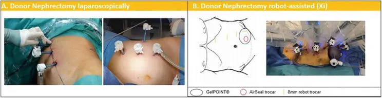
Figure 1.
Trocar placement and patient positioning for nephrectomy during RAKT on living donors.
(A) Laparoscopically: patient positioned in lateral decubitus, linear port configuration along the pararectal line, with the camera placed at the most cephalic position (at the 12th rib level).
(B) Robot-assisted: patient positioned in lateral decubitus; GelPOINT device at the level of the ipsilateral fossa through a 6 cm Pfannenstiel incision; a 15-mm AirSeal trocar is placed in the device to introduce endovascular stapler and the 15-mm EndoCatch bag for organ extraction. An additional trocar is used to raise the kidney during the section of the vessels.
2.2.2 Back table preparation
- After robot-assisted/laparoscopic living donor nephrectomy, the preparation of the kidney is performed at the back table close to the operating bed. First, the graft is placed in a basin with slushed ice and perfused with 1 liter of storage solution (Celsior®, or Custodiol®, or Institut Georges Lopez-1®).
- Next, the graft vessels are carefully dissected. If the donor kidney has multiple arteries or veins, a vascular reconstruction can be performed. Siena et al. demonstrated that RAKT is possible using grafts with multiple vessels. The ureter can be pre-stented with a double-J if preferred. Subsequently, the kidney is wrapped in a gauze filled with slushed ice, with the artery and vein brought out through an opening in the gauze.
- The aim is to keep the donor kidney at a constant low temperature after insertion in the abdominal cavity, until the vascular anastomoses are finished, and the kidney is reperfused. In addition, the gauze can prevent potential graft injury from manipulation with the robot arms. To keep the graft temperature below 20°C intracorporeally, ice is added through the GelPOINT® (Applied Medical, Rancho Santa Margarita, CA, USA) every 15 min.
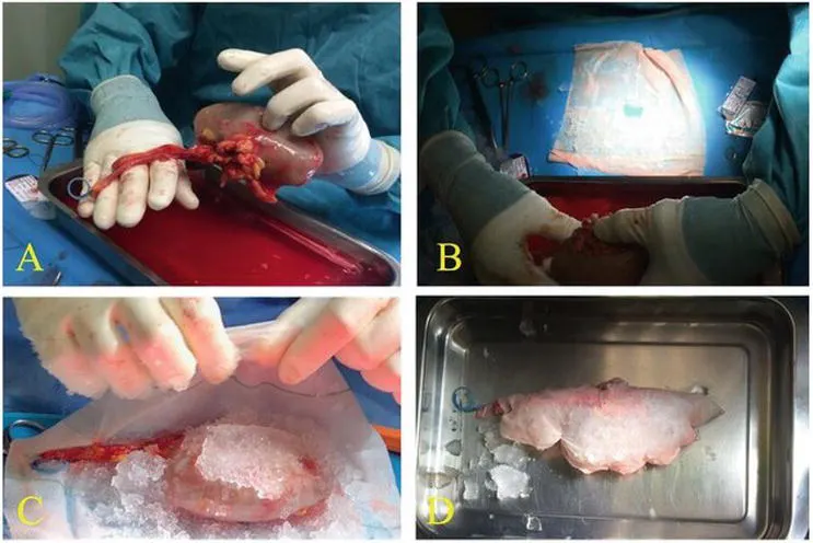
Figure 2.
Preparation of the kidney graft after nephrectomy during RAKT from living donors.
(A) Ureteral double J stent is placed in the graft.
(B) A central hole in the gauze from which the artery and vein are outside.
(C and D) The graft is wrapped in a gauze jacket filled with ice slush.
2.2.3 Robot-assisted living donor kidney transplantation
2.2.3.1 Patient and trocar positioning
- When using the da Vinci Si® or X® system, the patient is positioned in lithotomy position according to the Vattikuti-Medanta technique. When the da Vinci Xi® system is used, the patient is positioned in dorsal decubitus. A 20–30° Trendelenburg position is recommended. The required robotic instruments are monopolar scissors, Potts scissors, bipolar forceps, prograsp forceps, large needle driver, black diamond micro-forceps, and bulldog clamps.
- A 12 mm camera port is inserted in the supra-umbilical area, and a pneumoperitoneum is created. Veres's needle puncture, optical trocar access, or the Hasson technique can also be used for access to the abdomen and creation of a pneumoperitoneum. The open approach (Hasson technique) has been reported to result in fewer complications. Three extra robotic 8 mm ports are placed under vision, and the robot is docked. Minimal changes in port placement are related according to the robotic system used.
- A GelPOINT® device replaces the camera trocar through a 6–8 cm (four fingers) periumbilical incision once the transplant bed preparation has been performed. Alternatively, the GelPOINT® device can be introduced from the beginning through a 6–8 cm periumbilical incision, containing the camera and an assistant port.
- This GelPOINT® device is used to introduce the graft in the abdominal cavity and allows for insertion of slushed ice (± 200 ml) via modified Toomey syringes into the abdominal cavity, surrounding the graft surface with the intent to achieve regional hypothermia (i.e., low constant temperature (<20°C) of the graft).
- Additionally, GelPOINT® is a useful device for fast hand introduction if needed (i.e., in case of massive bleeding). In selected cases, the graft can be introduced transvaginal as described by few authors. The AirSeal® (Conmed, Utica, NY, USA) system might be used in order to maintain a stable and low-pressure pneumoperitoneum at 8 mmHg.
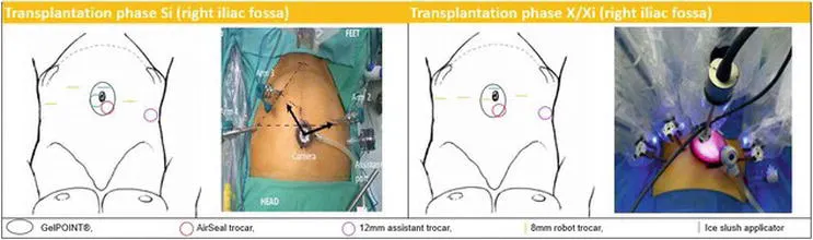
Figure 3.
Trocar placement and patient positioning for RAKT for Si/X/Xi in the right iliac fossa. Patient repositioned in dorsal decubitus, legs in Allen stirrups, table in 20–30° Trendelenburg; GelPOINT® device at the level of the umbilicus through a 6–8 cm vertical peri-umbilical incision; camera trocar and ice applicator in the GelPOINT® (eventually with 12 mm AirSeal® port); 3–8 mm robotic trocars in the lower abdomen, 2 in the left fossa and 1 in the right iliac fossa.
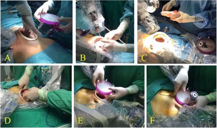
Figure 4.
Introduction of the kidney and ice through the Gel-POINT®.
(A) The GelPOINT® device is placed through a 6 cm (four fingers) incision.
(B) Ice slush is introduced in the abdominal cavity using modified Toomey syringes.
(C and D) The graft is introduced into the abdominal cavity.
(E and F) Once the graft is inside, Gel-POINT® cup is inserted to close the abdomen.
2.2.3.2 Transplant bed preparation
- Accurate dissection of the external iliac vessels is performed. Subsequently, the bladder is prepared for ureteral reimplantation. A retroperitoneal pouch is created by incision of the peritoneum following a transverse line above the level of the appendix and mobilization of the peritoneal flaps.
- These will be used to cover (retroperitonealize) the graft once the vascular anastomosis is completed. Although RAKT is a transperitoneal approach, retroperitonealization of the kidney is performed to avoid pedicle torsion and to enable future graft biopsies.
2.2.3.3 Venous and arterial anastomosis
- After clamping of the external iliac vein with robotic bulldog clamps and the distal clamp followed by the proximal clamp, a longitudinal venotomy using cold scissors is performed. An end-to-side anastomosis between the graft renal vein and the external iliac vein is created, using a 6/0 Gore-Tex® CV-6 TTc-9 or THc-12 needle (W.L. Gore and Associates Inc., Flagstaff, AZ, USA) continuous suture. At the proximal angle, the suture is tied to secure the posterior wall of the anastomosis watertight and to avoid stenosis, and then the continuous suture is completed until the distal angle.
- Prior to finishing the anastomosis, the lumen is flushed with heparinized solution using a 4.8 Fr ureteric catheter. The catheter may be pulled out by the assistant from outside the abdomen while the surgeon tightens the knot to secure the anastomosis. Next, the graft vein is clamped, and the bulldog clamps are removed from the external iliac vein and positioned on the external iliac artery, first proximally and then distally.
- The artery may be incised with the cold scissors or a scalpel at the 1–2 o’clock position. Arteriotomy may be completed using a laparoscopic aortic punch to transform the linear arteriotomy into a circular one. In both arterial and venous anastomosis, the anastomosis is started by passing the needle in the external iliac vessel in an outside-inside direction and then inside-outside through the graft vessel. For the venous anastomosis, the knot is tied now, and the needle is then passed outside-inside through the renal vein to start the running suture.
- For the arterial anastomosis, the suture is not tied yet (as for the venous anastomosis), and the needle is passed through the graft artery outside-inside before tying the suture to a loop that is left outside. This is done to prevent a difficult first needle passing in a small arterial lumen. After completing the arterial anastomosis, a clamp is positioned on the graft artery while the external iliac artery is declamped. If no sign of leakage (bleeding) is observed, the graft vein and artery are declamped.
- The evaluation of the graft perfusion is primarily visual: pink colorization, a pulsatile graft artery, filling of the renal vein, and small bleedings from the renal capsule and urine output are signs of perfusion. Doppler ultrasound evaluation (drop-in ultrasound probe linked to TilePro™ function of the da Vinci Surgical System) is recommended to verify adequate perfusion of the graft.
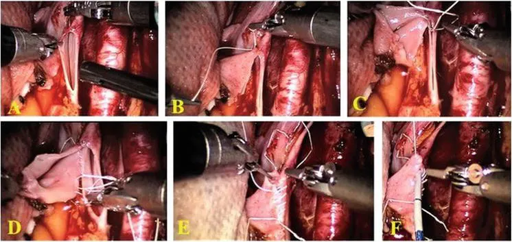
Figure 5.
Overview of the main steps for venous anastomosis during RAKT from living donors.
(A) The graft renal vein is anastomosed in an end-to-side continuous fashion to the external iliac vein using a 6/0 Gore-Tex®.
(B and C) At cranial angle, the suture is knotted to fix the posterior wall of the anastomosis.
(D and E) The running suture is completed at the caudal angle.
(F) Before completing the anastomosis, the lumen is flushed with heparinized solution using a 4.8 Fr ureteral catheter.
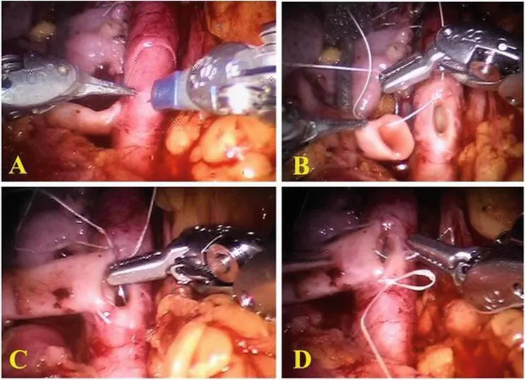
Figure 6.
Overview of the main steps for arterial anastomosis during RAKT from living donors.
(A) The robotic scalpel is used to make a linear incision on the iliac artery, converting it in circular hole with a laparoscopic vascular punch.
(B) The running suture is carried out using a 6/0 Gore-Tex®; particularly in the caudal tying of an arterial anastomosis, the needle is passed in the external iliac vessel in an outside-inside direction, then outside-inside through the graft vessel.
(C and D) The running suture is completed at the caudal angle.
2.2.3.4 Ureteroneocystostomy
- After flipping the kidney on the psoas and retroperitonealization of the graft, the ureteroneocystostomy is performed according to the Lich-Gregoire technique using a Monocryl or PDS 5/0 (Ethicon Inc., Cincinnati, OH, USA) continuous suture.
- Care is taken to construct an adequate detrusor tunnel as anti-reflux mechanism. A double-J stent is inserted to protect the anastomosis. The stent can be removed after 3 weeks.
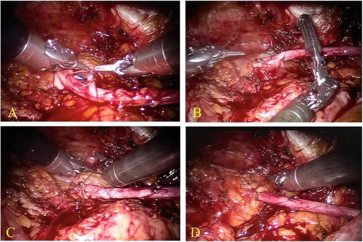
Figure 7.
Ureteroneocystostomy performed according to the Lich-Gregoir technique.
In (A) and (B) running suture between ureteral and bladder mucosa using 5-0 Monocryl.
In (C) and (D) the details of the anti-reflux tunnel.
2.3 Results
- After reporting a single center experience on 17 cases of RAKT from living donation the European Experience in RAKT was published in 2018. One hundred twenty cases were prospectively collected in eight centers across Europe. The authors demonstrated the low complication rate (at 1 month follow-up) while maintaining excellent graft function (median eGFR at 30 days was 58 ml/min) and cosmetic results.
- Three cases of graft loss due to arterial thrombosis during the first postoperative week were reported in these series (2.5%). This complication might be associated with technical errors during the learning curve. Territo et al. demonstrated that the functional results at 1-year follow-up were not statistically different from the functional results at 1-month follow-up.
- The complication rate remained low. To date, there are no studies available comparing RAKT with the conventional open approach. However, an increasing body of evidence confirms that RAKT is at least non-inferior to open KT regarding both patient and graft survival.