Project 2
For Project (a) (from 7 to Module 9) : Student can consider Gastrology system , Urinary System and Homeostasis condition (150mks)
Gastroenterologists
- A gastroenterologist is a medical professional who specializes in conditions that affect the organs and structures of the digestive system.
- Gastroenterologists diagnose, treat, and care for people with a range of gut-related illnesses. These include long-term conditions, such as inflammatory bowel disease (IBD) and celiac disease
- Gastroenterology is a subspecialty of internal medicine.
- It relates to the study of the function and diseases of the gastrointestinal tract and the digestive system.
- According to the American College of Gastroenterology, gastroenterology involves a detailed understanding of:
- the gastrointestinal organs
- the movement of material through the st
- omach and intestines
- the digestion and absorption of nutrientsthe removal of waste from the body
- the liver
- A gastroenterologist is a medical professional who specializes in diseases and conditions that affect the digestive organs and structures.
- These parts of the body include the:
- esophagus, or food pipe
- stomach
- small intestine
- colon
- rectum
- pancreas
- gallbladder
- bile ducts
- liver
- To become a gastroenterologist, a doctor must complete a 3 year internal medicine residency after medical school. Following this residency, they will undertake 2–3 years of intense, specialized training called a fellowship.
- Once they become qualified, gastroenterologists often work closely with other healthcare professionals to provide care to a wide range of people.
- A gastroenterologist may work with:
- hepatologists, who specialize in the liver
- radiologists, who specialize in imaging
- oncologists, who specialize in cancer
- In some cases, doctors need to refer people to medical professionals with specialist knowledge.
- A doctor may refer an individual to a gastroenterologist if they suspect that a problem with the digestive system is causing the person’s symptoms.
- The symptoms of such issues can vary, but they may include:
- difficulty swallowing
- stomach pain
- nausea and vomiting
- heartburn
- diarrhea
- constipation
- jaundice
- The gastroenterologist will use their specialist knowledge to assess the person and recommend treatment and management if necessary.
- Gastroenterologists play a role in the diagnosis, treatment, and management of people with conditions such as:
- colon polyps
- colon cancer
- celiac disease
- hepatitis
- heartburn, or gastroesophageal reflux (GERD)
- gallbladder disease
- irritable bowel syndrome (IBS)
- pancreatitis
- Gastroenterologists carry out several procedures to diagnose, treat, and manage conditions.
- These procedures include:
Endoscopy
- During an endoscopy, a gastroenterologist uses a camera that attaches to a long, thin tube called an endoscope to look inside the body.
- They insert the endoscope through the mouth, down the throat, and into the esophagus. It sends images back to a screen for monitoring.
- A gastroenterologist might perform an endoscopy to investigate symptoms such as:
- persistent heartburn
- nausea and vomiting
- bleeding
- problems with swallowing
- stomach pain
- unexplained weight loss
Colonoscopy and sigmoidoscopy
- These procedures are similar to an endoscopy, but they require the gastroenterologist to insert the tube into the rectum, rather than the mouth.
- A gastroenterologist can see the entire colon and rectum during a colonoscopy. A sigmoidoscopy only allows them to examine the rectum and lower colon.
- During these procedures, gastroenterologists might be looking for:
- early signs of colon or rectum cancer
- causes of bowel habit changes
- causes of specific symptoms, such as abdominal pain, rectal bleeding, or unexplained weight loss
Polypectomy
- During a polypectomy, a gastroenterologist will remove one or more polyps from the lining of the bowel.
- Polyps are noncancerous growths that can develop on the colon. They are very common, affecting 30%Trusted of those over the age of 50 years in the United States.
Esophageal dilation
- During esophageal dilation, a gastroenterologist will stretch out a narrowed area of the esophagus.
- Acid reflux can scar this muscular tube, narrowing it and making it hard for a person to swallow food.
- Layers of excess tissue, cancer of the esophagus, and scarring from radiation treatment can all lead to the same problem.
- The gastroenterologist stretches the tube by using a plastic dilator or inflating a balloon. They will usually carry this out during an endoscopy.
- The gastroenterologist may sedate the person for the procedure. Alternatively, they may apply a local anesthetic spray to the back of the person’s throat.
- A gastroenterologist is a medical professional who specializes in diseases of the digestive system and digestive tract.
- A doctor might refer someone to a gastroenterologist if they have symptoms that suggest a problem with the digestive system.
- Gastroenterologists can diagnose, treat, and manage a wide range of gut-related conditions.
- They also carry out certain medical procedures, such as endoscopies and colonoscopies.
colonoscopy
- A colonoscopy is a type of imaging study in which a doctor uses a particular scope called a colonoscope to examine the colon’s lining. The doctor can also remove polyps or tissue for a biopsy at the same time.
- A doctor may recommend a colonoscopy to evaluate the colon for cancerous lesions, identify areas of bleeding, or diagnose other gut-related disorders.
- Before the procedure, a doctor will prescribe a colonoscopy preparation. There are various approaches, but they all aim to empty the colon of fecal matter so that a doctor can view it clearly. The preparation may start up to a week before the colonoscopy.
- The colonoscopy procedure involves inserting a thin scope with a light on the end into the rectum. The doctor will advance the scope through the colon to examine it.
- In this article, we look at how to prepare for a colonoscopy appointment and what to expect before, during, and after the procedure.
- In the week before the procedure, the person will follow several steps. It is essential to prepare as recommended because incomplete preparation can affect trusted the results.
- It may mean canceling the procedure. If the colonoscopy goes ahead, the doctor may miss a lesion, or the person may need to repeat the procedure.
One week before
- A doctor is likely to recommend specific dietary changes in the week leading up to a colonoscopy.
- While canned or cooked vegetables are acceptable to eat, people should avoid the following foods during this week:
- grains, including brown rice, oatmeal, and wheat bread
- nuts
- seeds, including poppy or sesame seeds
- whole kernel corn
- raw fruits and vegetables
- A doctor may also recommend that people stop taking certain medications that could increase the risk of bleeding during a colonoscopy. Examples include blood thinners, such as warfarin and clopidogrel, and nonsteroidal anti-inflammatory drugs (NSAIDs), such as ibuprofen.
- The gastroenterologist will work with the individual during the consultation to decide how long they should suspend using a specific medication before and after the colonoscopy.
- The consultation also is a good time to ask any questions about the procedure, including the risks and benefits.
Two days before
- At this point, people should drink plenty of water or other liquids to ensure that they do not become dehydrated when they begin their bowel prep. Drinking around eight 8-ounce glasses of water a day can help reduce the risk of dehydration.
- A doctor may also recommend avoiding red or orange foods at this point, as they could make a person’s colon look like it is bleeding.
24 hours before
- It is crucial to consume only clear liquids in the 24 hours leading up to the colonoscopy.
- Examples of acceptable clear liquids include:
- clear broth or bouillon
- coffee or tea, but with no milk or creamer added
- electrolyte-containing beverages, such as sports drinks
- gelatin
- popsicles
- strained fruit juices without pulp
- In general, if it is possible to see through the food or drink, it should be acceptable within a clear liquid diet. However, a person should always consult their doctor if they have any questions about specific foods.
- A doctor will often prescribe an oral solution to drink to help clean out the colon. This solution will cause the person to have frequent bowel movements until there is no fecal matter left in their colon. Examples of oral solutions include trusted sources GoLytely, Colyte, NuLytely, TriLyte, Suprep, and Clenpiq.
- These solutions are likely to have different dosage methods. Sometimes, a doctor will prescribe drinking the entire solution the night before the colonoscopy. Other times, they will recommend drinking one portion of the solution the night before and the remainder on the day of the examination.
- The solution is intended to make a person go to the bathroom. Their bowel movements will turn into diarrhea.
- The bowel prep can sometimes make a person feel nauseous, bloated, thirsty, or dizzy. If this occurs, the individual should give themselves a break of at least 30 minutes from drinking the prep and slowly sip on clear fluids. If they can tolerate returning to drinking the prep, they should.
- With a doctor’s permission, a person potentially can add a powdered drink solution that is not red, blue, or purple to the prep to improve its taste.
- During the bowel prep, a person may benefit from:
- staying near a bathroom so that they can go to the toilet easily
- wearing elastic-waist pants that are easier to pull down
- drinking clear liquids after swallowing the prep to remove the unpleasant taste
- applying petroleum jelly around the anal opening or using baby wipes to reduce irritation to the anus
- The bowel movements will eventually turn clear or yellow. This change signals that a person’s colon is likely to be clear of fecal matter.
- In case of problems with constipation, a doctor may also recommend using a stool softener to make stools easier to pass.
- By the time of the colonoscopy appointment, a doctor will expect a person to be passing only clear to yellow-tinted liquid from the bowel.
- The individual’s doctor will explain the procedure. The person will also meet the nursing staff who will be in the room with them during their colonoscopy, as well as anesthesia representatives who will be providing sedation.
- A member of the medical staff will insert an intravenous (IV) catheter into the person’s vein so they can receive medications during the procedure.
- The person will then go to the colonoscopy suite. An anesthesiologist who is part of the procedure team will administer sedatives to the person via the IV and be responsible for keeping their airway safe throughout the procedure.
- Once the person is sedated, a doctor will insert the colonoscope through the rectum and advance it to begin the examination. They will look for irregularities in the colon’s lining.
- The gastroenterologist will evaluate each polyp individually. Based on its size, shape, and location, the doctor will determine the best technique to remove the polyp. They may also take a sample of tissue for testing in the laboratory, known as a biopsy.
- Ideally, a person’s colon prep will be so effective that a doctor can advance the colonoscope far enough into the colon to see where the small and large intestines join.
- When the doctor has completed the examination, they will remove the scope. The anesthesia professional will stop administering medication, and the person will wake up.
- It is not unusual to feel sleepy or groggy after the procedure. For that reason, a person should always have someone else available to drive them home from the appointment.
- After a colonoscopy, the person may have to attend another appointment to discuss the results or any findings of tests.
- During the appointment, the doctor will explain any findings. This might include reviewing images from the colonoscopy.
- Possible results may be:
- Negative: There are no growths or polyps.
- Growths or polyps: The doctor has removed polyps and may wish to follow up after testing.
- Bowel cancer: The doctor may have taken a biopsy. If they suspect cancer, they will talk to the individual about what to do next.
- Other conditions: If there are signs of Crohn’s disease, diverticulitis, ulcerative colitis, or another condition, the doctor will discuss the next steps with the person.
- If a colonoscopy is positive, meaning cancerous or precancerous cells are present, the doctor may recommend further interventions. These can include additional colonoscopies or even surgery.
- There are some risks involved in a colonoscopy. The doctor should explain these to the person before the procedure.
- They include
- complications relating to anesthesia
- rectal tears
- bleeding
- pain
- bloating
- infection
- post-polypectomy electrocoagulation syndrome (PPES)
- colon wall perforation
- Severe complications are rare, and experts consider the procedure relatively safe.
- PPES can cause severe abdominal pain several hours after the procedure. Treatment usually involves bowel rest and antibiotics.
- Colonoscopy preparation can be unpleasant but is vital for ensuring a successful procedure.
- If the results are not clear, the person may need to repeat the procedure.
- For this reason, a person should follow the preparation plan. A doctor will make follow-up recommendations if the colonoscopy shows that further investigation or treatment is necessary.
polypectomy
- Polypectomy is the removal of a polyp. The most common types of polypectomy are uterine polypectomies and colon polypectomies.
- However, a doctor can perform a polypectomy on any area of the body that develops polyps, such as the nose or stomach. A polypTrusted Source is a growth that begins in the tissue and extends into the hollow space around it.
- A polypectomy involves removing polyps that may be causing symptoms, may be cancerous, or require examination.
FAST FACTS ON POLYPECTOMIES
- Most polyps are benign growths, which means they do not contain cancer.
- In most cases, a doctor can remove a polyp while the person is awake.
- The right technique for polyp removal depends on the size and location of the polyp.
- Polypectomies are safe, with a low rate of complications.
- The two most common types of polypectomy are uterine polypectomies and colon polypectomies.
- A uterine polypectomy is a procedure to remove polyps in the endometrial tissue, the tissue that lines the inside of the uterus. A colon polypectomy involves removing polyps in the colon.
- Polyps may also develop in other regions of the body. Some other relatively common sites of polypectomy include:
- the nose
- the cervix
- the vocal cords
- the stomach
- Polyps grow out of tissue into hollow spaces in the body. For example, colon polyps begin in the tissue of the colon then extend into the hollow space inside it.
- Most are benign, but some are cancerous or precancerous. It is not possible to rule out cancer without examining a polyp, so many doctors recommend routine removal to test for cancer.
Are they common?
- Polyps are common, and most people do not know they have them. Approximately 20–30% of people in the United States have colon polyps.
- Uterine polyps are most common in people aged 40–49 years trusted Source. About 1–5% are cancerous, with the risk of cancerous uterine polyps being higher in people who are postmenopausal.
How big do they get?
- Even when they are not cancerous, some polyps can grow very large. When they do, they may alter blood flow, put pressure on organs, and cause a range of other symptoms.
- Preparation for a polypectomy depends on the surgical technique and the location of the polyps.
- People undergoing removal of uterine polyps with local anesthesia may need no special preparation.
- However, colon polypectomy requires the colon to be empty. People may need to use enemas, laxatives, or both in the 12–24 hours before surgery.
Surgical techniques and what to expect
- Many people prefer to be awake for the procedure. Others may want to receive general anesthesia. In this case, the doctor will give the person a sedative, usually through an IV needle. This makes them feel drowsy and relaxed.
- Doctors can remove most polyps with an endoscope. This is a tube the doctor inserts into the body, eliminating the need for a large incision. For a uterine polypectomy, a doctor inserts a hysteroscope through the vagina into the cervix and uterus. A camera helps guide the scope to the polyp.
- During a colon polypectomyTrusted Source, the doctor inserts the colonoscope through the rectum. As with a uterine polypectomy, a camera helps the doctor locate the polyp.
- Doctors use different surgical techniques for removing polyps. The specific removal technique is unlikely to affect the person’s experience of the surgery.
- Some options include:
- Hot snare polypectomy: Hot snare polypectomy (HSP) is the preferred treatment for most polyps and employs a heated snare. A snare is a loop that can grab and remove the polyp. The surgeon may also use electrocautery to burn away any remaining polyp tissue.
- Cold snare polypectomy: Cole snare polypectomy (CSP) uses a cold snare and is preferred for diminutive (1-5 mm.) polyps.
- Cold or hot forceps polypectomy: A more outmoded technique uses forceps to pull the polyp loose, to remove small polyps. The surgeon will also use a wire to remove the portion of the polyp that extends into the tissue, or if hot, a procedure called electrocautery to burn away any remaining polyp tissue and prevent bleeding.
- A person may feel pressure or pulling during the procedure, but they should feel no pain. Depending on the location of the polyp and other factors, a doctor may give the person pain-blocking medication before, during, or after the procedure.
- When polyps are very large, removing them through an incision may be necessary. This procedure is more complicated and typically requires general anesthesia.
- Recovery from a colon polypectomy is fairly quick, and most people can get back to their usual diet immediately. People may feel some mild pain, gassiness, or cramping following the procedure, particularly immediately afterward, but these symptoms are typically short-lived.
- Taking the pain-relieving medication the doctor prescribes can help.
- It is normal to experience some bleeding for a few days following a uterine polypectomy, but call a doctor if the bleeding is very heavy, stops and then starts again, or has a foul odor that could indicate an infection.
- After a surgeon removes the polyp, they will send it to a lab for testing. If the polyp is not cancerous, the person may undergo an exam following the removal but will likely need no further treatment.
- Most people with polyps do not know they have them.
- When they cause symptoms, a doctor may recommend screening tests. In some cases, doctors discover polyps as part of screening for something else. This means that people are more likely to undergo a polypectomy when they also undergo a screening test that can detect polyps.
- People who have symptoms related to their polyps, such as pain or bleeding, usually need to have the polyps removed.
- Like all surgeries, polypectomies carry some risks. These include:
- Organ perforation: This occurs when a surgeon punctures the organ they are operating on. This can be fatal, but is rare.
- Infection: A polypectomy creates a wound, and any wound can become infected. Infection is more likely when people do not follow doctor’s advice after surgery or are already not in optimal health.
- Excessive bleeding: Sometimes the wound does not properly heal, causing excessive bleeding.
- Incomplete removal: In some cases, the first procedure may leave some tissue behind, necessitating a second polypectomy.
- A polypectomy is a routine procedure that can eliminate symptoms of polyps and test for cancer. As with any procedure, people should weigh the benefits and risks with the help of a doctor.
- However, for most polypectomy recipients, the procedure is a minor inconvenience that can offer peace of mind and relief from troublesome symptoms.
Radiologist
- A radiologist is a type of doctor who uses medical imaging to support the diagnosis and treatment of various conditions and injuries.
- A radiologist is a type of doctor who specializes in medical imaging. Radiologists analyze images, such as X-rays, to help diagnose, monitor, and treat various conditions or injuries.
- Radiologists are different than radiographers. Although both of these professionals work with medical imaging, radiographers are the people who operate the machinery.
- There are different types of radiologists, including diagnostic radiologists and medical physicists.
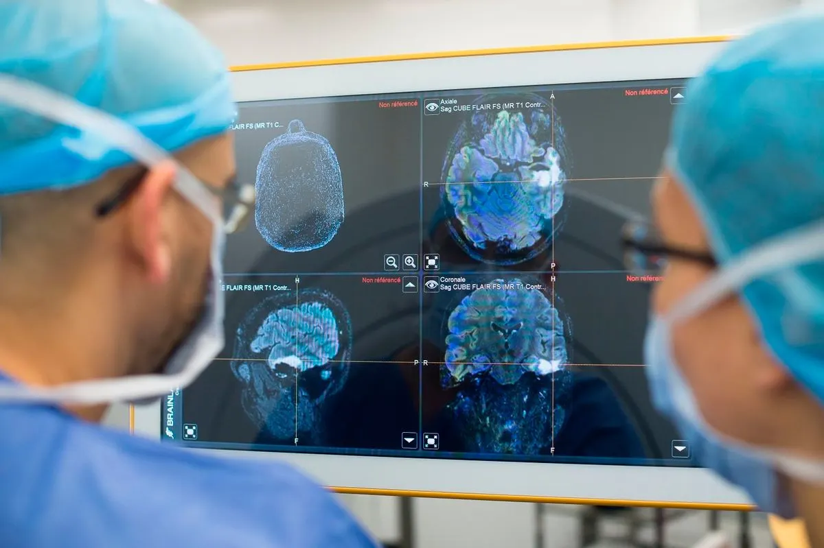
- There are several different specialties of radiology, including:
- Diagnostic Radiology
- Diagnostic radiologists use medical imaging to diagnose and treat diseases. They can use a variety of different imaging methods, such as:
- X-rays
- radionuclides
- ultrasounds
- electromagnetic radiation
- Diagnostic radiologists use medical imaging to diagnose and treat diseases. They can use a variety of different imaging methods, such as:
Interventional radiology
- Interventional radiologists use medical imaging to provide therapy to people with noncancerous conditions.
- For example, an interventional radiologist might use medical imaging to support a surgical procedure.
- This imaging can make surgical procedures safer and lead to faster recovery times. Interventional radiologists typically work on keyhole surgery.
- Keyhole surgery involves making small cuts instead of larger ones and using tiny cameras to see inside the body.
Radiation oncology
- A radiation oncologist uses radiation-based therapy to treat cancer. This therapy involves the use of high energy radiation to damage cancer cells, which stops them from spreading further.
- It can help reduce symptoms or, in some cases, cure the condition entirely.
Medical physics
- Medical physicists use their understanding of physics to support the practice of medicine in different ways.
- For example, they can advise on and deliver the technical aspects of medical imaging to ensure the safety of patients and the effectiveness of the results.
- Some medical physicists are also researchers and play a role in developing new medical technology. Medical physicists have developed many devices that doctors commonly use today, such as magnetic resonance imaging (MRI).
- Radiologists can work in clinical practices, hospitals, or universities. The job of radiologists varies depending on their specialty.
- All radiologists work with medical imaging methods, which include:
- computed tomography (CT) scans
- MRI scans
- positron emission tomography (PET) scans
- X-rays
- ultrasounds
- nuclear medicine
- fusion imaging
- Most of these techniques involve the use of radiation. Radiologists are highly trained in keeping people safe from the harmful effects of radiation.
- These professionals can help other doctors decide on the right imaging method to use and understand what the results mean for treatment. They can also help interpret different images and other test results to make a diagnosis or monitor whether current treatments are working.
- Certain types of radiologists, including interventional radiologists, are more actively involved in the treatment process. Others, such as diagnostic radiologists, might provide support to other healthcare professionals.
- Some radiologists rarely work with patients and instead work in labs doing research. For example, some clinical studies might include a radiologist to help with the analysis of medical images.
- Radiologists are medical doctors, so they follow a similar path to those working in other specialties.
- All radiologists need a medical degree, which involves 4 years of training and education from a medical school.
- Most medical schools require students to have an undergraduate degree and pass a Medical College Admission Test before entering.
- After finishing medical school, radiologists do a year of clinical training. They may spend a preliminary year focusing on one area of medicine, such as internal medicine, or it may be a transitional year that involves several rotations through different specialties.
- Following the clinical year, radiologists usually complete 4 years of paid residency. Residency is a combination of further medical education and on-the-job training in different areas of radiology.
- After a residency, most radiologists do a fellowship. A fellowship is an additional 1 or 2 years of training in a specialized area of radiology, such as nuclear radiology. Interventional radiologists must undertake a 2 year fellowship.
- Radiologists are medical doctors. They share some of the same duties as a family doctor, such as performing diagnoses or monitoring treatment, but most radiologists do not work directly with patients.
- Radiologists are different than radiographers. Radiographers operate medical imaging equipment, but they do not interpret the results.
- Radiologists are medical doctors who work with medical imaging techniques, such as MRIs or X-rays.
- There are several different specialties of radiology, each of which plays a different role in medicine.
- For example, a diagnostic radiologist helps support diagnosis and treatment, while an interventional radiologist uses imaging to guide surgical procedures.
- Most radiologists work with other doctors to diagnose and treat a wide range of conditions and injuries. They receive an education similar to that of other medical doctors, which takes about 8 to 10 years.
URINARY SYSTEM
- The urinary system's function is to filter blood and create urine as a waste by-product. The organs of the urinary system include the kidneys, renal pelvis, ureters, bladder and urethra.
- The body takes nutrients from food and converts them to energy. After the body has taken the food components that it needs, waste products are left behind in the bowel and in the blood.
- The kidney and urinary systems help the body to eliminate liquid waste called urea, and to keep chemicals, such as potassium and sodium, and water in balance. Urea is produced when foods containing protein, such as meat, poultry, and certain vegetables, are broken down in the body. Urea is carried in the bloodstream to the kidneys, where it is removed along with water and other wastes in the form of urine.
- Other important functions of the kidneys include blood pressure regulation and the production of erythropoietin, which controls red blood cell production in the bone marrow. Kidneys also regulate the acid-base balance and conserve fluids.
Kidney and urinary system parts and their functions
-
Two kidneys. This pair of purplish-brown organs is located below the ribs toward the middle of the back. Their function is to:
-
Remove waste products and drugs from the body
-
Balance the body's fluids
-
Release hormones to regulate blood pressure
-
Control production of red blood cells
-
- The kidneys remove urea from the blood through tiny filtering units called nephrons. Each nephron consists of a ball formed of small blood capillaries, called a glomerulus, and a small tube called a renal tubule. Urea, together with water and other waste substances, forms the urine as it passes through the nephrons and down the renal tubules of the kidney.
-
Two ureters. These narrow tubes carry urine from the kidneys to the bladder. Muscles in the ureter walls continually tighten and relax forcing urine downward, away from the kidneys. If urine backs up, or is allowed to stand still, a kidney infection can develop. About every 10 to 15 seconds, small amounts of urine are emptied into the bladder from the ureters.
-
Bladder. This triangle-shaped, hollow organ is located in the lower abdomen. It is held in place by ligaments that are attached to other organs and the pelvic bones. The bladder's walls relax and expand to store urine, and contract and flatten to empty urine through the urethra. The typical healthy adult bladder can store up to two cups of urine for two to five hours.
Upon examination, specific "landmarks" are used to describe the location of any irregularities in the bladder. These are:
-
Trigone: a triangle-shaped region near the junction of the urethra and the bladder
-
Right and left lateral walls: walls on either side of the trigone
-
Posterior wall: back wall
-
Dome: roof of the bladder
-
-
Two sphincter muscles. These circular muscles help keep urine from leaking by closing tightly like a rubber band around the opening of the bladder.
-
Nerves in the bladder. The nerves alert a person when it is time to urinate, or empty the bladder.
-
Urethra. This tube allows urine to pass outside the body. The brain signals the bladder muscles to tighten, which squeezes urine out of the bladder. At the same time, the brain signals the sphincter muscles to relax to let urine exit the bladder through the urethra. When all the signals occur in the correct order, normal urination occurs.
Facts about urine
-
Normal, healthy urine is a pale straw or transparent yellow color.
-
Darker yellow or honey-colored urine means you need more water.
-
A darker, brownish color may indicate a liver problem or severe dehydration.
-
Pinkish or red urine may mean blood in the urine
urologists
- A urologist is a physician who specializes in diseases of the urinary tract and the male reproductive system.
- Patients may be referred to a urologist if their physician suspects they may need treatment for a condition relating to bladder, urethra, ureters, kidneys, and adrenal glands.
- In men, urologists treat disorders related to the epididymis, penis, prostate, seminal vesicles, and the testes.
- A urologist will help with medical situations concerning the urinary tract.
- A patient may be referred to a urologist for treatment of a range of conditions:
- Urinary tract infections (UTIs): These often arise when bacteria migrate from the digestive tract to the urethra. Symptoms include abnormal urination, pain, incontinence, nausea, vomiting, fevers, and chills. It mostly affects women.
- Incontinence: A malfunction in the urinary system can lead to involuntary loss of bladder control. In women, this may result from a weakening of the pelvic floor muscles during pregnancy.
- Male infertility: This can result from damage to the male reproductive tract and a variety of sperm disorders. One common cause is varicoceles, an enlarged vein in the sac beneath the penis. Surgery can sometimes help.
- Kidney disease: Damage to the kidneys can lead to swelling in the hands and ankles, high blood pressure, and other symptoms. If the kidneys no longer work effectively, this is kidney failure. Ultimately, it can be fatal.
- Renal transplantation: A person may require kidney transplants following kidney failure.
- Urologic oncology: Treatment of cancers that relate to the urological or male reproductive system, such as bladder cancer and prostate cancer.
- Bladder prolapse: when the tissues and muscles of the pelvic floor are no longer able to support the organs in the pelvis, the organs can drop from their usual position.
- Cancers: the bladder, kidneys, prostate gland, testicles, and any other cancer that affects the urinary system or, in men, the reproductive system.
- Enlarged prostate: Benign prostatic hyperplasia (BPH) affects around 1 in 3 men over the age of 50 years. An overgrowth of cells in the prostate gland causes the urethra to constrict, leading to problems with urinatination. A urologist will often take a urine sample to ascertain the diagnosis.
- Erectile dysfunction: The penis is unable to attain sufficient rigidity to fully participate in sexual intercourse. This is often a symptom of an underlying condition.
- Peyronie’s disease: A fibrous layer of scar tissue develops beneath the skin of the penis. This can lead to bending or curving in the penis (phimosis) during an erection that can cause pain and lead to difficulties with sexual intercourse.
- Interstitial cystitis or painful bladder syndrome: A chronic inflammatory bladder condition can produce discomfort ranging from mild to severe.
- Kidney and ureteral stones: Small, hard deposits made from mineral and acid salts form in the kidneys but can pass through into the ureters. They can affect urination and cause pain, nausea and vomiting.
- Prostatitis: Infection or inflammation of the prostate can cause painful urination or ejaculation. It can be acute or chronic.
- Undescended testes, or cryptorchidism: Normally, the testicles form inside the abdomen of a fetus and descend into the scrotum before birth. If one or both does not descend, sperm production can be impaired, and there is a risk of complications.
- Urethral stricture: scarring of the urethra can narrow or block the path of urine flowing from the bladder. Causes include infection, inflammation or injury. Symptoms include painful urination and reduced output. It can lead to complications such as prostatitis and urinary tract infections.
- Pediatric urology: This includes the treatment of urological problems in children that are too complex for non-specialized pediatricians.
- The urologist will normally have notes from the referring doctor, but they will ask questions about the patient’s medical history and carry out a physical examination.
- They may also order some tests.
- Imaging tests, such as CT scans, MRI scans, or ultrasound can help the doctor locate the problem.
- A cystoscope is a long, thin probe with a camera on that can be used to see inside the urinary tract, and, sometimes, to take a sample of tissue for testing.
- A urine test can check for bacteria or other signs of disease.
- A biopsy can check for cancer and other disorders.
- Urodynamic testing can check how fast the urine leaves the body, how much urine remains in the bladder after urinating, and how much pressure there is in the bladder.
- Following diagnosis, the treatment will depend on the condition. Both medical management and surgery are options.
- Treatment will vary according to the diagnosis. It includes the use of medications and surg
- Urologists can treat men’s reproductive health issues, such as erectile dysfunction.
- Types of medication include:
- antibiotics for infections
- hormone treatment for prostate cancer
- phosphodiesterase 5 (PDE5) inhibitors for erectile dysfunction, such as Cialis, or tadalafil
- drugs that act on the smooth muscle of the bladder to reduce urinary incontinence
- chemotherapy drugs for cancer
- Surgery can be:
- open surgery
- laparoscopic, or minimally invasive “keyhole” surgery
- laser therapy to treat BPH, cancer, and kidney stones, among others
- A urologist might perform surgery to:
- remove a tumor or the entire bladder, prostate, or other parts, in cases of cancer
- carry out repairs after a trauma
- relieve strictures in the urethra, caused by scar tissue, known as urethral dilation
- relieve stress incontinence, for example, with a sling procedure
- break up or remove kidney stones
- remove part of a kidney
- transplant of a kidney
- Urologists also perform circumcisions. This procedure is done to remove the skin from the tip of the penis, for cultural, religious, or medical reasons.
- A vasectomy, a permanent form of male contraception, is also carried out by a urologist.
Hemostasis
- Hemostasis is your body’s natural reaction to an injury that stops bleeding and repairs the damage.
- This capability is usually for your benefit, conserving blood and preventing infections. In rare cases, the process doesn't work as it should, and this can cause problems with too much or too little clotting.
- Hemostasis is your body’s normal reaction to an injury that causes bleeding. This reaction stops bleeding and allows your body to start repairs on the injury. This capability is essential to keep you alive, particularly with significant injuries.
- However, in uncommon cases, the processes that control hemostasis can malfunction, causing potentially serious — or even dangerous — problems with bleeding or clotting.
How does hemostasis work?
- Hemostasis combines the terms “hemo” (meaning “blood”) and “stasis” (meaning “standing still”). In this context, it’s the term for how your body stops bleeding.
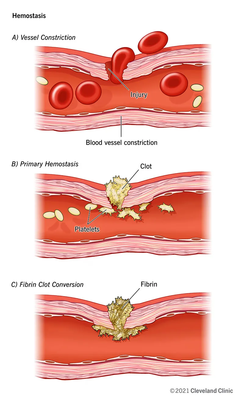
- Rather than being just a single process, hemostasis is actually a collection of several processes. Though they look like separate processes, these all happen at the same time when your body forms a blood clot.
Primary hemostasis (platelet clotting)
- Primary hemostasis is when your body forms a temporary plug to seal an injury. To accomplish that, platelets that circulate in your blood stick to the damaged tissue and activate. That activation means they can “recruit” more platelets to form a platelet “plug” to stop blood loss from the damaged area.
- That clot works much like a cork or bottle stopper, keeping blood in and debris or germs out. Primary hemostasis may also involve constriction (narrowing) of the damaged blood vessel, which can happen because of substances that activated platelets release.
Secondary hemostasis (coagulation cascade)
- The platelet plug is the first step to stop bleeding, but it isn’t stable enough to stay in place without help. The next step, which stabilizes the plug, is secondary hemostasis. This step, sometimes called coagulation, involves molecules in your blood called “coagulation factors.”
- Those factors activate in sequence, the “coagulation cascade,” which amplifies clotting effects as the sequence continues. Ultimately, the coagulation cascade forms a substance called fibrin. During this step, the platelet plug acts like bricks and the fibrin acts like mortar. Together, they form a solid, stable clot.
Fibrin clot remodeling
- The last stage of hemostasis is when your body remodels the existing clot into a fibrin clot. Your body does that because blood clots are a temporary patch, not a permanent solution.
- That removal involves a process called fibrinolysis. During fibrinolysis, your body remodels the clot into the same kind of tissue that was there before the injury.
POSSIBLE CAUSES
What causes hemostasis?
- Your body naturally monitors itself for injuries, and when it detects one, it reacts quickly to take control of the situation. Without normal hemostasis, even minor injuries could cause dangerous blood loss. An example of this is hemophilia, a condition where hemostasis doesn't work properly and blood can't clot effectively.
- Any break in your skin is also a risk for germs to enter your body. Clots help reduce that risk by sealing the injury.
What potential health problems can happen with hemostasis?
- Hemostasis refers to normal blood clotting in response to an injury. However, your body can also have too much clotting, known as hypercoagulability. That can cause many blood clots to form spontaneously and block normal blood flow.
- When blood clots form inside your blood vessels, this is known as thrombosis. When you have thrombosis that happens repeatedly, that’s a condition called thrombophilia.
Thrombophilia (hypercoagulability or too much clotting)
- Hypercoagulability is when your blood clots too much or too easily. This is dangerous because those clots can develop or get stuck in different places in your body and cause severe, life-threatening problems. Examples of these problems include:
- Stroke.
- Deep vein thrombosis (DVT), which can then cause a pulmonary embolism.
- Heart attack.
- Many types of cancer can cause hypercoagulability, and some rare conditions that cause hypercoagulability are also genetic. That means they are either inherited from your parents or happen because of a random mutation in your DNA. People with these conditions have thrombophilia. A few examples of conditions like this include:
- Protein C deficiency.
- Prothrombin gene mutation.
- Factor 5 Leiden mutation.
- Inherited disorders that cause thrombophilia aren’t as common as “acquired” conditions, which you usually develop later in life. Examples of acquired conditions include:
- Antiphospholipid syndrome.
- Inflammatory bowel disease.
- Severe reactions to infections, such as sepsis.
- Most medications that treat thrombophilia make it harder for your blood to clot in some way. Examples of these include antiplatelet, anticoagulant and fibrinolytic (fibrin-breaking or clot-busting) drugs.
Hypocoagulability (not enough clotting)
- When your blood doesn’t clot well, any injury becomes a much more dangerous event. Without proper clotting, even minor injuries can cause you to lose a lot of blood. It also means you’re at greater risk for injuries to organs and blood vessels inside your body, which can then cause internal bleeding.
- Certain types of cancer like leukemia can cause you to bleed too easily. That’s because they often involve a lack of platelets in your body or anti-clotting activity. Conditions that keep your blood from clotting are often genetic, also. Some examples of genetic conditions include:
- Hemophilia.
- Von Willebrand disease.
- Inherited thrombocytopenia (low platelet count).
- Treating conditions that keep your blood from clotting usually involves medications that slow down or block your body’s anti-clotting processes, that boost your body’s ability to make platelets or that add more of certain clotting factors to your blood. You can also receive transfusions of platelets to add more if your body needs them.
How are hemostasis problems diagnosed?
- A healthcare provider — such as a hematologist — can diagnose blood clotting problems based on your symptoms and blood tests that analyze the clotting-related components in a sample of your blood.
When should I call a doctor about problems with clotting?
- Hypercoagulability can happen with a variety of different symptoms depending on where and when an abnormal clot develops. Examples of this include:
- Brain: Stroke symptoms, including weakness or paralysis on one or both sides of your body, slurring speech, inability to move one side of your face and headache.
- Heart: Heart attack symptoms, including chest pain, shortness of breath, weakness, fast or irregular heartbeat, heart palpitations (feeling your own heartbeat) and passing out.
- Lungs: Pulmonary embolism symptoms, including wheezing or sudden problems breathing, pain in your chest, coughing blood, fingertips or lips that are pale or blue-tinted and passing out.
- Deep vein thrombosis (DVT): This can often lead to pulmonary embolism. Symptoms include pain in the calf or elsewhere in the leg, as well as swelling or redness. However, blood clots in your leg can sometimes have relatively minor symptoms, and may just feel like a pulled muscle.
- Kidneys: Blockages in your kidney can cause blood in your urine (pee), needing to pee less often and pain in your lower back on either side of your spine.
- Hypocoagulability will cause you to bleed more easily. Most often, this causes the following symptoms:
- Nosebleeds that are hard to stop.
- Bleeding around your gums when you brush your teeth.
- Wounds that may seep or ooze blood for a long time and take much longer to heal. However cuts in certain areas, such as the face, fingertips or scalp, can take a long time to stop bleeding because there are many more blood vessels in those areas than in other places in your body.
- Finding bruises and you can’t remember how you got them or bruising much more easily and visibly (bruises are bigger and more colorful).
- Hemostasis is a natural part of your daily life. It helps your body protect itself from bleeding and infection, and recover from injuries big and small. Understanding how it works can help you better care for yourself. It also means you can recognize potential problems with your body’s natural recovery abilities and get help sooner rather than late
For Project (b) (from Module 10 to Module 12): Students can consider the Respiration system, cardiovascular system, or Nervous system (150mks)
Respiratory System
- The respiratory system is the organs and other parts of your body involved in breathing when you exchange oxygen and carbon dioxide.
Parts of the Respiratory System
Your respiratory system includes your:
- Nose and nasal cavity
- Sinuses
- Mouth
- Throat (pharynx)
- Voice box (larynx)
- Windpipe (trachea)
- Diaphragm
- Lungs
- Bronchial tubes/bronchi
- Bronchioles
- Air sacs (alveoli)
- Capillaries
How Do We Breathe?
- Breathing starts when you inhale air into your nose or mouth. It travels down the back of your throat and into your windpipe, which is divided into air passages called bronchial tubes.
- For your lungs to perform their best, these airways need to be open. They should be free from inflammation or swelling and extra mucus.
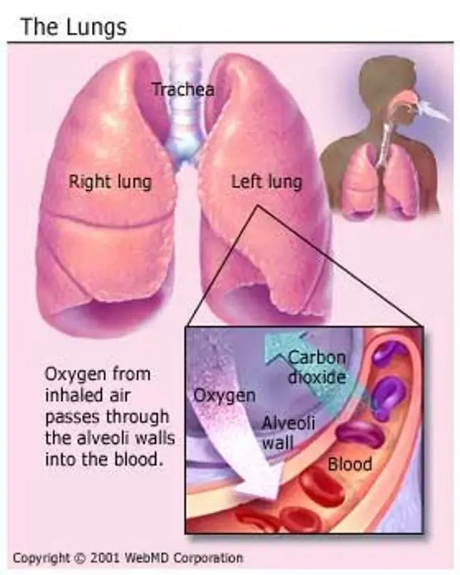
- As the bronchial tubes pass through your lungs, they divide into smaller air passages called bronchioles. The bronchioles end in tiny balloon-like air sacs called alveoli. Your body has about 600 million alveoli.
- The alveoli are surrounded by a mesh of tiny blood vessels called capillaries. Here, oxygen from inhaled air passes into your blood.
- After absorbing oxygen, blood goes to your heart. Your heart then pumps it through your body to the cells of your tissues and organs.
- As the cells use oxygen, they make carbon dioxide that goes into your blood. Your blood then carries the carbon dioxide back to your lungs, where it’s removed from your body when you exhale.
Inhalation and Exhalation
- Inhalation and exhalation are how your body brings in oxygen and gets rid of carbon dioxide. The process gets help from a large dome-shaped muscle under your lungs called the diaphragm.
- When you breathe in, your diaphragm pulls downward, creating a vacuum that causes a rush of air into your lungs.
- The opposite happens with exhalation: Your diaphragm relaxes upward, pushing on your lungs, allowing them to deflate.
the Respiratory System Clean the Air
- Your respiratory system has built-in methods to keep harmful things in the air from entering your lungs.
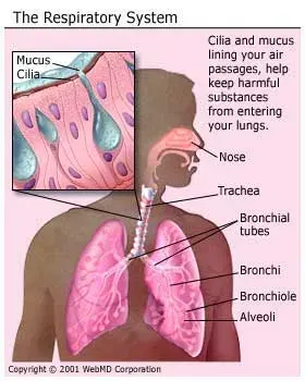
- Hairs in your nose help filter out large particles. Tiny hairs, called cilia, along your air passages move in a sweeping motion to keep the passages clean.
- But if you breathe in harmful things like cigarette smoke, the cilia can stop working. This can lead to health problems like bronchitis.
- Cells in your trachea and bronchial tubes make mucus that keeps air passages moist and helps keep things like dust, bacteria and viruses, and allergy-causing things out of your lungs.
- Mucus can bring up things that reach deeper into your lungs. You then cough out or swallow them.
Respiratory System Diseases
- Common diseases of the respiratory system include:
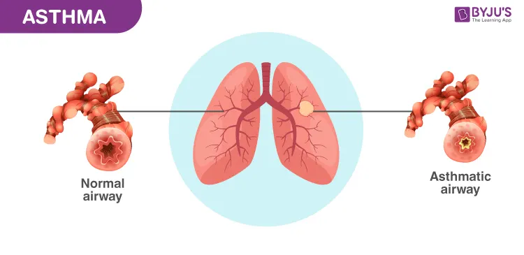
- Asthma. Your airways narrow and make too much mucus.
- Bronchiectasis. Inflammation and infection make your bronchial walls thicker.
- Chronic obstructive pulmonary disease (COPD). This long-term condition gets worse over time. It includes bronchitis and emphysema.
- Pneumonia. An infection causes inflammation in your alveoli. They might fill up with fluid or pus.
- Tuberculosis. A bacterium causes this dangerous infection. It usually affects your lungs but might also involve your kidney, spine, or brain.
- Lung cancer. Cells in your lung change and grow into a tumor. This often happens because of smoking or other chemicals you’ve breathed in.
- Cystic fibrosis. This disease is caused by a problem in your genes and gets worse over time. It causes lung infections that don’t go away.
- Pleural effusion. Too much fluid builds up between the tissues that line your lungs and chest.
- Idiopathic pulmonary fibrosis. Your lung tissue becomes scarred and can’t work the way it should.
Pulmonary Diagnostic Tests & Procedures
- Choose Essentia for a wide range of diagnostic tests, treatments, and procedures for your lung conditions, including:
- Bronchoscopy – Examines the airways of the lungs through a thin, lighted tube (bronchoscope) to diagnose or treat lung conditions, biopsy tissue, and detect lung cancer
- Chest X-ray – Takes pictures of your heart, lungs, and bones in your chest
- Lung biopsy - Removes lung tissue samples for examination under a microscope; includes needle, transbronchial, thoracoscopic, and open biopsy methods
- Low-dose CT lung cancer screening – Detects lung cancer
- Mediastinoscopy – Uses a long, thin, flexible tube with a light and tiny camera (mediastinoscope) to examine the space between and in front of your lungs and collect tissue to examine in a lab
- Methacholine challenge testing – Evaluates your lung function and looks at narrowing or tightening of your airways after inhaling a methacholine mist; helps diagnose asthma
- Peak flow measurement – Measures air flow from your lungs when you forcefully exhale
- Pulmonary function tests – Assesses your breathing and how well your lungs are functioning with normal and deep breathing tests
- Pulse oximetry – Measures the oxygen level in your blood by clipping a device on your finger
- Thoracentesis – Removes excess fluid or air from around the lungs using a needle inserted through your chest wall
Minimally Invasive Lung Procedures
- Count on the pulmonary team at Essentia to use advanced techniques and minimally invasive procedures to treat your lung condition. That means you’ll benefit from:
- Shorter recovery time
- Less pain
- Lower risk of complications
- Your care team will answer your questions and help you understand your treatment options.
Cryotherapy
- When you have a lung tumor, cryotherapy freezes and destroys diseased tissue and cancer cells.
- An interventional radiologist performs this treatment using an image-guided needle-like probe. The probe inserts nitrogen or argon into your tissue.
Electromagnetic Navigational Bronchoscopy (ENB)
- This procedure uses GPS-like technology to create a 3-D roadmap of your lungs. The map helps your pulmonologist guide catheters through your lungs’ most complex airways. ENB can diagnose, biopsy, and treat damaged lung tissue in a single procedure.
- If you have lung cancer, your doctor can also use this procedure to place markers that help your cancer care team target radiation to your tumors.
Endobronchial Ultrasound (EBUS)
- During your EBUS, your doctor will insert a bronchoscope through your mouth and trachea with an ultrasound probe to perform a needle aspiration on your lymph nodes.
- Using this procedure, your doctor can view hard-to-reach tumors and access smaller lymph nodes for biopsy than with other methods.
- EBUS is used to diagnose lung cancer and other diseases causing enlarged lymph nodes in your chest.
Bronchial Thermoplasty for Asthma
- If inhalers and other medications don’t control your severe asthma, your pulmonologist may recommend bronchial thermoplasty (BT).
- During BT, your pulmonologist inserts a flexible bronchoscope through your mouth or nose and into your lungs. Through the bronchoscope, a small catheter delivers mild heat to your lung’s airway to shrink the amount of smooth muscle.
- Following BT, your airway won’t constrict as much and you will experience fewer asthma attacks.
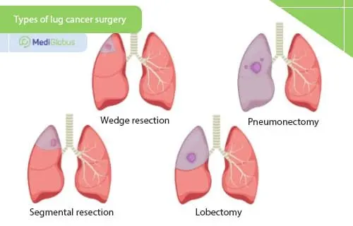
Thoracic Surgery
- If your doctor recommends surgery for your lung condition, you can trust the highly skilled and experienced cardiothoracic surgeons at Essentia Health
Interventional Radiology
- Trust Essentia’s interventional radiologists to diagnose and treat a wide array of conditions using image-guided procedures that are minimally invasive.
- These procedures have less risk, less pain and less recovery time compared to open surgery.
- This service is available in Duluth, Minnesota as well as Fargo, North Dakota.
What's an Interventional Radiologist?
- An interventional radiologist is a radiologist with specialized training in the use of image guidance methods to gain access to vessels and organs. They perform minimally invasive procedures to diagnosis and treat conditions throughout the body.
Minimally Invasive Procedures
- Essentia's interventional radiologists may perform the following procedures:
- Angioplasty and stenting – Opens narrowed or blocked arteries
- Atherectomy – Removes plaque or a clot from your blood vessels
- Biopsy – Removes tissue samples for examination under a microscope
- Catheter embolization – Blocks blood vessels to control or prevent bleeding, close off blood flow to a tumor, or treat aneurysms
- Central venous catheter (central line) placement – Inserts a long, thin, flexible tube that can be used to give medications, nutrients and other fluids
- Chemoembolization – Delivers chemotherapy directly into a cancerous tumor through a narrow catheter
- Radioembolization – Places tiny beads filled with radioactive material inside the blood vessels that feed a cancerous tumor
- Radiofrequency ablation – Destroys cancerous tissue by thermal heat
- Sympathetic nerve blocks – Relieves chronic pain
- Vertebroplasty and kyphoplasty – Treats certain types of spine fractures
Cardiothoracic Surgery
- If you have a condition that requires heart, chest, or lung surgery, trust the skilled and experienced surgeons at Essentia Health to provide exceptional care before, during, and after your surgery.
- Our surgeons use the latest techniques and technology to help you achieve the best possible health outcome.
Cardiothoracic Surgeon
- A cardiothoracic surgeon is a surgeon with specialized training in the diagnosis and surgical treatments of conditions related to the heart, lungs, and other thoracic (chest) organs.
Conditions Treated
- See the full list of conditions we treat, and select a condition to find providers and locations near you.
Types of Heart Surgery