Project 2
- Detailed description of the clinical condition selected, Points to be considered are
Normal function of the organ
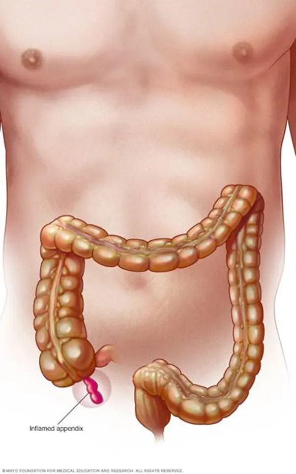
The appendix usually resides in the lower right abdomen. The appendix's purpose is uncertain. According to one explanation, during diarrheal infections, the appendix serves as a reservoir for healthy bacteria, "rebooting" the digestive tract. Some specialists hold the opinion that the appendix is just a worthless holdover from our evolutionary history.
Current pathological state in detail
One of the most prevalent inflammatory illnesses of the digestive tract is acute appendicitis. The combination of an obstruction, a decreased blood supply, ischemic damage to the mucosa, and bacterial infection is the primary cause of acute appendicitis. other abdominal sites inflamed
Reason for the pathological condition
 (source: internet)
 (source:internet)
The appendix, a finger-shaped pouch that emerges from your colon on the bottom right side of your abdomen, becomes inflamed when you have appendicitis.
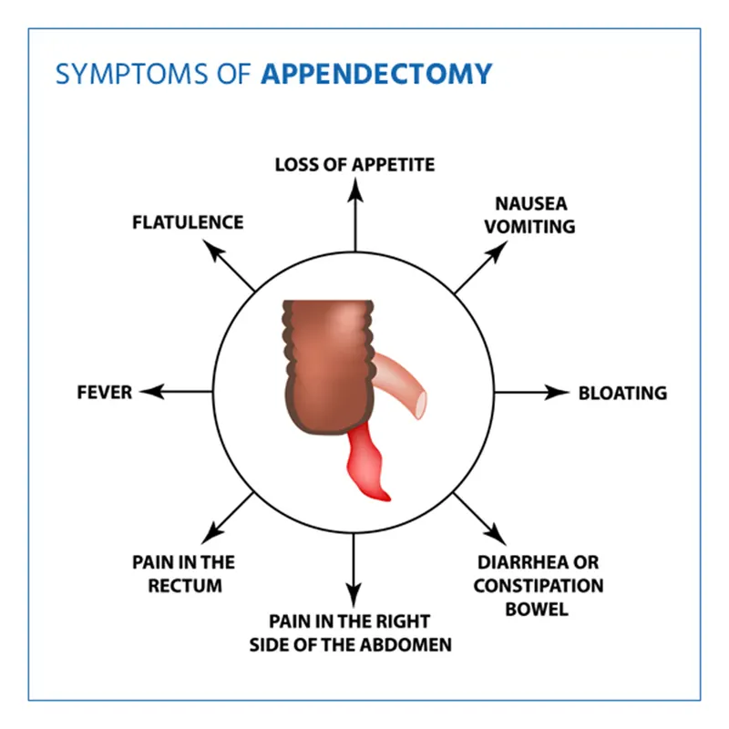
The lower right abdomen is painful if you have appendicitis. However, for the majority of patients, the pain starts near the navel and spreads. Appendicitis pain often intensifies and finally becomes unbearable as the inflammation develops.
Appendicitis can affect anyone, however it most frequently affects adults between the ages of 10 and 30.
Effect of homeostasis imbalance on human body
Copper (Cu) and zinc (Zn) are involved in inflammatory process. This study was to investigate the clinical significance of Cu and Zn homeostasis alterations in acute appendicitis (AA). One hundred twenty-two AA patients and 102 healthy controls were enrolled in this study. Of which, 85 patients' appendixes were collected after appendectomy. Another six appendixes from colon cancer patients were collected as tissue controls. The contents of Cu and Zn in serum or appendix were detected, and the Cu to Zn ratio (CZr) was calculated. The concentrations of serum ceruloplasmin (CP), Cu/Zn superoxide dismutase (SOD1), interleukin-6 (IL-6), and interleukin-22 in serum were measured, as well as the activity of CP and SOD1. The serum Zn concentration and SOD1 activity, appendix contents of Cu and Zn significantly decreased in AA patients, compared with those of controls, while serum CZr, concentrations of CP, SOD1, and IL-6, as well as CP activity increased significantly in AA patients. Additionally, serum concentrations of Zn, CP, CZr, or SOD1 activity varied in different pathological types of AA. Indicators such as serum SOD1 activity might serve as predictors for pathological classification before surgery. The serum Zn and CZr may be helpful for diagnosis of pure AA. The Cu and Zn homeostasis was altered in AA patients, which might contribute to inflammatory process of AA.
Source: https://pubmed.ncbi.nlm.nih.gov/30771140/
- Description of the diagnostic methods selected by the surgeon for assessing the condition. Points to be considered are
Diagnostic method
How is appendicitis determined to exist?
- Your healthcare professional will do a physical examination and inquire about your prior health. The following tests might also be administered by him or her:
- Blood tests: To check for signs of infection, such as having a high white blood cell count.
- Urine tests: To see if you have a urinary tract infection.
- You may also have some imaging tests, including:
Abdominal ultrasound: Lets the doctor see internal organs as they work and checks how blood is flowing through different blood vessels.
- CT scan: Shows detailed images of any part of the body, such as the bones, muscles, fat, and organs.
- MRI: Sometimes used to diagnose appendicitis, especially in a pregnant woman, instead of CT scan
Data acquisition method
Data acquisition method of appenicities could be done by the diagnosis method of it like the image data of the organ from CT or MRI
Further data could be taken from blood report, urine report or advance imaging and pattern recognition.
How the doctor identifies the problem from data, parameters he considered
Imaging exams
To help diagnose appendicitis or rule out other possible reasons of your pain, your doctor might also advise an abdominal X-ray, abdominal ultrasound, computed tomography (CT) scan, or magnetic resonance imaging (MRI).
Result to the patient
What outcome does appendix produce?
Pain from an inflamed appendix may come and go. Or it could rupture, resulting in an instantaneous, excruciating pain. Bacteria can enter the abdominal cavity through a burst appendix. Peritonitis is a dangerous, occasionally fatal infection brought on by these bacteria.
- What are all the available clinical solutions to the pathology condition. Consider worst case also
- Minor severity of the condition - Medication or lifestyle change
- Major severity condition – Surgery
Acute appendicitis is characterised as an inflammation of the appendix vermiformis's internal lining that later extends to the rest of the organ. There are many potential causes of this clinic-pathologic illness, but luminal blockage is thought to be the most important one because it starts the inflammatory response. Venous outflow obstruction results from lumen obstruction, which happens when intraluminal pressure is higher than that in the appendiceal veins.
Finally, the appendiceal wall experiences ischemia, which erodes the integrity of the epithelium and raises the danger of bacterial invasion of the organ. The most frequent causes of luminal obstruction are lymphoid hyperplasia and fecaliths, but other, less common causes have also been linked to the condition, including enterobiasis, endometriosis, tuberculosis, amebiasis, actinomycosis, adenovirus, granulomatous diseases, eosinophilic granuloma, neurogenic appendicopathy, foreign body melano Using appendectomy specimens from individuals who had undergone surgery to address an initial diagnosis of acute appendicitis, this study was created to look into the frequency and distribution of unexpected pathologic findings.
Patients with uncommon histopathologic features are evaluated.
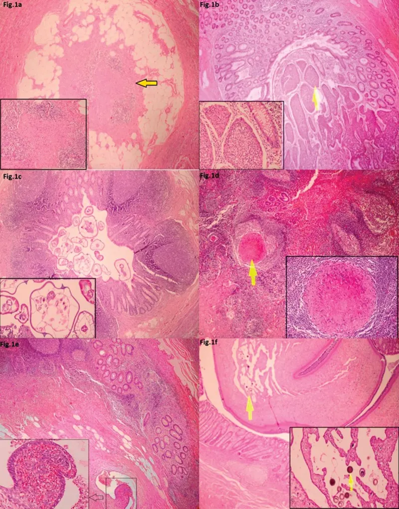
Of the patients who underwent appendectomy, 88 (or 7.0%) had unusual histopathologic findings. Table 2 summarises the clinicopathologic features of these patients. These patients were divided into 52 female and 36 male patients. This group's average age was 34.5 15.9 years (range, 17–72 years). Fibrous obliteration, commonly known as neurogenous hyperplasia or an appendiceal neuroma, was present in 57 of the specimens with atypical pathology (Fig. 1a). In addition, 8 of the specimens had an infection with the parasite Enterobius vermicularis, 8 had a carcinoid tumour, 6 had granulomatous inflammation, 2 had appendiceal endometriosis, and 1 specimen each had an infection with the parasite Taenia saginata, eosinophilic infiltration, and appendicular diverticulitis.
 (source:internet)
Most patients can receive a differential diagnosis of acute appendicitis using abdominal ultrasonography (US), computed tomography (CT), or diagnostic laparoscopy. US is a cost-effective addition to the clinical assessment and useful in the diagnosis of appendicitis cases that are questionable. For adult patients whose diagnosis is difficult to ascertain from their medical history, physical examination, and other radiologic tests, CT has emerged as the preferred imaging modality. An operator-free, rapid, and accurate test, CT produces simple-to-understand pictures and is relatively simple to execute. The differential diagnosis of other appendicular diseases using CT is also very effective.
Surgical solution in detail
Appendectomy If you have appendicitis, you'll probably need to have your appendix removed. That implies that your appendix will be surgically removed. An emergency treatment called an appendectomy is performed to stop appendicitis' potentially fatal side effects. Laparoscopic and open appendectomy procedures are both available. The ideal kind for you will be decided by your surgeon.
What is an appendectomy?
A surgical operation to remove the appendix is called an appendectomy. Appendicitis, a painful inflammation of the appendix, is typically treated surgically with an appendectomy. Appendicitis is regarded as a medical emergency because an inflamed appendix may rupture. An appendix rupture can be quite deadly. On the other hand, a healthy appendix doesn't seem to serve any vital purposes for the digestive system. You won't miss your appendix if it needs to be removed.
Describe the appendix.
The appendix is a tiny, tubular pouch that emerges like a tiny tail from the base of your colon. Your pointer finger is about how long it is. Your abdomen's bottom right side is where it is. (If you have appendicitis, the painful area will be acutely felt.) The specific function of the appendix is unknown to scientists. There are hypotheses, for instance, that it could be a location where your body stores specific good gut flora. Nobody has ever seen any significant consequences of removing it, though.
What makes appendicitis an urgent situation?
The appendix is so small that it expands fast from inflammation and can rupture with little effort. An appendix rupture can cause peritonitis, an infection of the abdominal lining, by dispersing infectious organisms throughout the abdominal cavity. Sepsis may result if the infection enters your bloodstream. Sepsis and peritonitis both carry a high risk of serious disease and possibly death. Time is of the essence when you seek treatment because the appendix can rupture within 36 hours of the onset of your first appendicitis symptoms.
What makes the appendix obsolete?
The safest course of action for appendicitis is typically an appendectomy. There are several exceptions, such as those who may not be suitable candidates for surgery. If the appendicitis is simple and detected early enough, some patients may recover completely on antibiotics. However, these persons are susceptible to recurrent appendicitis. In the end, the chance of rupture outweighs the surgery's relatively low risk by a wide margin. It's best to have your appendix removed if it is so inflamed that it could explode.
How frequently are appendixes removed surgically?
For 120 years, appendicitis has been treated mostly with an appendectomy. Every year, 300,000 appendectomies are carried out in the United States.
Laparoscopic appendectomy
 (source:internet)
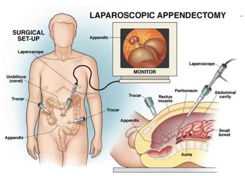
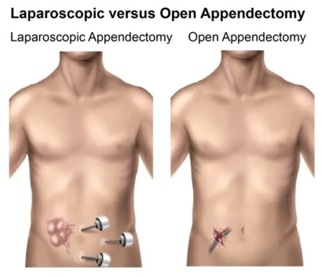
Your doctor will make a small incision close to the belly button to start the laparoscopic appendectomy procedure. A tiny port will be inserted into the incision, and via the port, a tiny tube known as a cannula will be inserted. Your abdominal cavity is inflated with carbon dioxide gas via a cannula. This provides more space for the procedure and enhances the visibility of the abdominal cavity and its contents in photographs.
The cannula will then be removed, and a laparoscope—a lengthy, thin tube with a small light and high-resolution camera attached—will be inserted.
The surgeon can find the appendix and direct the instruments through one to three tiny incisions while watching the procedure on a video screen thanks to the camera. On occasion, a laparoscopic procedure may need to switch to open surgery to address issues that the laparoscope revealed that weren't anticipated.
Open appendectomy
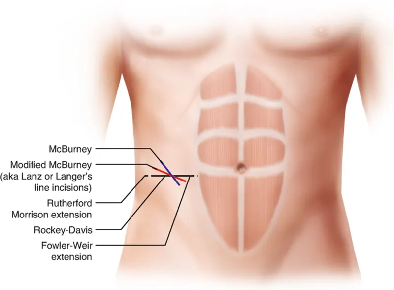 (source:internet)
Your surgeon will make one larger incision in your lower right abdomen to perform an open appendectomy. To find the appendix underneath, they will open up your abdomen and separate your abdominal muscles. Before physicians can conduct the appendectomy if your appendix has ruptured, they may need to drain an abscess or fluid from the abdominal cavity. They will then use a saline solution to flush out the abdominal cavity.
Your appendix is tied off with stitches, separated from the intestine, and removed in both surgeries. Your incisions will be used to drain extra fluid and gas. Your surgeon might leave a drainage tube in your belly to continue draining fluids if you had peritonitis, and then later remove it. Your breathing tube will be taken out, and your wounds will be cleansed, treated, and stitched up. The recovery room will be your temporary home till you awaken.
A picture showing both procedures:
 (source:internet)
What potential side effects could an appendectomy have?
Although uncommon, complications are possible. They consist of:
Bleeding.
infection of a wound.
bowel blockage.
organs nearby are hurt.
What should one do and not do after having an appendectomy?
Follow these recommendations as you heal at home:
Do:
To avoid infection, keep your incision(s) clean and dry.
Until your bowels can handle more solid things, eat soft foods. Move slowly.
Reduce your level of exercise. Your abdominal muscles may pain after prolonged standing if you underwent open surgery.
If you experience any strange symptoms, contact your doctor.
Don't:
Take medication without a prescription from your doctor. You may bleed more readily if you take some painkillers.
Do not bathe unless instructed. Wait till your stitches are out before going swimming.
your abdominal muscles to a strain. Avoid carrying heavy goods and ascending stairs.
Stay perfectly still.
To avoid blood clots and to help your digestive system function, it's vital to periodically get up and move about.
Sources:
https://my.clevelandclinic.org/health/treatments/21922-appendectomy
https://www.hopkinsmedicine.org/health/treatment-tests-and-therapies/appendectomy
cardiovascular system
Detailed description of the clinical condition selected, Points to be considered are
- Normal function of the organ
- Current pathological state in detail
- Reason for the pathological condition
- Effect of homeostasis imbalance on human body
Heart Attack:
A heart attack occurs when the blood flow to the heart is severely restricted or blocked. The obstruction is often brought on by the build up of fat, cholesterol, and other substances in the heart's (coronary) arteries. The fatty, cholesterol-containing formations are known as plaques. The process of plaque accumulation is known as atherosclerosis. On rare occasions, a plaque may rupture, causing a clot to form and obstruct blood flow. A reduction in blood flow can damage or even destroy a portion of the heart muscle. A heart attack is also known as a myocardial infarction.
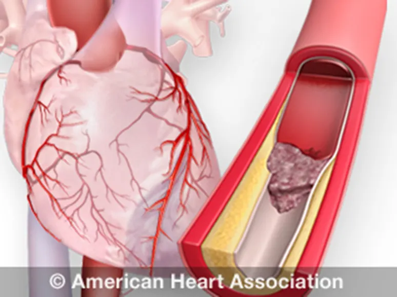
(picture taken from internet)
When the blood flow that supplies oxygen to the heart muscle is significantly lowered or entirely stopped, a heart attack happens.
These words are frequently used interchangeably, however they refer to distinct events.
When the heart's blood supply is cut off, a heart attack occurs. It has to do with circulation.
In cases of sudden cardiac arrest (SCA), the heart experiences a malfunction and unpredictably stops beating. An electrical issue causes sudden cardiac arrest.
A cardiac arrest may result from a heart attack. Death occurs in cardiac arrest, also known as sudden cardiac death (SCD), when the heart abruptly ceases functioning properly. Arrhythmias, or abnormal cardiac rhythms, are the root cause of this.
Ventricular fibrillation is the most typical arrhythmia in cardiac arrest. The bottom chambers of the heart suddenly begin to pulse erratically and stop pumping blood at this time. Within minutes of the heart stopping, death happens.
If CPR (cardiopulmonary resuscitation) is carried out and a defibrillator is utilised within minutes to shock the heart and re establish a normal heart rhythm, cardiac arrest may be reversed.
Causes of a Heart Attack
Plaque accumulation in the arteries is the root cause of the blockage (atherosclerosis). Deposits, cholesterol, and other materials make up plaque. Immediately after a plaque ruptures, a blood clot forms. The heart attack is actually brought on by a blood clot.
Heart muscle cells start to incur damage and begin to die if the blood and oxygen supply is cut off. Within 30 minutes of obstruction, irreversible harm starts. The upshot is that oxygen-deprived cardiac muscle ceases to function properly.0
The heart's four primary functions include: Pumping oxygen-rich blood to other bodily components.
delivering hormones and other essential nutrients to various bodily areas.
receiving and transporting metabolic waste products from the body to the lungs where it is oxygenated.
keeping blood pressure constant.
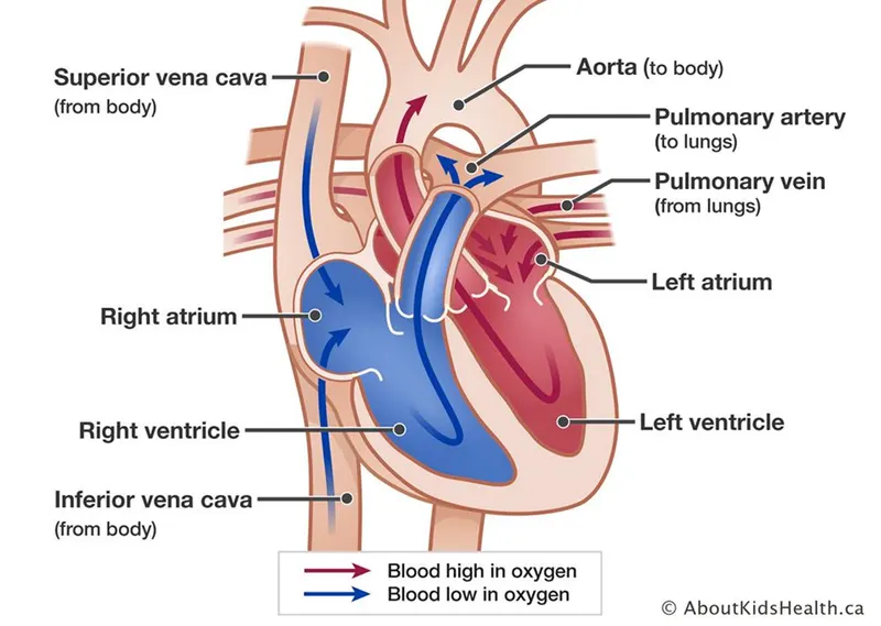 (picture showing normal parts/functions of heart, source: internet)
What is the heart's homeostasis?
The regulation of blood flow to satisfy the needs of the various organ and tissue systems is referred to as hemodynamic homeostasis.
Throughout our lifetime, the heart's output is kept in a homeostatic balance to ensure that it always meets the needs of the body's tissues. The heart may not be able to sustain an adequate flow when it suffers damage, like after a heart attack. Blood pressure drops as a result, setting off homeostatic feedback loops to try to restore normal blood pressure (and cardiac output).
A heart attack is one method in which the heart may be harmed. This happens when coronary blood arteries, which provide oxygen and nutrients to the heart's cells, dramatically narrow or are entirely stopped by a buildup of plaque and thrombus (clot). The lack of oxygen during a heart attack causes the cells to perish. The heart weakens so that it can no longer pump as much blood, which causes the various side effects linked to this condition if enough heart muscle cells perish.
An additional prevalent ailment that affects cardiac output is congestive heart failure. It could appear as a result of a heart attack or as
a result of numerous other heart ailments. The issue with congestive heart failure is that the heart muscle is so weak that it struggles to effectively pump out the blood that is entering it (the blood entering the heart is referred to as venous return). In actuality, one of the primary feedback loops that controls blood pressure alters the body's blood supply.
This is due to the fact that, given a healthy heart, the more blood we have, the higher our venous return and cardiac output will be. More venous return may actually result in reduced cardiac output due to the weakened heart caused by congestive heart failure. The body responds to a drop in blood pressure by increasing blood volume, which raises venous return and lowers cardiac output even further. It is a positive feedback loop, and I don't want to be in one where the issue with cardiac output and blood pressure keeps getting worse.
Description of the diagnostic methods selected by the surgeon for assessing the condition. Points to be considered are
- Diagnostic method
- Data acquisition method
- How the doctor identifies the problem from data, parameters he considered
- Result to the patient
- Angiography:
With a focus on the arteries, veins, and heart chambers, angiograms are a type of medical imaging treatment used to observe the interior, or lumen, of the body's blood channels and organs. Angiography is an X-ray method used to evaluate blood vessels. A particular dye must first be injected into your blood because a regular X-ray does not properly show blood vessels. Your blood vessels will be easier to see, which can help your doctor identify any problems. The X-ray images created during an angiography are known as angiograms. Your blood vessels' health and how blood flows through them are both assessed. It can help with the investigation or diagnosis of several conditions involving blood vessels, such as heart attack, peripheral arterial disease, blood clots, etc.
The angiography procedure
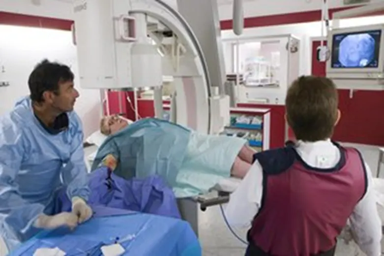
(picture taken from internet)
While having angiography you'll be asked to wear a hospital gown and lie on an X-ray table
To be tested:
Although you'll typically be awake, general anaesthesia, which puts you to sleep, may be used on very young infants
Local anaesthetic is applied to numb the area where a small cut is made in the skin over one of your arteries, generally near your groyne or wrist, so it won't hurt.
The artery is carefully guided to the area being inspected as a long, thin, flexible tube (catheter) is inserted. When this is done, you could feel some pulling and pushing, but it shouldn't hurt.
The artery is carefully guided to the area being inspected as a long, thin, flexible tube (catheter) is inserted. When this is done, you could feel some pulling and pushing, but it shouldn't hurt.
Through the catheter, a specific dye (contrast agent) is administered. After this is completed, a series of X-rays are taken as the dye travels through your blood vessels, which may cause you to feel warm, flushed, and as though you need to urinate for a short while.
Treatment can occasionally be administered simultaneously, such as when a balloon or tiny tube is inserted to widen a constricted artery. Angioplasty is the term used for this.
The catheter is taken out after the procedure, and pressure is applied to the wound to halt any bleeding. There's no need for stitches.
- Magnetic Resonance Imaging:
Can cardiac issues be found via an MRI?
Because MRI gives medical professionals a more detailed view of the heart, they can provide patients with more precise diagnosis and customised treatments. Dr. Brian Shapiro: Since you can clearly see the location and severity of the heart attack.
What exactly is a heart MRI?
A huge magnet, radio signals, and a computer are used in the test known as magnetic resonance imaging (MRI) to create images of the body's organs and tissue. The heart is visualised in this instance.
The MRI machine is a sizable, cylindrical device. A powerful magnetic field is produced around the body. Some MRI units are more transparent.
Within your body, the hydrogen protons are aligned by the magnetic field. The protons are then moved out of place by the radio waves. They transmit radio signals as they reposition themselves in their right location. The signals are gathered and transformed into photographs of the body by a computer. This picture can be seen on a monitor.
When examining soft tissues or organs, MRI may be used instead of a CT scan.
Why may I require a heart MRI?
An MRI of the heart may be performed to examine any indications or symptoms that could mean:
Atherosclerosis. This is the result of fatty compounds and other substances in the blood stream gradually blocking the arteries. It takes many years to develop.
Cardiomyopathy. This occurs as a result of the heart muscle becoming weak and thick.
congenital cardiomyopathy These are cardiac abnormalities that develop as the foetus grows. An illustration is a hole in the heart's two bottom chambers' wall (ventricular septal defect).
a heart attack. Because to this illness, the heart's muscle is frail and unable to adequately supply the body with blood.
Aneurysm. This results in the aorta or a portion of the heart muscle expanding and weakening.
heart valve illness Heart blood flow can be obstructed when heart valves are damaged.
heart tumour. Heart tumours can develop either inside or outside of the organ.
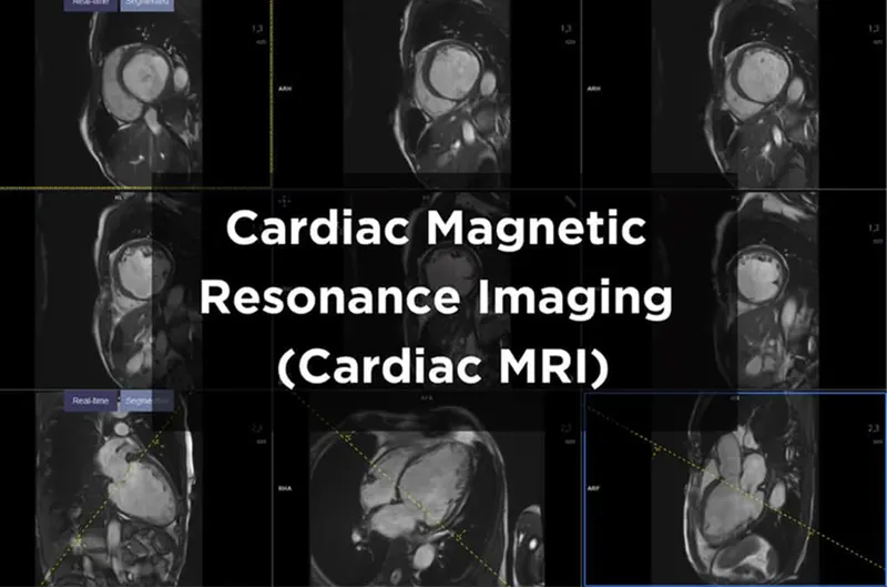
(picture taken from internet)
- Echocardiogram:
An echocardiography, often known as an ultrasound examination of the heart, is performed to look at the composition and functionality of the organ. An echo can be used to diagnose a number of conditions, including cardiomyopathy and valve failure. An echocardiogram (echo) is a visual representation of your heartbeat. You will undergo an echo test so that the physician can use ultrasonography (high-frequency sound waves) from a hand-held wand implanted on your chest to capture images of the chambers and valves of your heart. This helps the doctor determine how efficiently your heart is pumping. Medical experts routinely combine echo, Doppler ultrasonography, and colour Doppler treatments to evaluate blood flow across your heart's valves. Echocardiography uses no radiation. This sets an echo apart from other tests, such X-rays and CT scans, which use almost any radiation.
How come I could require an echocardiogram?
An echocardiography may be performed for a more thorough assessment of indications or symptoms that could indicate:
Atherosclerosis. a condition when fatty molecules and other substances in the bloodstream gradually block the arteries. It may result in issues with your heart's pumping or wall movements.
Cardiomyopathy. a cardiac enlargement brought on by thick or frail heart muscle
congenital cardiomyopathy defects that appear in one or more cardiac components while the foetus is still developing, like a ventricular septal defect (hole in the wall between the 2 lower chambers of the heart.
a heart attack. a condition when blood cannot be pumped effectively because the heart muscle has grown weak or tight due to cardiac relaxation. Edema (swelling) in the feet, ankles, and other areas of the body may result from this, as well as fluid accumulation (congestion) in the blood vessels and lungs.
Aneurysm. an enlargement and weakening of the aorta or a portion of the heart muscle (the large artery that carries oxygenated blood out of the heart to the rest of the body). There's a chance the aneurysm will burst.
heart valve illness failure of one or more heart valves, which could result in irregular blood flow within the heart. Blood cannot flow through the heart or out to the lungs and body when the valves are constricted. Blood might seep backwards through the defective valves as well. The tissue of the heart valves can be examined for infection using an echocardiography.
heart tumour. a cardiac tumour that can develop on the heart's exterior, inside one or more of its chambers, or within the muscular tissue (myocardium) of the heart.
Pericarditis. the sac that surrounds the heart being inflamed or infected.
Pericardial tamponade or effusion. Blood, fluid, or infection can fill the sac that surrounds the heart. The heart muscle may get compressed as a result, failing to beat and pump blood normally. This may result in symptoms such as feeling faint or dizzy or a potentially dangerous drop in blood pressure.
flaws in the septum or atrial wall. Uneven pathways between the right and left sides of the heart might exist from birth, result from trauma, or develop during a heart attack. These flaws might be found in the lower pumping chambers or the top filling chambers (atria) (ventricles). This could result in poor blood flow, cardiac failure, or an elevated risk of stroke.
Shunts. Shunts are a common finding in atrial and ventricular septal abnormalities, as well as when the liver and lungs drive erratic blood flow through the circulatory system.
In order to evaluate the general structure and function of the heart, an echocardiography may also be performed.
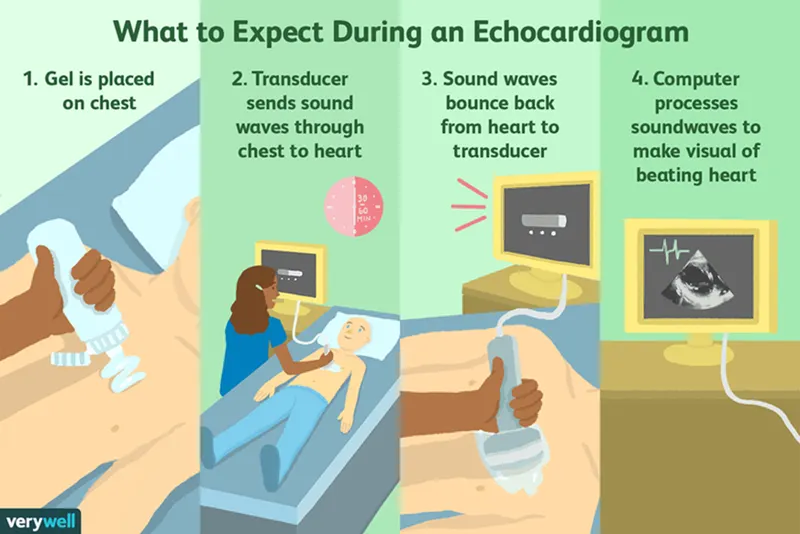
(picture taken from internet)
. What are all the available clinical solutions to the pathology condition. Consider worst case also
- Minor severity of the condition - Medication or lifestyle change
- Major severity condition - Surgery
Following a cardiac attack, more heart tissue is harmed or dies every minute. Blood flow needs to be fixed immediately in order to increase oxygen levels. Oxygen is administered right away. Whether there is a partial or total blockage of blood flow will determine the specific heart attack treatment.
MEDICATIONS
Medications to treat a heart attack might include:
- Aspirin reduces blood clotting. It helps keep blood moving through a narrowed artery. If you called 911 or your local emergency number, you may be told to chew aspirin. Emergency medical providers may give you aspirin immediately.
- Clot busters (thrombolytics or fibrinolytics).These drugs help break up any blood clots that are blocking blood flow to the heart. The earlier a thrombolytic drug is given after a heart attack, the less the heart is damaged and the greater the chance of survival.
- Other blood-thinning medications.A medicine called heparin may be given by IV or injection. Heparin makes the blood less sticky and less likely to form clots.
- This medication widens the blood vessels. It helps improve blood flow to the heart. Nitroglycerin is used to treat sudden chest pain (angina). It's given as a pill under the tongue, as a pill to swallow or as an injection.
- This medicine is given to relieve chest pain that doesn't go away with nitroglycerin.
- Beta blockers.These medications slow the heartbeat and decrease blood pressure. Beta blockers can limit the amount of heart muscle damage and prevent future heart attacks. They are given to most people who are having a heart attack.
- ACEThese drugs lower blood pressure and reduce stress on the heart.
- These drugs help lower unhealthy cholesterol levels. Too much bad (low-density lipoprotein, or LDL) cholesterol can clog arteries.
Surgical and other procedures
If you've had a heart attack, a surgery or procedure may be done to open a blocked artery. Surgeries and procedures to treat a heart attack include:
- Coronary angioplasty and stenting.This procedure is done to open clogged heart arteries. It may also be called percutaneous coronary intervention (PCI). If you've had a heart attack, this procedure is often done during a procedure to find blockages (cardiac catheterization).
During angioplasty, a heart doctor (cardiologist) guides a thin, flexible tube (catheter) to the narrowed part of the heart artery. A tiny balloon is inflated to help widen the blocked artery and improve blood flow.
A small wire mesh tube (stent) may be placed in the artery during angioplasty. The stent helps keep the artery open. It lowers the risk of the artery narrowing again. Some stents are coated with a medication that helps keep the arteries open.
- Coronary artery bypass surgery (CABG). This is open-heart surgery. A surgeon takes a healthy blood vessel from another part of the body to create a new path for blood in the heart. The blood then goes around the blocked or narrowed coronary artery. CABGmay be done as an emergency surgery at the time of a heart attack. Sometimes it's done a few days later, after the heart has recovered a bit.
Cardiac rehabilitation
Cardiac rehabilitation is a personalized exercise and education program that teaches ways to improve heart health after heart surgery. It focuses on exercise, a heart-healthy diet, stress management and a gradual return to usual activities. Most hospitals offer cardiac rehabilitation starting in the hospital. The program typically continues for a few weeks or months after you return home.
People who attend cardiac rehab after a heart attack generally live longer and are less likely to have another heart attack or complications from the heart attack. If cardiac rehab is not recommended during your hospital stay, ask your provider about it.
Surgical solution in detail
Heart Procedures and Surgeries
If you've had a heart attack, you may have already had certain procedures to help you survive your heart attack and diagnose your condition. For example, many heart attack patients have undergone thrombolysis, a procedure that involves injecting a clot-dissolving agent to restore blood flow in a coronary artery. This procedure is administered within a few (usually three) hours of a heart attack. If this treatment isn't done immediately after a heart attack, many patients will need to undergo coronary angioplasty or coronary artery bypass graft surgery (CABG) later to improve blood supply to the heart muscle.
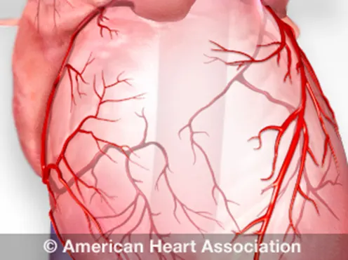 (source: internet)
Heart Procedures and Surgeries
Angioplasty
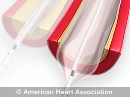 (sourrce: internet)
Also known as Percutaneous Coronary Interventions [PCI], Balloon Angioplasty and Coronary Artery Balloon Dilation.
What the Procedure Does
Special tubing with an attached deflated balloon is threaded up to the coronary arteries. The balloon is inflated to widen blocked areas where blood flow to the heart muscle has been reduced or cutoff. Often combined with implantation of a stent (see below) to help prop the artery open and decrease the chance of another blockage. Considered less invasive because the body is not cut open. Lasts from 30 minutes to several hours. May require an overnight hospital stay.
Reason for the Procedure
- Greatly increases blood flow through the blocked artery.
- Decreases chest pain (angina).
- Increases ability for physical activity that has been limited by angina or ischemia.
- Can also be used to open neck and brain arteries to help prevent stroke.
Medications That Your Doctor May Prescribe Post-Procedure
Learn more about cardiac medications, including dual antiplatelet therapy, that you may need to take after your procedure to prevent complications and to put you on the path for the best recovery.
Angioplasty, Laser
What the Procedure Does
Similar to angioplasty except that the catheter has a laser tip that opens the blocked artery. Pulsating beams of light vaporize the plaque buildup.
Reason for the Procedure
- Increases blood flow through blocked arteries.
Artificial Heart Valve Surgery
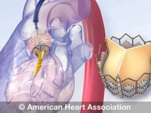 (source: internet)
(Also known as Heart Valve Replacement Surgery)
What the Procedure Does
Replaces an abnormal or diseased heart valve with a healthy one.
Reason for the Procedure
- Restores function of the heart valves.
Atherectomy
What the Procedure Does
Similar to angioplasty except that the catheter has a rotating shaver on its tip to cut away plaque from the artery.
Reason for the Procedure
- Increases blood flow through the blocked artery by removing plaque buildup.
- May also be used in carotid arteries (major arteries of the neck leading to the brain) to remove plaque and reduce risk for stroke.
Bypass Surgery
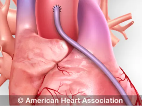 (source: internet)
(Also known as CABG, pronounced "cabbage," Coronary Artery Bypass Graft done via Open-Heart Surgery)
What the Procedure Does
Treats blocked heart arteries by taking arteries or veins from other parts of your body — called grafts — and using them to reroute the blood around the clogged artery to supply blood flow to your heart muscle. A patient may undergo one, two, three or more bypass grafts, depending on how many coronary arteries are narrowed. Requires several days in the hospital.
Reason for the Procedure
- One of the most common and effective procedures to manage blockage of blood to the heart muscle.
- Improves the supply of blood and oxygen to the heart.
- Relieves (angina).
- Reduces risk of heart attack.
- Improves ability for physical activity that has been limited by angina or ischemia.
Medications That Your Doctor May Prescribe Post-Procedure
Cardiomyoplasty
What the Procedure Does
An experimental procedure in which skeletal muscles are taken from a patient's back or abdomen. Then they're wrapped around an ailing heart. This added muscle, aided by ongoing stimulation from a device similar to a pacemaker, may boost the heart's pumping motion.
Reason for the Procedure
- Increases the pumping motion of the heart.
Heart Transplant
What the Procedure Does
Removes a diseased heart and replaces it with a healthy human heart when a heart is irreversibly damaged. Uses hearts from organ donation.
Reason for the Procedure
- Recognized as a proven procedure to restore heart health in appropriately selected patients.
Minimally Invasive Heart Surgery
(Also known as Limited Access Coronary Artery Surgery and includes Port-Access Coronary Artery Bypass (PACAB or PortCAB) and Minimally Invasive Coronary Artery Bypass Graft (MIDCAB or minimally invasive CABG)
What the Procedure Does
An alternative to standard bypass surgery (CABG). Small incisions ("ports") are made in the chest. Chest arteries or veins from your leg are attached to the heart to "bypass" the clogged coronary artery or arteries. The instruments are passed through the ports to perform the bypasses. The surgeon views these operations on video monitors rather than directly. In PACAB, the heart is stopped and blood is pumped through an oxygenator or "heart-lung" machine. MIDCAB is used to avoid the heart-lung machine. It's done while the heart is still beating. Requires several days in the hospital.
Reason for the Procedure
- Manages blockage of blood flow to the heart and improves the supply of blood and oxygen to the heart.
- Relieves
- Reduces risk of heart attack.
- Improves ability for physical activity.
Radiofrequency Ablation
(Also known as Catheter Ablation)
What the Procedure Does
A catheter with an electrode at its tip is guided through the veins to the heart muscle with real-time, moving X-rays (fluoroscopy) displayed on a video screen. The catheter is placed at the exact site inside the heart where cells give off the electrical signals that stimulate the abnormal heart rhythm. Then a mild, painless radiofrequency energy (similar to microwave heat) is transmitted to the pathway. This destroys carefully selected heart muscle cells in a very small area (about 1/5 of an inch).
Reason for the Procedure
- Preferred treatment for many types of rapid heartbeats (arrhythmias) especially supraventricular tachyarrhythmias.
What precautions should be taken after heart attack?
Then, follow the tips below for a healthy lifestyle.
- Stop smoking. Tobacco use is a major risk factor for heart disease. ...
- Control your blood pressure. ...
- Control your cholesterol levels. ...
- Check for diabetes. ...
- Regular cardio exercise can make your heart stronger. ...
- Eat a heart-healthy diet. ...
- Control your stress level.
Surgical solution in detail
Despite the advantages, surgery can trigger cardiac events including heart attacks, heart failure, heart rhythm disturbances, and death. Previous research has shown that nearly three-quarters of patients who die after surgery were never admitted to critical care,3 suggesting that their risk was unrecognised.
Each year, cardiac complications occur within 30 days after major noncardiac surgery in more than 10 million people worldwide; postoperative mortality is 1.5%. Enhanced patient monitoring and measurement of natriuretic hormone and troponin levels may improve outcomes.
INTRODUCTION
Cardiac complications occurring in the postanesthetic care unit (PACU) are typically due to hypotension, hypertension, and dysrhythmias. Patients with known coronary artery disease or congestive heart failure are more prone to these complications after surgical procedure.
HYPOTENSION
Decreased intravascular volume, or hypovolemia is due to inadequate intravenous fluid administration or blood loss. Patients can be resuscitated with crystalloids, colloids, and various blood products. If fluid resuscitation is inadequate to perfuse end organs, then vasopressors and inotropes should be added.
Myocardial ischemia with acute heart failure and ventricular or valvular dysfunction can also lead to hypotension. This may be associated with tachycardia and ST segment changes on electrocardiogram. A history of coronary artery disease predisposes patients to these complications and should be noted on preoperative evaluation. Drug-eluting stents typically require antiplatelet therapy for surgical procedures; if antiplatelet therapy is halted, patients may be at increased risk for acute coronary events. Suspected coronary thrombosis requires immediate evaluation for cardiac catheterization.
Decreased systemic vascular resistance in the PACU setting is usually iatrogenic and leads to hypotension. Disease states that cause decreased SVR include sepsis, spinal shock from spinal cord injury, and histamine release during anaphylactic reactions. While supportive measures are instituted, the underlying cause should be identified and treated. Residual effects of anesthetics, including inhalational, intravenous, and neuraxial agents, also produce hypotension. Treatment is indicated if mean arterial pressure is 20% less than baseline.
HYPERTENSION
Pain is a common cause of hypertension in the PACU. Surgical trauma and pain cause increased sympathetic tone leading to hypertension and tachycardia. Multimodal pain management strategies are preferable.
Hypercarbia from respiratory failure also leads to hypertension. Treatment includes promoting effective gas exchange via invasive or noninvasive, positive pressure ventilation.
Urinary retention and bladder distention are a common cause of hypertension in the PACU. It is more common after inguinal hernia repair, neuraxial anesthesia, and in elderly men with prostatic obstruction. Patients may require bladder catheterization. Patients who remain intubated in the PACU, if not adequately sedated, may become hypertensive from irritation of the endotracheal tube.
ARRHYTHMIAS
Arrhythmias occur often in the PACU and some can be life threatening. If cardiac arrest should occur, PACU treatment may have to be tailored to accommodate surgical incisions. Thorough review of current Advance Cardiac Life Support (ACLS) algorithms should be reviewed.
Bradycardia in the PACU can be due to vasovagal reflexes, residual effects of anticholinesterases, β-blockers, or opioids. Bradycardia may also result from severe myocardial infarction with complete heart block. The ACLS algorithm should be consulted for unstable bradycardia. Anticholinergic medications and pacing options must be readily available.
Sinus tachycardia can be due to pain, hypovolemia, fever, sepsis, or certain drugs such as albuterol or anticholinergics.
Atrial.
BYPASS SURGERY OVERVIEW
Coronary artery bypass graft surgery, also known as CABG or bypass surgery, can help to restore blood flow to an area of the heart. However, surgery does not stop the progression of atherosclerosis (coronary heart disease), which deposits fatty material into artery walls, narrowing them and eventually limiting blood flow at other sites in the bypassed arteries or in previously normal coronary arteries.
Patients and healthcare providers must work together after surgery to treat the underlying atherosclerosis and the factors that can cause progression of heart disease.
This topic review discusses treatments that are recommended after coronary artery bypass graft surgery. These treatments can help to:
- Reduce the risk of developing complications of coronary heart disease, including having a subsequent heart attack or dying.
- Help a person to feel better and have more energy.
An overview of coronary artery bypass graft surgery is discussed in detail separately.
CARE AT HOME AFTER BYPASS SURGERY
Care after bypass surgery aims to reduce the risk factors for heart disease and includes strategies to help patients and family members stop smoking, control high blood pressure, improve cholesterol levels, begin exercising regularly, reduce weight if necessary, and reduce stress. Some of these changes can be made by adjusting lifestyle habits through diet and exercise. However, lifestyle changes alone may not be adequate and medications are often needed.
Discharge from the hospital — Patients with an uncomplicated CABG usually go home after about five days in the hospital. In some cases, the hospital stay is longer. If complications have occurred, discharge is delayed until the person's condition is stable.
Before leaving the hospital, it is important for the patient and family to participate in and understand the discharge plan. Make sure all questions are answered and obtain written directions for how to take all medications (new and old). After bypass surgery, it is common to start new medications and stop or adjust the doses of previous medications.
Medications — Most people who have had bypass surgery are sent home with prescriptions for several medications, most of which are taken every day. Some of these drugs improve survival, decrease the risk of complications, and help to prevent or treat recurrent chest pain.
- Antiplatelet therapy– Aspirin is an antiplatelet medication that is given to help prevent the formation of blood clots that can block either your coronary arteries or coronary bypass graft. It is usually recommended indefinitely.
- Beta blockers– Beta blockers slow the heart rate, lower blood pressure, and decrease the heart's demand for oxygen. They are given to some patients with high blood pressure, heart failure, some rhythm changes or a heart attack, and to some patients in whom bypass surgery is not expected to relieve all symptoms of angina. If a person cannot tolerate a beta blocker, a calcium channel blocker may be substituted.
- Nitrates– A nitrate, either as short-acting nitro glycerin, or as a long-acting preparation (isosorbide mononitrate or dinitrate). These drugs dilate coronary blood vessels, bringing more blood to the heart muscle. Nitrates also reduce the amount of blood returning to the heart, which decreases the heart's demand for oxygen. Nitrates are often given to treat or prevent further episodes of chest pain. Nitrates may be given to patients after bypass surgery if some of the coronary blood vessels could not be bypassed.
- ACE inhibitor– Angiotensin converting enzyme (ACE) inhibitors are often used to treat high blood pressure.
Examples of ACE inhibitors include captopril (brand name: Capoten), enalapril (brand name: Vasotec), lisinopril (sample brand names: Zestril or Prinivil), and ramipril (brand name: Altace).
Some patients who cannot tolerate an ACE inhibitor (often because of a chronic cough) may be prescribed an angiotensin II receptor blocker (ARB). These related drugs are satisfactory replacements.
Examples of ARBs include losartan (brand name: Cozaar), valsartan (brand name: Diovan), and irbesartan (brand name: Avapro).
- Lipid-lowering therapy– Almost all patients are given a medication to lower lipids after CABG. Cholesterol lowering can be beneficial both before and after CABG because it can reduce the progression of atherosclerosis in both native and graft vessels.
- Other medications– Other medications may be given on a short-term basis to prevent the development of an irregular heart, to manage discomfort associated with healing incisions, or to allow for regular bowel movements.
Lipid therapies are recommended even for patients who have values that are in the "normal" range. The goal level for "bad" cholesterol (called LDL or low density lipoprotein) is less than 70 mg/dL (1.8 mmol/L).
Statins are the most common medications used to lower cholesterol levels. Other drugs may be used as well
Wound care — After discharge from the hospital, the patient is usually given instructions about how to care for their chest and/or leg wounds. It is important to follow these instructions closely and to notify a healthcare provider immediately if there are questions or concerns.
- Avoid heavy lifting and extremes of shoulder movement (eg, as in tennis, baseball, and golf) for six to eight weeks after surgery to allow for complete healing of the breast bone (sternum)
When to seek help — If the patient develops any of the following signs or symptoms of wound infection, a healthcare provider should be contacted immediately. Most wound infections develop within 14 days of the surgery.
- Fever greater than 100.4º F (38º C)
- New or worsened pain in the chest or around the incision
- A rapid heart rate
- Reddened skin, bleeding or pus-like drainage from the incision
Sources:
View an illustration of coronary arteries.