Project 1
PART A
Clinical-Gastric ulcer
These are sores that devlops on the lining of the stomach.
Stracturally, the Stomach is a large sac like organ located in the left hypochondriac and epigastric region and it is usually J -shaped with two ends that is esophageal and duodenal and it has two curvetures that is the greater and lesser curveture.It also has two surfaces and three parts that is the fundus,body and pylorus.
The fundus is dome-shaped and is filled with gas in an upright position.The body is the largest part of the stomach and it presents the lesser and greater curveture .The pylorus is the part that connects the stomach to the duodenum and the phyloric sphincter marks the end of the stomach and is the entrance to the duodenum.The wall of the stomach consist of four layers that is the Mucosa ,submucosa,muscularis externia and the serosa.When empty the stomach is contracted and its mucosa and submucosa are thrown into distinct folds called a rugae and when distended with food,the rugae are ironed out and flat.It is in the stomach where chemical digestion takes place with the helps of enzymes .
Microscopically,the stomach is covered with numerous small holes which are the openings of gastric pits that extends into the mucosa as straight and branched tubules forming gastric glands.There are four major types of secretory epithelial cells covering the surface of the stomach and extends down into the gastric pits and glands:-
Mucous cells which secrete alkaline mucous that protects the epithelium against shear stress and acid,parietal cells that secrete Hydrochloric acid,chief cell that secrete pepsin and proteolytic enzyme and the G-cell that secrete the gastrin hormone.The distribution of this cell types along the stomach wall differs for example the parietal cells are more in the glands of the body and abscent in the pyloric glands.
The arterial supply to the stomach is derived primarly from the celiac axis which arises from the proximal abdominal aorta and typically branches into common hepatic,splenic and left gastric arteries.The veins of the stomach runs parallel to the arteries where the left and right veins drains into hepatic portal veins .The short gastric vein,left and right gastro-omental vein ultimately drains into superior mesenteric vein.
The stomach receive innervation from autonomic nervous system which includes, parasympathetic nerve supplies which arises from the anterior and posterior vagal trunks,derived from the vegus nerve.The sympathetic nerve supplies arises from T6 and T9 spinal cord segments and passes to coeliac plexus via the greater splanchnic nerve and it also carries some pain transmitting fibres.
Diagram showing the stracture of the stomach
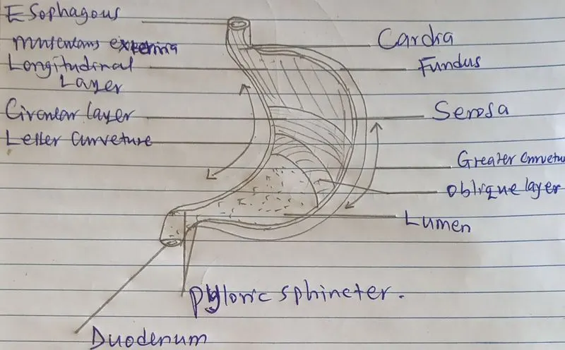
As defined earlier that gastric ulcers are sores that develps on the lining of the stomoch.Gastric ulcers occur when stomch acid damage the lining of the digestive tract and the common causes includes bacteria H.pylori,prolonged use of anti-inflamatory pain relivers such as asprin,ibuprofen and many others.The gastric ulcers are commonly located at the lesser curveture between the antrum and the fundus.
People with gastric ulcers will present with signs and symptoms of burning stomach pain,feeling of fullness,belching or bloating,intolarance to fatty foods,heart burns and Nausea.The mos popular symptom of gastric ulcer is the burning stomach pain.The stomch acid secreted by parietal cells makes the pain worse as does having an empty stomach.Less often some people may present with vomitting blood as a result of bleeding along the stomach wall ending up with a complication of gastrict perforation.
During the investigations for gastric ulcers ,the following techniques can be applied:-
- Laboratory test for helicobacter pylory which is for antigen antibody test.
- Upper gastrointestenial endoscopy and biopsy to study the nature of ulcerations and possible causes.This is a procedure done under general anaesthesia where a tiny camera on the end of a long flexible tube that uses the principles of total internal reflection is introduced into the gastrointestenial tract and the images are captured for intepretetion by the gastroenterologysts.
- Upper gastrointestinal series using x-rays following consumption of barium swallow as a radioactive tracer.
- Using computerised tomography scan following the consumption of barium tracer.
The treatment of Gastric ulcers is done by pharmachological mechanism using proton pump inhibitors and in severe case where there is a complication of gastric perforation,gastrectomy my be done inorder to prevent further complications of peritonitis.This surgical procedure is done to remove only the perforated area followed by repair of the area.
Clinical condition-Bronchitis
These refers to the inflamation of lining of the respiratoey bronchial tubes which carries air to and from the lung.The bronchioles are part of the respiratory system that plays a vitial role during respiration.They are approximately 1mm or less in diameter and their walls consist of ciliated cuboidal epithelium and a layer of smooth muscles.Bronchioles divides into smaller bronchioles called terminal bronchioles which are approximately 0.5mm or less in diameter.
The bronchioles consist of a bronchial vein which are small vessels that forms part of the venous drainage of the lungs.These vessel drains the larger pulmonary bronchi and the area of the lung closer to the hilum and it can be divided into two systems which are deep and superficial veins.The deep bronchial vein drains into the main pulmonary vein or left antrium while the superficial vein on the right side of the body drains into the azygos vein.
The blood supply to the bronchial always originate from the aorta or inercostal artaries entering the lung at the hilum,branching at the mainstream bronchus to supply the lower trachea,extrapulmonary air ways, and suppoerting stractures.This part of the bronchial vasculature drains into the right heart via systemic vein.
The walls of the bronchi and bronchioles are innervated by autonomic nervous system.The bronchial epithelium and smooth muscles consist of b2 adrenagic receptors which are responsible for bronchiodilation and secretion along the bronhioles and the a1 adrenagic receptor on the other hand inhibit secretion.
Illustration of the bronchial tree
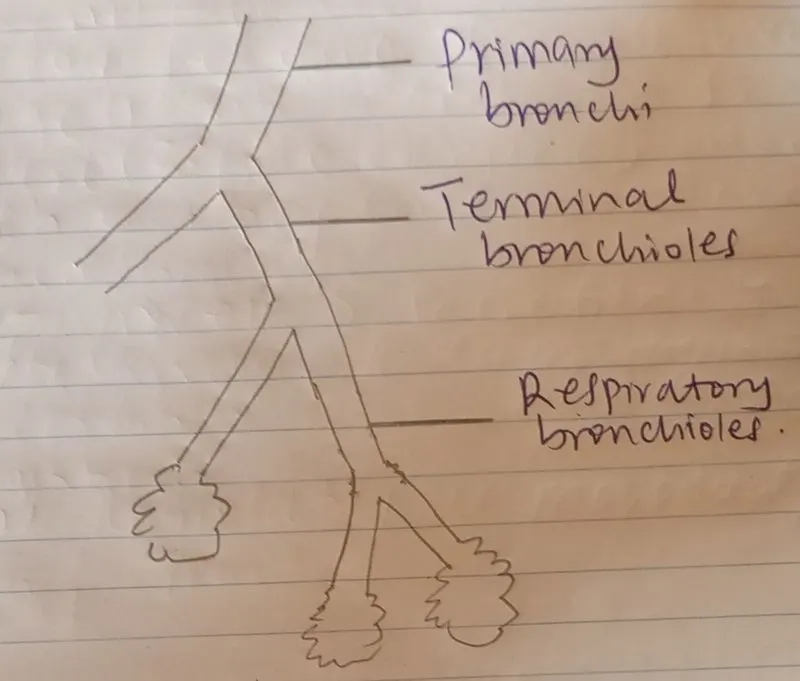
As defined earier that bronchitis is an inflamation of the respiratory bronchioles,this condition often make sufferers to cough thick mucous which can be discolored and pleurorent and this condition may either be chronic or acute.Bronchitiis may develop from cold or other respiratory infection and the acute form is very popular.The chronic type is a more serous condition which involve constant irritation or inflamation of the lining of the bronchial tube and it is often due to smoking.For either acute or chronic bronchitis,the signs and symptoms includes the follwing:-Cough,production of mucous,fatigue ,shortness of breath, and chest discomfort.
The following clinical investigations can be conducted to diagonise bronchitis:-
- Pulmonary functional test:-This is done using the spirometer where the patient is made to breath into the device that will inturn measure the respiratory volume of the patient where it will later on be compared with the normal reference range.
- Blood test by doing complete blood count that will enable the doctor to analyse the percentages of white blood cells available.
- Chest X-ray which is done to help confirm the diagnosis for chronic bronchitis and other lung conditions like cancer and pneumonia.
- Bronchoscopy:-This an endoscopic technique of visulizing the inside of the air way for diagonistic and theraputic purposes.The device is inserted through the nose or mouth or sometimes trachostomy down to the trachea and lungs.This procedure ia done under general anaesthesia and the tube is always soft and flexible and contains camera at the tip which is then used to capture the image of the area of intrest.
The treatment of bronchitis targets the symptoms which inlcudes the use of medications like antibiotics,anti-inflammatory and bronchiodilators which are the mucous clearing device that helps one to cough out the mucous easily.In very severe cases,oxygen theraphy may be necessary as the airways are compromised.
Clinical condition-Diabetes
This a chronic disease that occurs either when the pancreas does not produce enough insulin or when the body can not effectively use its insulid.It is the role of the beta cells found in the pancrease to produce insulin which is responsible for the regulation of body glucose.
To begin with the pancreas,it is an accessory organ that plays a role in digestion of food.It is an elongated tapered organ located accross the back of the belly behind the stomach.The right side of the organ called the head is the widest part and lies in the curve of the duodenum which is the first division of the small intestine.It is both endocrine and exocrine gland that secretes both hormones and enzymes.In adults,the pancreas measures about 15-25cm in length,3- 5cm in height,and 1.5-3.5cm in thickness and weigh 70-110g. It consist of cells which are the beta and alpha cells of islets of Langherans.The beta cells are responsible for synthesising,storage and release of insulin which is the ant-hyperglycermic hormone which antigonises glucagon produced by the alpha cells.
Corporocaudal segment of human pancreas is supplied with blood from splenic artery.The branches of splenic artery which extends to the body and the tail of the gland includes the dorsal pancreatic artery,inferior pancreatic artery,greater pancreatic artery,artey to the tail of the pancreas and pancreatic branches.
The venous drainage of the duodeno-pancreas is effected via two territories that is posteriosuperior and an anterioinferior where the later drains towards the portal vein and the second into the superior mesenteric vein.The borderline between the two is represented by the inferior posterior pancreato-duodenal vein.
The pancreas receives involutary innervation via autonomic nervous system .Its parasympathetic innervation originate from the vagus nerve and its sympathetic innervation originate from the greater and lesser splanchnic nerves.
Illustration of the pancreas
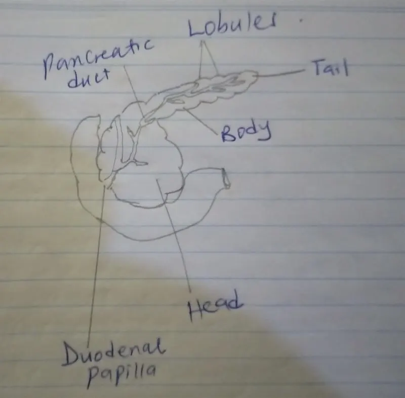
When the beta cells of islets of Langerhans fails to produce insulin,the sugar level will go abnormally resulting into type one diabetes which is sometimes called insulin dependant.Sometimes the insulin might be produced by the beta cells but the absorptive cells becomes non responsive to glucose resulting to type two diabetes which is the most commonest type.
Type two diabetes is caused by several factors including lifestyle, genes,obesity,insulin resistance,and hormonal diseases.The type one diabetes is thought to be caused by an autoimmune reaction that attacks and destroy the beta cells of the pancreas that produce,store and secrete insulin.This process can go on for months or years before the symptoms appear.The signs and symptoms of diabetes includes feeling more thirsty than usual,polyuria,polyphagia,weight loss especially in type one,blurrdness of vision,feeling tired and weak,frequent infection due to lowered immunity and many others.
The investigation carried out to confirm diabetes is by doing blood test for random blood sugar test or fasting blood sugar test and some time Oral glucose tolarance test (OGTT).The managment of the condition is pharmacological which is type specific involving the use of artificial insulin for the case of type one and hypoglycemic drugs for the case of type two diabetes.
PART B
CASE STUDY
The type of bones the skull belongs to are flat bones which are made up of layer of spongy bone between two thin layers of compact bones.They have a flat shape not rounded and they have marrow but do not have bone marrow cavity in them.They are joint together by sutures which is a type of fibrous immovable joint and they are connected by a fiber called sarphey's fibers.
The cranial meningeal artaries supplies the dura and leptomeninges and are multiple in the anterior,middle and posterior cranal fossae where they freely anastomose.These includes the ophthalamic,internal and external cerebral ,middle meningeal and vertebral arteries.This therefore nurishes the flat bones of the skull.The venous drainage takes place through nutrient and diploic vein which are the veins leaving juxtasutural foramina and capillaries leaving the inner and outer surfaces of the bone generally.There is free and extensive communication between dural meningeal vein,the vein of the skull vault and those of the pericarnium.
Bone remodeling is a life long processes where mature bone tissues is removed from the skeleton and new born tissues are formed.This process serves to adjust bone architecture to meet changimg mechanical needs and it helps to repair microdamages in bone matrix preventing the accumulation of old bones.It also plays a role in maintaining plasma calcium homeostasis and the regulation of bone remodeling is both systemic and local.
The remodeling cycle consist of three conservative phases which includes:-reabsorption during which osteoclasts digest old bone,reversal which occurs when mononuclear cells appear on the bone surface and formation occurs when osteoblasts lay down new bone until the reabsorbed bone is completely replaced. The process of remodeling also control reshaping or replacement of bone following injuaries like fractures and also microdamage that occurs during normal activities.
The anatomical basis for bone implant is very important as several factors determines how well an implant will bond with the native bone.They include physical and chemical factors like chemical composition of the implant and porosity of the native bone.Any implant that is to be used in the body must be compatible,safe due to the invasive nature to the body and must serve the purpose for which it was inserted.
The indication for bone implant into the body follows complete damage of the native bone that may not heal and unite completely and these damage may be due to an accident or pathological in the case of osteomylitis and cancer.Some peope may also go for bone implant just for cosmetic purposes.The contraindications to implant include hypersensensitivity reactions to the chemical composition of the implant and those who frequently goes for MRI examination.