Project 1
Project 1
Part A
Skeletal system
Components- Bones and joints
Function-Skeletal system gives the structure and support to the body. It forms the framework of our body.
Common disorders
- Fractures
- Bone cancer
- Osteoporosis
- Arthritis
Clinical Condition-Fracture of femur
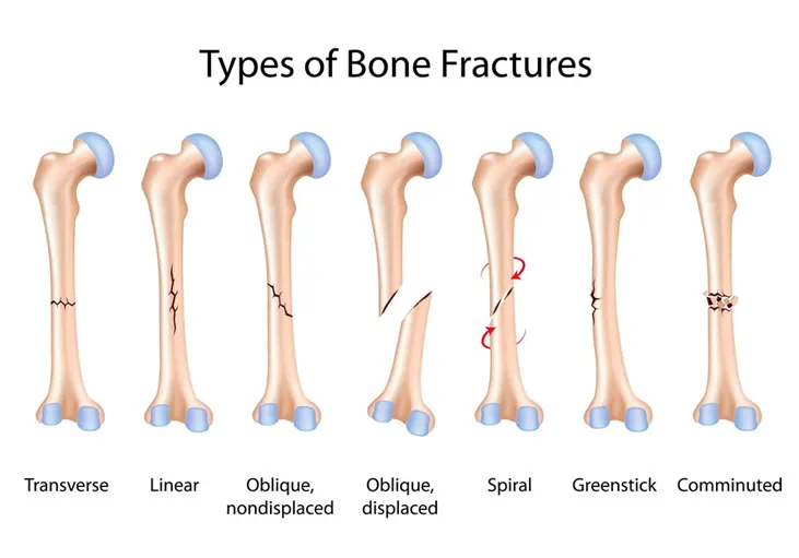
Fracture is a condition where there is a complete or partial break in the continuity of a bone.
Organ involved-Bone (Femur)
Gross anatomy-
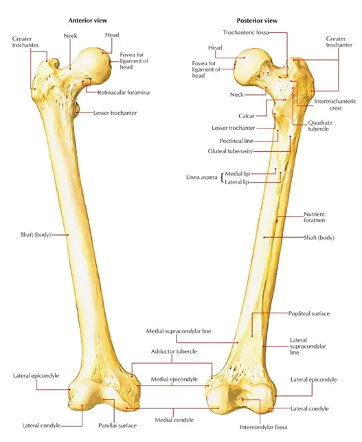
Femur is the longest and the only bone in the thigh. There are two, one on each side-namely right and left.
Femur is a long bone
The head of the femur articulates with pelvic bone to form hip joint and the distal part of femur articulates with tibia to form knee joint.
Femur consists of head, neck and greater and lesser trochanter.
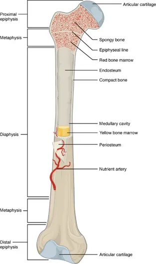
Long bone consists of outer cortical bone and inner trabecular (spongy) bone. Bone has 3 parts namely-
- Epiphysis- The two ends of the bone are called epiphysis
- Diaphysis- The long shaft is called as diaphysis
- Metaphysis-The part that is in between epiphysis and diaphysis is called metaphysis
- Epiphyseal plate-It is a layer of hyaline cartilage that is growing area of the bone.
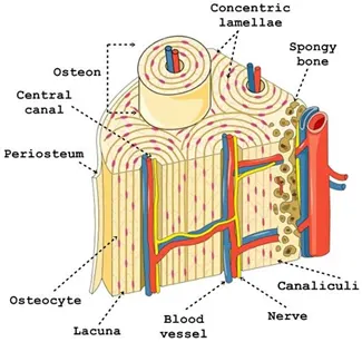
Microscopic anatomy of bone-Haversian system
The structural unit of bone is haversian system or osteon. It is a central canal with blood vessels and nerves that pass through it. Canal is surrounded by the concentric rings of matrix called as lamellae. Osteocytes are present on the lamellae.
Blood supply and nerve supply
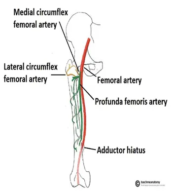
Femur is supplied by deep femoral artery and its branches medial and lateral circumflex branches of profunda femoris.
Femoral nerve, originating from lumbar plexus supplies the femur.
Clinical condition-Femoral fracture
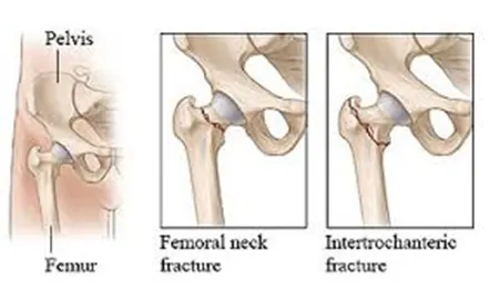
Femoral fracture is the fracture involving the femur. Fracture can take place at femoral head, femoral shaft and femoral neck.
Diagnosis
- Physical examination
- Imaging-X-ray
X-ray
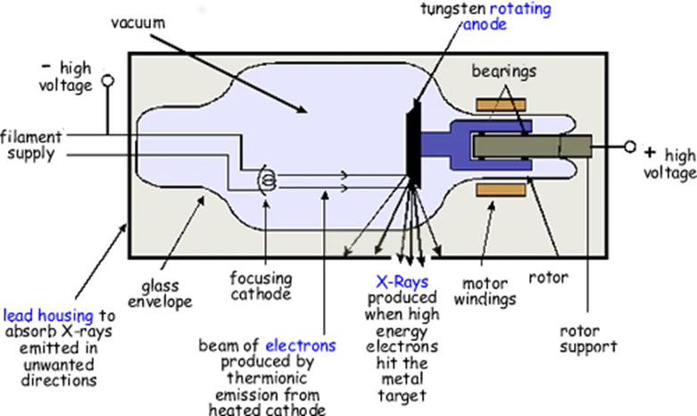
X-rays are produced when negatively charged cathode is charged by a high voltage emitting the beam of electrons that when directed onto the tissue gets absorbed or scattered depending on the density of the tissue.
Surgical procedure-Open reduction and internal fixation
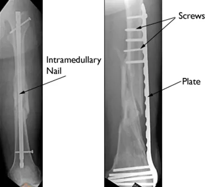
The most common way to repair the femur fracture is surgery. In the surgery, a large rod or nails or plates are inserted into the bone to support it.
Surgical management
Prior to surgery, CT or X-ray imaging of the bone is obtained.
Patient is anesthetized and monitored for vital signs. Then a surgeon makes a incision through the skin and muscle. Surgeon brings the broken femur pieces into the alignment and secure the pieces through plates, screws and nails.
Materials for fixators: Titanium and titanium alloys.
Recent advances
Recent advances in Arthroplasty (Joint replacement surgeries) involves use of additive manufacturing or 3D printing to obtain the custom-made prosthesis suitable for individual patient.
Respiratory system
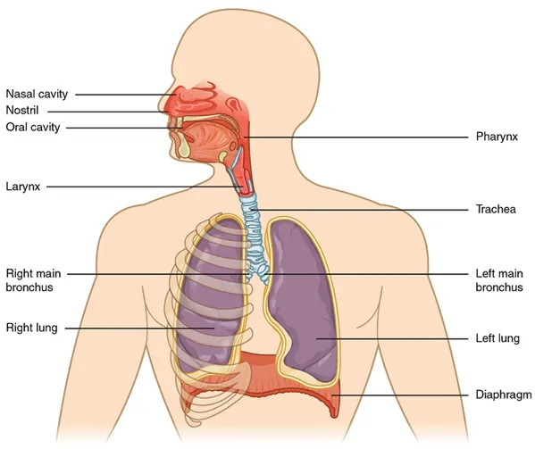
Components-Nose; pharynx, larynx, trachea, bronchi and lungs.
Function-The respiratory system is involved in exchange of gases. It helps us breathe in oxygen and exhale carbondioxide. The exchange of carbondioxide and oxygen is called as respiration.
Clinical condition-Pneumonia
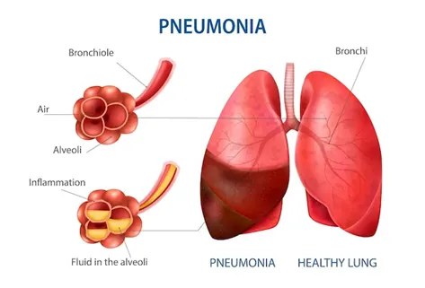
Pneumonia is infection that inflames the air sacs in the lungs that may fill them with fluid or pus.
Organ involved-Lungs
Gross anatomy of lungs
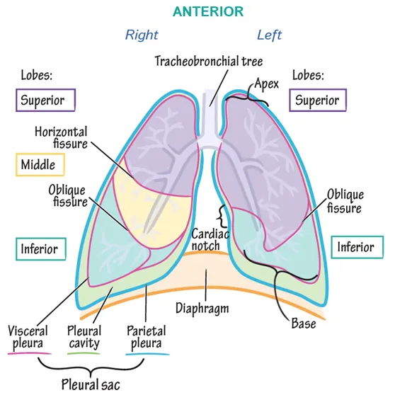
Lungs are located in the thoracic cavity. The apex of the lung is at the first rib while the base of the lung reaches the diaphragm. The space in between the lungs gives place for heart.
Right lung has 3 lobes namely-superior, medial and inferior
Right lung is divided into lobes by two fissures-horizontal and oblique
Left lung has 2 lobes namely-superior and inferior
Left lung is divided into two lobes by one fissure-oblique
Microscopic structure
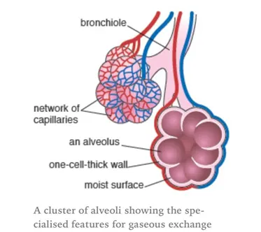
The structural unit of lung is alveoli. These are the primary site of gas exchange. Each alveoli is made up of flattened epithelial cells, numerous capillaries and supporting tissue.
Blood supply and nerve supply
Blood supply to the lungs is provided by bronchial arteries. They are branched from thoracic aorta. Bronchial arteries supply oxygenated blood to lungs and deoxygenated blood is collected into bronchial veins.
Nerve supply-Pulmonary plexus
Common clinical conditions associated
- Pneumonia
- Lung cancer
- Tuberculosis
- Asthma
- Pleural effusions
Pneumonia
Infection that causes inflammation in the lungs that fills the air sacs with fluid or pus. It is caused by virus, bacteria.
Diagnosis
- Blood tests- Blood is collected and is used to detect the micro-organism causing the infection
- Chest X-ray- This is used to determine the extent of damage caused to the lung
- Pulse oximetry-It is used to determine the oxygen level in the blood. Pneumonia prevents oxygen moving into our lungs, thereby decreasing saturated oxygen levels.
- Sputum test-Fluid from our lung(sputum) is taken after deep cough and analyzed for the micro-organism causing the infection.
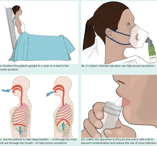 Sputum culture test
It is a culture test that checks for bacteria or any other organism causing infection. The dark yellow or green sputum often indicates the bacterial infection.
Treatment/ Medical management
Treatment for pneumonia involves curing the infection. This could be achieved by using
- Antibiotics- To treat bacterial pneumonia
- Cough syrups- To relax the muscles as coughing moves the fluid out and it causes discomfort. This is relieved by cough syrups.
- Pain/Fever relievers- To calm the discomfort such as high fever or pain.
Recent advancements in pneumonia handling
Diagnosis-CT is being widespread means of detection and very recently ultrasound is also being explored for pneumonia detection.
Treatment-Pneumococcal vaccine for prevention
Digestive system
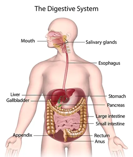
Components-Mouth, pharynx, esophagus, stomach, small intestine, large intestine, rectum, anus, salivary glands, liver and pancreas.
Function-To ingest the food, digest and absorb the digested food and egest the waste after digestion.
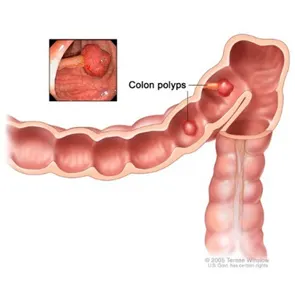
Clinical condition-Polyps
Polyps are the benign growths in the colon that have variety of shapes that grow eventually as cancer.
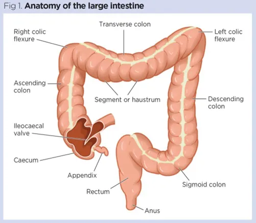
Gross anatomy of colon (large intestine)
Large intestine is present next in line with small intestine. It is divided into –
- Cecum-The joining that connects small intestine with large intestine
- Ascending colon-The ascending part of the colon
- Transverse colon-After the right turn inferior to the liver, colon turns to the left to form the horizontal bend called transverse colon
- Descending colon- The vertical portion continuing from transverse colon
- Sigmoid colon-S-shaped colon
- Rectum- The part continuing from sigmoid colon
- Anus- The part that excreted feces.
Microscopic anatomy
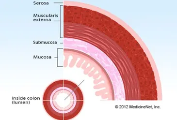
Colon is made up of 4 layers
- Mucosa
- Submucosa
- Muscularis interna
- Serosa
Blood supply and nerve supply
Blood supply to large intestine is provided by superior and inferior mesenteric artery
Nerve supply to large intestine- Sympathetic and parasympathetic nerve supply from superior mesenteric plexus.
Common clinical conditions
- Ulcers
- Polyps
- Hemorrhoids
- Colon cancer
Polyps are the benign growth in the colon.
Diagnosis
- Colonoscopy
- CT colonography
- Stool based tests
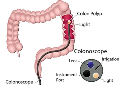
Colonoscopy
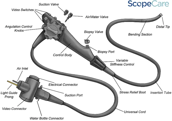
It is an endoscopic procedure where a flexible hollow tube with camera attached to its end is inserted through the rectum to acquire the images of the colon.
Parts of colonoscope
Surgical management-polypectomy
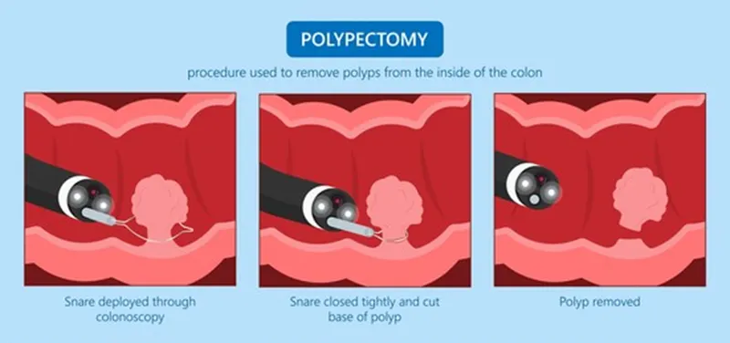
The snare is deployed through colonoscopy and it is closed tightly to hold the base and it is removed.
Recent advances
Transanal submucosal endoscopic resection
Part B
Case study:
Type of bone-Skull is a flat bone. A flat bone consists of two compact bone plates with a layer of spongy bone marrow in between them.
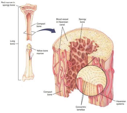
Microscopic anatomy of bone- Haversian system
Compact bone is made up of Haversian canals. The microscopic structural unit of bone is called osteon or haversian canal.
Haversian canal consists of cylindrical central canal that allows the blood vessels and nerves to pass through it. The haversian canal is surrounded by concentric rings of lamellae. These haversian canals communicate through Volkman’s channels.
Blood Supply of bone
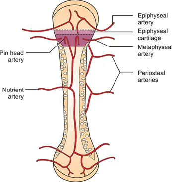
Blood supply to skull: Carotid arteries
Bone is supplied by nutrient artery that passes through the haversian canal.
The periosteal arteries, the branches of the muscular arteries supply the periosteum and epiphyseal arteries supply the epiphysis.
Bone remodeling and new bone formation
Bone remodeling refers to the removal of mature bone tissue by osteoclast and new bone is formed by osteoblasts. Bone remodeling occurs in 4 stages
- Activation: osteoclasts are attracted to remodeling sites
- Resorption: osteoclasts remove the mature bone called resorption and these cells disappear.
- Reversal: The undigested bone matrix is removed and the surface is prepared for bone formation
- New bone formation: The stem cells mature and differentiate into osteoblasts that form the new bone tissue.
Anatomical basis for bone implant
Skull-Cranioplasty
Prior to cranioplasty, MRI or CT is acquired to know the anatomical position of the injury. The scalp and temporalis muscle are reflected to reveal the defect and any soft tissues are cleaned and fixed to surrounding skull by the means of standard titanium screws or plates.
Indications- Protection, Functional improvement; Aesthetics
Contraindications-Osteoporosis, Uncontrolled diabetes, Blood disorders