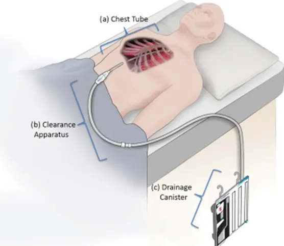
Project 2
Project 2
Anatomy & Physiology
Project A
Gastrointestinal System
1.Pathological condition: Gastric Ulcers in stomach
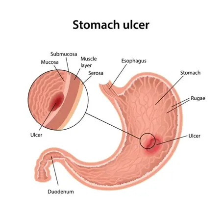
- Normal function of the stomach is partially digesting the food with gastric secretions with pepsin and hydrochloric acid, act as reservoir of food, enzyme secretion and acid secretion.
- Pathology stage: Stomach ulcers can block the passage of food, cause bleeding, anaemia.
- Reasons: Infection with bacterium (H.pylori); long-term usage of non-steroidal anti-inflammatory drugs like aspirin.
- Homeostasis change: A bleeding stomach ulcers cause loss of blood leading to low blood pressure or anaemia.
- Diagnostic methods:
X-rays (Barium swallow); Endoscopy; Endoscopy biopsy
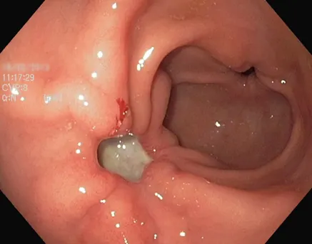
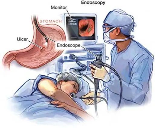
Image representing stomach ulcers detected by endoscope. Image reference:https://www.amboss.com/us/knowledge/Peptic_ulcer_disease
- During the endoscopy procedure, doctor examines the stomach for ulcers
- After detection of ulcers, a small tissue is taken for biopsy to check for the bacteria H.pylori in the stomach lining
- The presence of H.pylori bacteria confirms the stomach ulcer caused due to bacterial infection.
- In absence of H.pylori, the stomach ulcer is caused due to pain killers.
3.Medication-
- If gastric ulcer is caused by the presence of H.pylori bacteria-
- Treat with antibiotics
- Acid-suppression medication- Ranitidine, Cimetidine
- If ulcer is not caused by H.pylori bacteria-
- Acid-suppression medication for 4-8 weeks and follow up by endoscopy- Antacids like Gaviscon, sucralfate
4.Surgical solution
Failure of medical treatment is dealt by surgery. Surgical management for stomach ulcers includes-
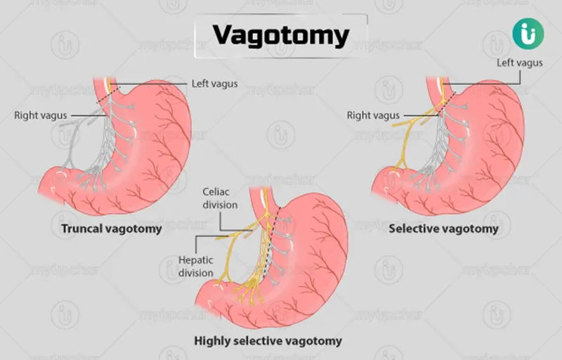
Vagotomy-Vagus nerve, that supplies the stomach is cut so as to interrupt the signals from brain. This helps in reduced acid secretion.
Antrectomy- The lower part of the stomach is called antrum. This secretes digestive juices. Removal of antrum is also usually accompanied with vagotomy to help in reduced secretions.
Pyloroplasty- Enlarging the pylorus to enable the passage of food. Here, the surgeon widens the pylorus by cutting to ensure that pylorus is kept open.
1.Vagotomy
- Patient is in supine position
- Procedure
Laproscopic procedure is a minimally invasive procedure that uses a laparoscope to visualize the internal of the stomach. The laparoscope is inserted through the mouth, passing into the esophagus and then stomach.
At the junction of esophagus and stomach, a cut is made to the tissue.
For truncal vagotomy, surgeon removes segment of each trunk and for highly selective vagotomy, surgeon cuts only the specific nerve branch.
4.Instruments required during vagotomy
- Endoscope
- Monitor
5.Post-surgical care
- Patient starts off with a liquid diet then gradually progress to solid diet depending on the condition
- Monitoring of vitals
- Check for diarrhoea
- Monitoring of stomach pain
6.Devices
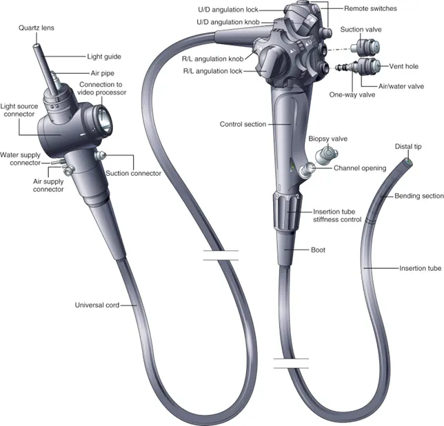
Endoscope-To visualize the internal structures
Urinary system
1.Pathological condition-Kidney failure
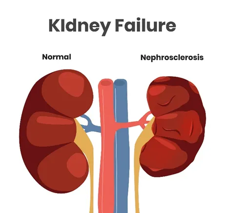
- Kidneys remove the wastes and extra fluid, control blood pressure, acid-base balance, aid in production of red blood cells.
- Pathological stage: Kidney failure causes fluid build-up, muscle weakness, inflammation of the pericardium of the heart and ultimately death
- Reasons: Impaired blood flow to kidneys, damage to the kidneys, urine blockage in the kidneys
- Homeostasis change: Damage to kidneys affects most of the organ systems as it is responsible for removal of waste. Kidney failure may cause disruption in acid-base balance, blood production.
2.Diagnostic methods:
Urine output tests, urinalysis, blood tests, ultrasound, CT, biopsy
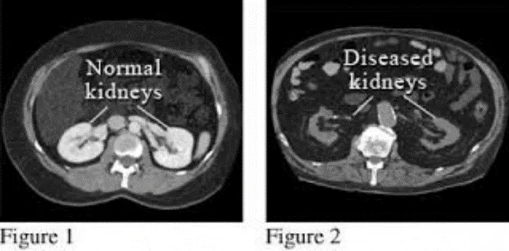
- Data provides the structural damage extent
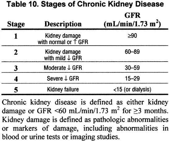
- GFR-Glomerular Filtration rate is the rate at which glomerulus (part of nephron) receives the blood per minute. The normal value is greater than 90ml/min.
- Medication:
Kidney failure complicates many systems. The following medications are used while kidney recovers-
Treatment to maintain fluid balance- Diuretics are used
Treatment to control blood potassium-Calcium, glucose are used to prevent accumulation of high levels of potassium in blood.
To restore calcium-Infusion of calcium
4.Treatment options
There are broadly two namely-
- Dialysis
- Kidney transplant
Surgical Solution
- Kidney Transplantation
- Anatomical position-Supine position
- Procedure-
- Patient is prepared by giving anaesthesia
- New kidney is placed on the lower right or left side of abdomen and it is surgically connected to nearby blood vessels
- The vein and artery of new kidney are attached to that of patient’s.
- New kidney ureter is attached directly to the bladder
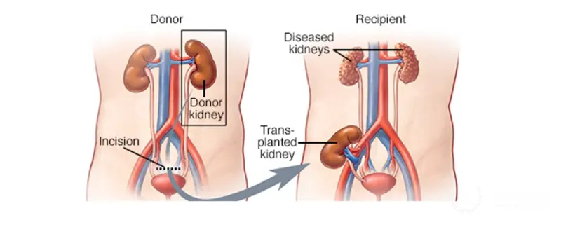
Kidney transplantation surgery- https://www.youtube.com/watch?v=iWSewFlU6o8&ab_channel=ClevelandClinic
5.Post operative care
- Avoid lifting heavy object
- You might need temporary dialysis until transplanted kidney functions
- Be aware of organ rejection-carefully observe for allergies, use anti-rejection medications
- Be watchful about Infection
- Monitor vitals
- Follow a healthy diet
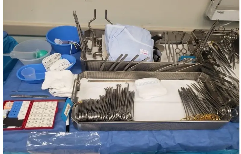
6.Surgical tools used
- Forceps- For grasping of tissue or objects
- Retractors-To hold the tissue in place
- Scalpel- To make incisions
- Debakey Bulldog clamp-for clamping and occluding
- Knife Handle- To cut and dissect
- Scissor-To cut and dissect
- Suctioning pump
- Needle holder- to aid in suturing
- Sutures- To close the incision
Project B
Respiratory System
1.Pathological condition: Chronic obstructive pulmonary disease
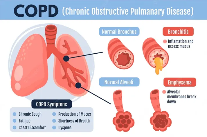
- The normal function of the lungs is exchange of gases. In specific, inhale oxygen and exhale carbondioxide.
- Pathological stage:
- Stage 1-Mild: Forced expiratory volume (FEV)->80%
- Stage 2-Moderate FEV50-79%
- Stage 3-Severe FEV 30-49%
- Stage 4-Very Severe FEV less than 30%
- Reasons: Tobacco smoking, people exposed to fumes from burning fuel (pipe smoke), air pollution.
- Effect on homeostasis: COPD causes imbalance of pro and anti-inflammatory mechanisms that ultimately affect immune homeostasis
2.Diagnostic methods:
Pulmonary function tests (Spirometry); Chest X-ray; CT scan; Arterial blood gas analysis.
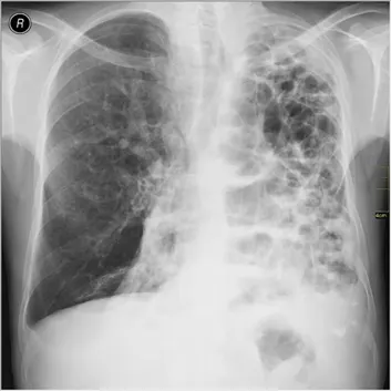
- Determination of COPD -Firstly pulmonary function tests to determine FEV
- Once, FEV less than normal is confirmed, X-ray is taken
- Data from X-ray determines the structural extent of damage.
3.Medications
- Bronchodilators-Levalbuterol, Albuterol
- Inhaled steroids-Fluticasone, Budesonide
- Oral steroids-Phosphodiestarase-4 inhibitors
- Antibiotics
Life-style change includes smoking cessation.
4.Surgical solution
Surgical solution is suggested for patients with severe emphysema
- Bullectomy- removal of large spaces called bullae from lungs
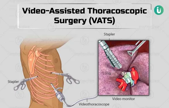
- Video assisted thoracoscopic surgery
- Surgical position- Full lateral decubitus position.
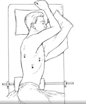
- Procedure-
Patient is prepared by giving anaesthesia.
During VATS, thoracoscope and surgical instruments are inserted into the chest through one or more incisions in the chest wall.
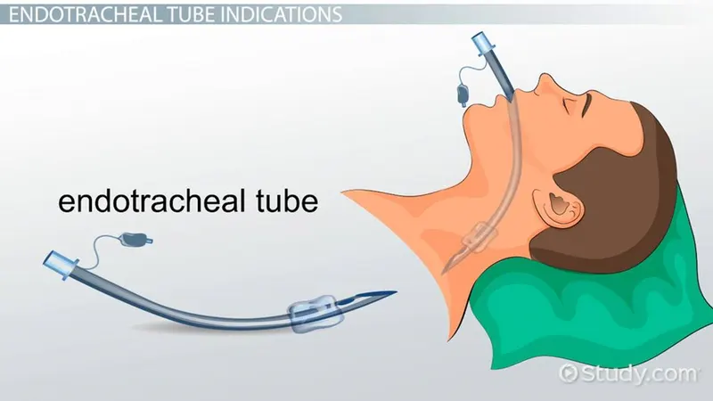
During the procedure, external breathing tube is inserted into the patient to facilitate breathing
Then surgeon makes incisions, inserts surgical instruments to perform the procedure.
4.
5.Link- https://www.youtube.com/watch?v=KoBw_-xj68E&ab_channel=MaimonidesHealth
5.Post-operative care
- Pain control
- Respiratory care
- Chest-tube management
- Restrictive fluid therapy
6.Devices used
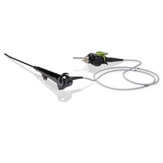
Thoracoscope- Helps in visualization of thoracic cavity
Cardiovascular system
1.Pathological condition: coronary artery disease
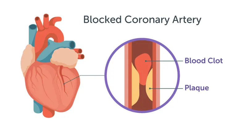
- Coronary arteries supply blood and nutrients to the heart. The main function of heart is to pump blood to various organs of the body.
- Pathological stage- Normal (No plaque), Mild, Moderate, and severe
- Reason: Atherosclerosis (gradual build-up of plaque), age factor, life style like sleep deprivation, lack of physical activity, smoking
- Effect of homeostasis: Blood pressure is affected as normal blood flow is disrupted.
2.Diagnostic methods:
ECG (electrocardiogram), CT scan, Angiogram, Blood tests
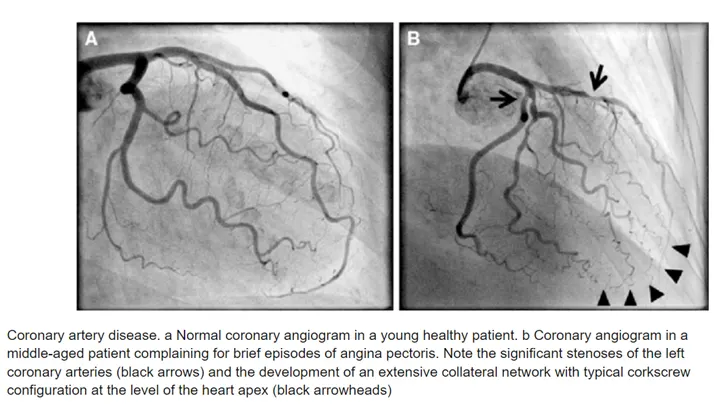
- Data from angiogram clearly shows the blockage in the coronary artery
- Data is acquired from CT scanner
3.Medications-
Life style changes like quitting smoking, getting regular exercise, healthy diet.
Pharmacological medications-
- Beta-blockers
- Calcium channel blockers
- Statins
4.Surgical Solution-
1.CABG-Coronary Artery Bypass Graft surgery
- Surgical position- Supine position
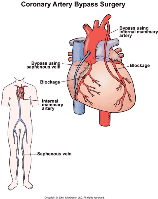
3.Procedure-
Patient is prepared by giving anaesthesia
Endotracheal tube is inserted and ventilation is started.
Chest is opened via sternotomy.
Grafts are harvested-A graft is taken from saphenous vein or artery in the chest
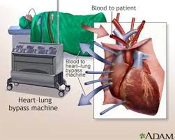
During the procedure, surgeon sutures the cannula into the heart and perfusion is started.
heart-lung machine is also used to facilitate the functioning of heart and lung externally.
Sutures are used to close the incisions.
4.Instruments used
- Heart-lung machine
- Ventilator
- Chest tubes
- Post-operative care
Sit in chair after a day, walks after 3 days and walk down and up the stairs after 5 or 6 days
- Pain control
- Monitor the vitals
- Dressing the wound
- Do not lift heavy objects
- Monitor for potential edema
- Infection risks
- Do not bend down
- Perform light activities like walking short distances, cooking, playing board games
6.Devices used
- Heart-lung machine-To facilitate the exchange of gases, oxygenation of blood and pumping of heart while heart is temporarily stopped working.
2.Endotracheal tube-To facilitate ventilation
3.Chest tubes-To drain the fluid and blood during surgery