Project 1
Part A
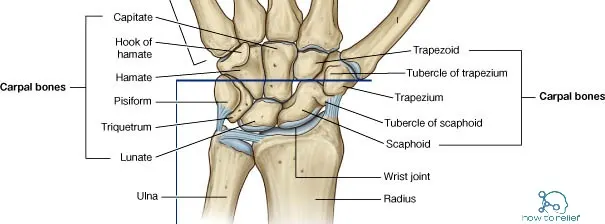
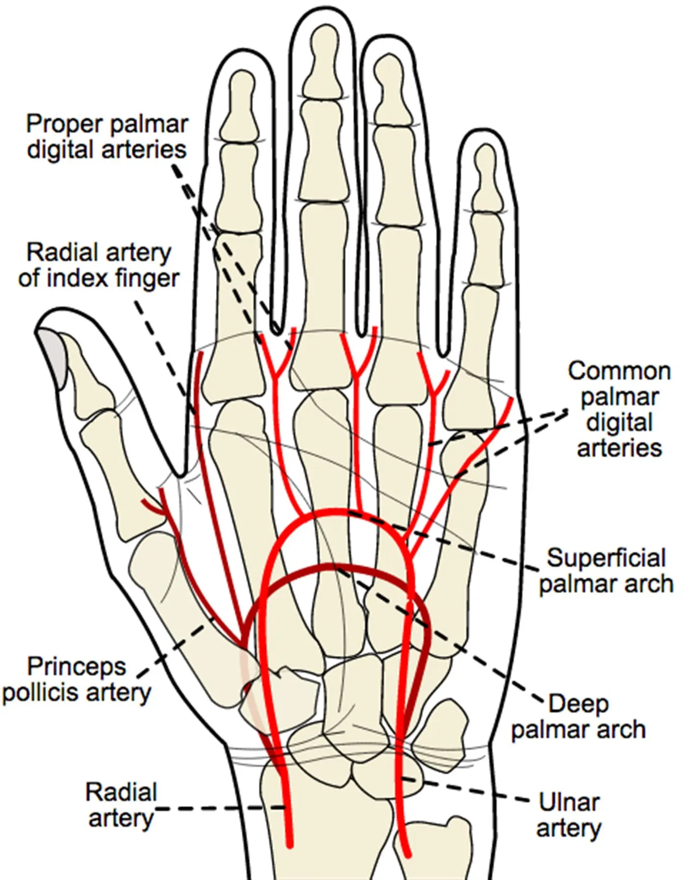
- Carpal Bones: Your index or middle finger meets the carpal bones at a point known as the carpometacarpal boss, or carpal boss. The eight little bones that make up your wrist are known as carpals or little fingers. Carpal bossing is another name for the ailment. A hard lump forms on the back of your wrist as a result of this overgrowth, and it is immobile. For the vast majority of sufferers of carpal boss, there are no symptoms. If the issue becomes uncomfortable or restricts your wrist's range of motion, you should seek medical attention.
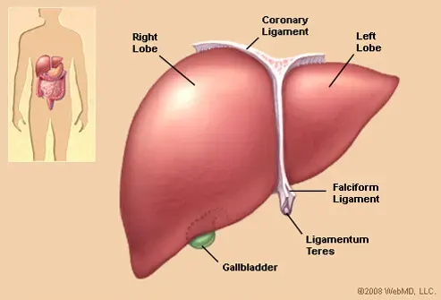
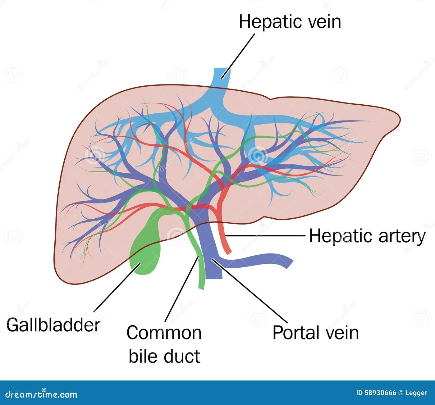
- Liver: Hundreds of metabolic, energy-storage, and waste-filtering processes are carried out by your liver. It aids in the digestion of food, the production of energy from it, and the storage of that energy for later use. It also aids in the removal of harmful toxins from your blood. All conditions that damage your liver might be referred to as liver disease. All of these disorders, no matter how they develop, can harm your liver and impair its normal function.
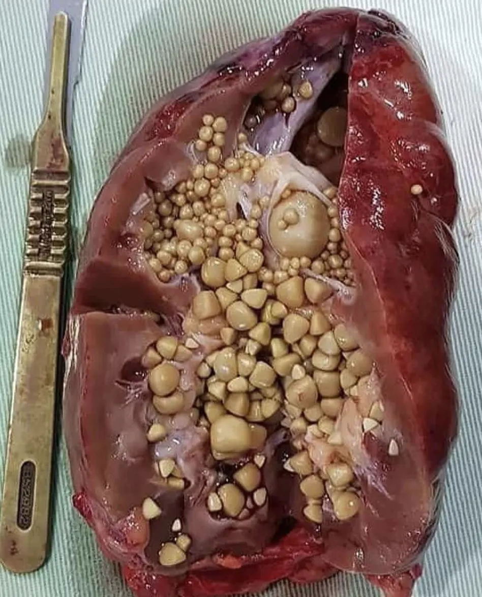
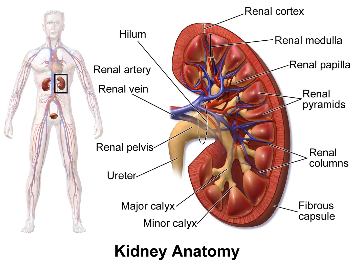
- Chronic kidney disease: Chronic renal disease is frequent. Chronic renal disease never improves. High blood pressure causes it. High blood pressure increases pressure on the kidneys' glomeruli. Glomeruli purify blood in the kidneys. Increased pressure destroys these arteries and reduces renal function. Kidney function deteriorates till the kidneys can't do their job. Dialysis would be needed. Dialysis removes waste from the blood. Dialysis can treat but not cure renal disease. Kidney transplant may be another possibility. Diabetes causes chronic kidney dysfunction. Diabetes raises blood sugar. High blood sugar gradually destroys renal blood vessels. The kidneys can't clear the blood. Toxin excess causes kidney failure.
- Kidney Stone: Stones in the kidney are yet another typical complication of the kidneys. They are the result of minerals and other chemicals in the blood crystallising and producing mass in the kidneys, which leads to kidney stones (stones). Urination is the normal route for the passage of kidney stones out of the body. The passing of kidney stones can be an excruciatingly painful process, but they rarely result in serious complications.
- Glomerulonephritis: An inflammation of the glomeruli is referred to as glomerulonephritis. Glomeruli are incredibly minute structures that are located within the kidneys and are responsible for filtering the blood. Infections, medications, or conditions that manifest themselves during or immediately after birth are some of the potential triggers for glomerulonephritis (congenital abnormalities). The majority of the time, it gets better on its own.
- Polycystic kidney disease: Polycystic kidney disease is a hereditary condition that leads to the development of a large number of cysts, which are defined as small sacs filled with fluid. These cysts can disrupt the normal function of the kidneys, which can lead to renal failure. It is essential to keep in mind that individual cysts in the kidney are not only reasonably common but also nearly invariably innocuous. Polycystic kidney disease is a different condition that is far more severe.
- Urinary Tract Infections: Urinary tract infections, sometimes known as UTIs, can affect any region of the urinary system and are caused by bacterial infections. The majority of urinary tract infections originate in the bladder and the urethra. They are simple to cure, and they almost seldom result in further health complications. In the event that these infections are not treated, however, they may progress to the kidneys and result in renal failure.
- Blood Testing: Blood tests could show that your blood has too much calcium or uric acid. The results of your blood test help your doctor check on the health of your kidneys and may lead him or her to look for other health problems.
- Urine Testing: The 24-hour urine test may show that you're getting rid of too many minerals that cause stones or not enough substances that stop stones from forming. For this test, your doctor might ask you to give two urine samples on two different days.
- Imaging: X-rays might be able to show that you have kidney stones in your urinary tract. Computerized tomography (CT) that moves quickly or uses two kinds of energy can show even tiny stones. Simple abdominal X-rays are used less often because small kidney stones can be missed by this type of imaging test.
- Ultrasound: Noninvasive kidney ultrasounds show size, shape, and location. Sonography measures kidney blood flow. High-frequency ultrasound employs a transducer. Ultrasonic waves travel from the skin to organs and structures. Organs echo sound to the transducer. Computers turn reflected waves into images of organs or tissues. Bone conducts sound quicker than air. The transducer converts sound wave speed and volume into tissue types. Ultrasound gel is applied to the transducer and skin to improve sound conduction. Doppler ultrasound displays chest blood flow speed and direction. Unlike ultrasound, Doppler waves are audible. Ultrasound can be used during pregnancy without radiation or contrast dyes. An ultrasound can be used to evaluate the kidneys, ureters, and bladder. Ultrasound detects kidney cysts, tumours, abscesses, blockages, fluid accumulation, and infection. Ultrasound detects kidney and ureter stones. An ultrasound can help put needles needed to biopsy the kidneys, drain a cyst or abscess, or place a drainage tube. This method can also measure renal artery and vein blood flow. Kidney ultrasonography is used to examine a transplanted kidney. Doctors may order a renal ultrasound for different reasons.
- Dietary management: Fluid consumption and dietary adjustments prevent kidney stone recurrence. Increasing urine volume to at least 2 L/day can prevent stone recurrence by 40–50%. Water should dominate fluid consumption. Tea and coffee contain oxalate; milk binds free oxalate. Increasing urine volume reduces citrate. Cucumber, green peppers, beetroot, spinach, soya bean, chocolate, rhubarb, popcorn, and sweet potato are oxalate-rich foods that should be avoided. Calcium restriction increases stone disease risk, hence it's not suggested. Hypocitraturia is present in 16–63% of calcium stone formers. Oral potassium citrate increases urine citrate and reduces stone recurrence. High citrate has been explored as a Kcit alternative. Lemonade treatment increased urinary citrate. Orange juice increased Odvina's urine pH and citrate. Both Kcit and lemonade treatment increased urine citrate, according to Kang et al. Kcit worked better than lemonade. Yilmaz et al. analysed tomato, orange, lemon, and mandarin juice. Fresh tomato juice has high citrate and low oxalate. Calcium oxalate and uric acid stone formers should limit animal protein.
Stone-specific therapies
- Calcium oxalate stones: Thiazide diuretics reduce idiopathic hypercalciuria recurrence rates by 70%. It's the only treatment for urinary calcium. Citrate supplements are helpful. Pyridoxine can help in primary hyperoxaluria, but not idiopathic. Oxalobacter formigenes degrades oxalate in the gut. Increased gut colonisation may reduce oxalate absorption and urine excretion. A prospective, randomised, placebo-controlled, double-blind trial contradicted the benefits of O. formigenes colonisation.
- Uric acid stones: Treatment for uric acid stones is to make uric acid more soluble in urine. Alkali treatment and increasing the amount of urine excreted are two ways to do this. The medication allopurinol is a helpful addition to treatment.
- Struvite stones: Struvite stones occur in alkaline urine from infection with urea-splitting bacteria. Antibiotics are the mainstay of the therapy with occasional use of acetohydroxamic acid.
- Cystine stones: This is an uncommon sort of stone. The goal of treatment is to decrease the free cystine concentration and enhance its solubility in urine. Desirable are a high fluid intake of up to 4-5 L/day OR 4-5 lit and alkalinization of urine with a target urine pH of >7. When the 24-hour urine cystine concentration surpasses 2,000 mol/l, chelating drugs such D-penicillamine or tiopronin are suggested.
- Shock Wave Lithotripsy: Patients most commonly use Shock Wave Lithotripsy for kidney stones. Small to medium-sized stones work best. Noninvasive surgery uses high-energy shock waves to break kidney stones without cutting the skin. In this surgery, external shock waves are used to fragment kidney stones. Stones shatter. Large portions can be treated again. Smaller-than-2-cm stones are suitable for SWL. Larger stones may not be treatable.
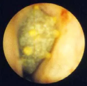
- Ureteroscopy: The doctor uses a narrow, flexible scope to locate and remove stones from the kidneys and ureters during this surgery. In this surgical treatment, there are no wounds made in your skin. In order to remove the stone from the ureter and bladder, a scope called a ureteroscope must be sent through these two passages. At this moment, the procedure will be performed. One to three hours are typical for a ureterscopy surgery under general anaesthesia. Small stones can be easily removed from the ureter if necessary. For large stones or ureters with narrower diameters, the stone would have to be shattered by laser, which is typically done. These bits are removed after the stone has been split into small pieces by the impact. Using a ureteroscope to remove large stones is impractical and impossible for individuals who have already had their urinary tracts reconstructed, which prevents a ureteroscope from passing through in the first place.
- Percutaneous Nephrolithotomy or Percutaneous Nephrolithotripsy: This method treats patients with large or irregular kidney stones, infections, or stones not broken up enough by SWL or who have not been treated by the above two modalities. This technique is for persons with larger than 2 cm (marble-sized) stones. Both procedures include back incisions to access the kidney. Once the surgeon reaches the kidney, a nephroscope and other devices are threaded in. If the stone is removed through the tube, it's called nephrolithotomy; otherwise, it's nephrolithotripsy. High-frequency sound waves break up stone, and a suction mechanism collects the dust.
- Open Surgery: Open kidney stone surgery is unusual. This approach is for huge stones or stones that can't be removed or crushed. Surgery helps when:
- Ureter stones.
- One hurts badly
- Stone blocks urine flow
- Bleeding or infection
Rigid ureteroscope specifications include the following:
-
Tip diameter - 4.5-9.5F (6.9F most common)
-
Optics - Fiberoptic bundles or digital imager
-
Working channels - One, 2, or 3 (2 channels preferred)
-
Accessory length - 40 - 60 cm
Flexible ureteroscope specifications include the following:
-
Tip diameter - 6.9-9.8F (7.5F most common)
-
Optics - Fiberoptic bundles or digital imager
-
Working channel - Single, 3.6F
-
Access - Guidewire (0.035 in nitinol or 0.038 in stainless steel)
-
Accessory length - 100 - 120 cm
Energy sources include the following:
-
Holmium:yttrium-aluminum-garnet (Ho:YAG) laser
-
Neodymium:yttrium-aluminum-garnet (Nd:YAG) laser
-
Thulium laser
-
Electrocautery
-
Electrohydraulic lithotripsy
-
Mechanical impactor (ie, Lithoclast)
Prophylaxis is as follows:
-
All patients receive a preoperative dose of a broad-spectrum parenteral antibiotic.
-
Patients with positive cultures should receive appropriate treatment course, based on sensitivity panel
- A High-powered Laser Facilitates Stone Removal: A new high-powered laser employs "Moses" to clear kidney stones. Faster, safer procedures. Moses reduces stone movement when the laser hits it, making it easier to hit moving stones as the patient breathes. This helps surgeons break apart stones without injuring surrounding tissues and creating bleeding. Newer high-powered lasers, like Moses', can "dust" kidney stones (breaking stones into a fine powder). "Dusting" patients may not need an unpleasant ureter stent after surgery. High-powered lasers can remove more stone per unit time, reducing operation time.
- A Miniaturized Procedure Could Reduce Bleeding: Surgeons use a tiny camera and equipment to break up and remove big kidney stones in an operation known as percutaneous nephrolithotomy (PCNL). Now, thanks to a procedure known as "mini PCNL" or "mini perc," incisions and devices can be reduced in size. This is part of a broader movement to reduce the amount of cutting and discomfort associated with various surgical procedures. Less bleeding and less discomfort during recuperation are theoretically possible as a result of this. Mini perc may have these effects, but further research is required before we can say for sure.
- Single-use Scopes Bring Treatment Closer to Home: These disposable ureteroscopes have the potential to increase the number of people who can benefit from ureteroscopy. Even in larger hospitals, reusable scopes are prohibitively expensive. In addition, the scopes are prone to breakage after a certain number of uses, necessitating costly repairs. Due to the fact that single-use scopes only need to be used once, this is not an issue. In the event that their reusable scope breaks, several hospitals maintain some single-use scopes on hand. In this way, patients are spared the inconvenience of postponing or cancelling their procedures and are given better access to care.
How a hematoma forms: How a Hematoma Forms When a bone breaks, blood vessels inside the bone break and bleed. This causes a hematoma, which is a collection of blood that has coagulated at the site of the break. The process of clotting makes sure that the blood vessels at the broken ends of the bone won't leak any more blood. When bone cells don't get enough food, cell death starts to happen.
Bone formation: After a break, capillaries start to grow into the hematoma and phagocytic cells start to eat the dead cells. This is where the process of making new bone starts. Even if there are still pieces of the blood clot left behind, fibroblasts and osteoblasts will move into the area and start to rebuild the bone. Fibroblasts are in charge of making collagen fibres, which are then used to connect the broken ends of a bone. Osteoblasts are in charge of starting the process of making spongy bone. A type of healing tissue called fibrocartilaginous callus grows between the two broken ends of the bone. It is made up of both hyaline and fibrocartilage. At this stage, bone spicules may also start to show up.
Formation of a bony callus: During this process, the fibrocartilaginous callus changes into a bony callus made of spongy bone. After a bone has been broken, it takes about two months for the two pieces to heal together and be strong again. During this process, osteoblasts, osteoclasts, and bone matrix are all present. This is similar to how bone is made when cartilage ossifies, which is called endochondral production of bone.
Bone remodelling: Osteoclasts and osteoblasts then rebuild the bone callus, getting rid of any extra material on the outside of the bone and in the medullary cavity. This process is called "remodelling the bones." First, compact bone must be added to make bone tissue that is the same as the original bone before it was broken. This may take a few months, and the bone may still be uneven for a few years after it is done.