Project 1
Ultrasound
How the Test is Performed
An ultrasound machine makes images so that organs inside the body can be examined. The machine sends out high-frequency sound waves, which reflect off body structures. A computer receives the waves and uses them to create a picture. Unlike with an x-ray or CT scan, this test does not use ionizing radiation.
The test is done in the ultrasound or radiology department.
- You will lie down for the test.
- A clear, water-based gel is applied to the skin on the area to be examined. The gel helps with the transmission of the sound waves.
- A handheld probe called a transducer is moved over the area being examined. You may need to change position so that other areas can be examined.
How to Prepare for the Test
Your preparation will depend on the part of the body being examined.
How the Test will Feel
Most of the time, ultrasound procedures do not cause discomfort. The conducting gel may feel a little cold and wet.
Why the Test is Performed
The reason for the test will depend on your symptoms. An ultrasound test may be used to identify problems involving:
- Arteries in the neck
- Veins or arteries in the arms or legs
- Pregnancy
- Pelvis
- Abdomen and kidneys
- Breast
- Thyroid
- Eye and orbit
Normal Results
Results are considered normal if the organs and structures being examined look OK.
What Abnormal Results Mean
The meaning of abnormal results will depend on the part of the body being examined and the problem found. Talk to your health care provider about your questions and concerns.
Risks
There are no known risks. The test does not use ionizing radiation.
Considerations
Some types of ultrasound tests need to be done with a probe that is inserted into your body. Talk to your provider about how your test will be done.
Alternative Names
Sonogram
PRODUCT/SERVICES AND QUALITY of ultrasound
Diagnostic ultrasound (US) images can be obtained from a quality device, in optimal working conditions, combined with the capable actions of the operator in tweaking the equipment’s characteristics. The quality assurance (QA) is the topic of this review article, and it is addressed in an US practice through proper selection of the equipment when purchasing, and through care and preventive maintenance of the machine for ensuring accurate performance. For optimal US system functioning, QA steps carried out on the basis of a scheduled program are needed in any US practice or department. It is critical to confirm in a semiannual or annual basis that the image quality is maintained according to standards and any subtle change in equipment’s functioning is detected and amended at an early stage. The use of test objects (also called US testing phantoms) is required above the basic level of QA testing. The scope of this review article is to inform the US user about necessary QA knowledge and at the same time, present the state of the art of the most suitable test methods for US QA. We referred to relevant publications, selected after performing a systematic literature search of the MEDLINE, EMBASE and COCHRANE databases, and also to the standards established by authoritative international societies.
General and practical care of the equipment
US systems contain delicate parts that can be physically damaged by improper use and handling. Conscious and diligent protection of the equipment during usage is a requirement for the operator who must avoid dropping or knocking the probe, or passage of the machine wheels over the probe cable during movement (the US machines are heavy, with some of them well above 150 kg). Frayed cords and cracked transducer casing represent an electrical hazard for the operator and the patient and, therefore, require immediate switch off of the device and the machine or transducer must be withdrawn from activity until specialized personnel has troubleshot the problem Cleanness of equipment (including transducers, control panel, monitor, vents and peripherals), environment, and also hygiene of transducers and other parts of the equipment are required. Dust that gathers in the electrical components can cause malfunctioning of the computer and of the components of the machine.
Daily optimal operation of the US system should be observed by the user and any deficiency or fault should be immediately reported and repaired . Principal concerns are presented by damaged cords or transducer housing, cracks or chips in the transducer face, failure of part of the transducer’s crystal elements as detected in the image, obvious caliper malfunctioning, irregular image or non-diagnostic poorly representative of anatomy, erratic depth adjustments, non-functioning keyboard or trackball, etc.
Technical support by phone offered by the manufacturer and the device’s user manual can be consulted for minor troubleshooting. Severe or complicated malfunctioning needs the onsite remedial action on the device by the service personnel or engineer.
Performance QA testing
Apart from these practical actions, regular QA testing and servicing should be scheduled and implemented on a routine basis. A well-planned and comprehensive preventive maintenance program for QA of US devices must be carried out and a record of each inspection, evaluation and/or calibration must be maintained .
The QA testing can be used for the device’s assessment at the moment of purchase, for proper selection of the machine, and also comparison of an US scanner against another scanner [1]. In this way, the imaging capabilities are evaluated when purchasing the equipment and thereafter routinely tested for assuring the maintaining the system’s accuracy .
To achieve the objectives of the quality assessment, the user’s judgment of the image and equipment performance is deemed subjective and scarcely reliable for a QA program. Furthermore, the operator, habituated with the obtained image of the US device used is largely incapable to percept gradual, slow, and subtle degradation of the sonographic image quality. In addition, the skill of the operator and patients’ characteristics are disturbing variables in quality evaluation .
Therefore, in response to these issues, an objective evaluation is mandatory, and quality control actions in the absence of the patient with the use of test objects (also called US phantoms) and measured variables . This quantifiable assessment is to be followed and reproduced over the timescale of years. In this way, a deterioration in performance is detected before it becomes perceptible to the operator and/or clinically relevant .
Resolution in the axial, lateral and elevational plane, beam penetration, contrast resolution (represented by the gray scale sensitivity and dynamic range), vertical and horizontal calibration, and overall registration of data are evaluated , and these issues will be addressed in this article.
A list of the main imaging characteristics to be evaluated is presented in Table 1.
Table 1
Description of the imaging capabilities of the US systems to be tested in a QA program
| Imaging characteristic tested | Details | Significance and comments |
|---|---|---|
| Axial resolution | Accuracy in identifying as separate two reflectors positioned parallel to the US wave | Spatial pulse length, beam former and transducer are in accurate and consistent conditions |
| Lateral resolution | Accuracy in identifying as separate two reflectors positioned perpendicular to the US wave in the imaging plane | Transducer elements and beam former with correct functioning |
| Elevational resolution (beam thickness) | Accuracy in identifying the details of reflectors positioned perpendicular to the US wave and perpendicular to the scanning plane | A thin US beam of a quality transducer, used with the adequate frequency and focusing, assures minimum partial volume artifacts and maximum margin details of lesions |
| Registration (calibration) | Accurate imaging of a pin in its size and location with correct representation of vertical and horizontal distances | Accurate size representation and measurements are critical components of US imaging |
| Dead zone | The problematic quality of the first centimeters from the transducer’s face, in the image, is tested | If the low-quality distance of the dead zone increases, the system has developed malfunctioning in pulsing or transducer elements |
| Sensitivity, contrast resolution and uniformity | The detection of weak echos, the uniform display of reflectors’ brightness throughout the image, and the correct differentiation of tissues with varying sound impedance | After proper adjustments of the time gain compensation, sensitivity and image uniformity failure represent transducer or output deficiencies |
Measuring against an affirmed quantifiable standard, these imaging capabilities offer an absolute evaluation which helps for a consistent objective testing ensuring that the performance does not fall short of diagnostic requirements deemed clinically relevant. International societies and accrediting agencies have produced QA guidelines also concerned with minimum standards to be achieved . A list of some of the principal organizations is presented in Table 2.
Table 2
List of some of the international societies that have established standards for US equipment and imaging QA
| Organization | |
|---|---|
| EFSUMB—European Federation of Societies for Ultrasound in Medicine and Biology | |
| IPEM—Institute of Physics and Engineering in Medicine (registered in England and Wales) | |
| WHO—World Health Organization | |
| AIUM—American Institute of Ultrasound in Medicine | |
| ACR—American College of Radiology | |
| IAC—Intersocietal Accreditation Commission (registred in USA) | |
| IEC—International Electrotechnical Commission (based in Geneva, Switzerland) |
Alternative to the absolute evaluation for QA mentioned above is the practical relative evaluation, which measures only the machine’s performance against that of the same machine at the time of purchase, or at any other previous time point. Not perceptible, developing faults are detected and less expertise and cheaper phantoms are required in this case .
Various test objects (Table 3) are designed for testing general and specific performance characteristics both in the absolute QA and relative one Unfortunately, the use of US phantoms and regular quality testing is not widespread despite the recommendations of authoritative guidelines . Only QA with phantom testing, on a regular basis during the equipment’s life, is capable of monitoring the optimal performance and early detecting malfunctioning .
Table 3
Test objects for US quality assurance, also called US phantoms
| Type of phantom | Architecture of construction | Use and measurements |
|---|---|---|
| General resolution phantom | Filaments, pins and tubes at varying depths, of varying size and impedance | Axial and lateral resolution, penetration, calibration, geometrical accuracy |
| Breast resolution phantom | Filaments or pins and tubes at varying depths, of varying but small, size, and different impedance | Axial and lateral resolution for breast application of high-end devices |
| Slice thickness phantom | Filament or tubes in diagonal position; or spheres | Elevational (azimuthal) resolution |
| Crystal control phantom | Simple simil-tissue material without internal elements | Detection of malfunctioning crystal elements and transducer artifacts |
| Color Doppler phantom | Motor moving a string, or a pump circulating in tubes a simil-blood fluid | Penetration of doppler signal imaging and velocity measurements |
| Elasticity phantom | Materials of varying rigidity | Evaluation of accuracy of US elastographic techniques |
The frequency of the quality control actions and/or servicing is slightly different in the various recommendations, with some authors advising QA up to four times per year More lenient and also clinically supportable intervals are recommended by the various organizations, such as the American Institute of Ultrasound in Medicine (AIUMB) ] that requires that US equipment must be tested, serviced, and calibrated at least yearly and more often if problems emerge, and that there must be routine inspection by the operator.
Easy-to-use methods
The US user can carry out some basic tests without phantoms, such as the air test . In the air test, the transducer is put in function in the air with a clean and dry face in its routine clinical pre-set with the frequency in its lowest value and the gain in the maximum level (both overall gain and time gain compensation) . The image obtained is one of a series of horizontal bands of reverberation due to the transducer–air impedance mismatch. These series of reverberations are used for a first line subjective evaluation assessing eventual faulty crystal elements (vertical line of reduced echos is seen), uniformity of image, transducer sensitivity and image noise . The US user can conserve a hard or soft copy of this image at the time of machine’s purchase and use it for relative testing and for future comparative images checking uniformity, noise, sensitivity and maximum depth at which the reverberations can be measured (penetration that can be measured with the calipers). Furthermore, localized bright or dark areas can be detected, which can be a sign of transducer lens failure.
Noise has to be distinguished from speckle in this test; noise is only the fluctuating echos that come and go between frames, whereas speckle is the unchanging pattern of echos. Noise is clearer in the depths where the last reverberations are weak and the noise predominates. The operator can then calibrate by reducing the overall gain to the point where noise is minimal and the reverberations are still clear. This position of the knob of the overall gain is then signed and used in the routine examinations
In the air test, the transducer is put in function in the air, with a clean and dry face. The image obtained is one of a series of horizontal bands of reverberation due to the transducer–air impedance mismatch. The measurement of the depth of “in-air” reverberations as a proxy for sensitivity is performed by positioning the caliper at the maximum depth in which the reverberations arrive.
(Courtesy of Institute of Physics and Engineering in Medicine)
The overall gain reduction to measure the noise threshold. The noise is only the fluctuating echos that come and go between frames, whereas speckle is the unchanging pattern of echos. Noise is clearer in the depths where the last reverberations are weak and noise predominates. On the right-side image, the operator can calibrate by reducing the overall gain to the point where noise is minimal and the reverberations are still clear. This position of the knob of the overall gain is then signed and used in the routine examinations
Another method for detecting faulty transducer elements is performed by applying a small amount of gel on the probe face, and sliding over an opened paper clip. In faulty elements, low brightness of the column of echos is shown [5] (Fig. 3).
Phantoms
Tissue-mimicking test objects for gray-scale evaluation and Doppler test objects are required for accurate QA, both absolute and relative (Fig. 4). Phantoms can be of general purpose testing a number of image characteristics, or specific purpose ones capable of assessing one or a few imaging parameters (Table 3). Gray-scale phantoms test the device’s resolution (spatial and contrast), sensitivity and calibration which evaluate the measurement accuracy of the machine (Figs. 4, ,5).5. Special tissue mimicking test objects for breast imaging are designed by most of the manufacturers of US phantoms as superior requisites have to be met for quality breast US. Doppler test phantoms perform assessment of the Doppler beam for depth penetration and sensitivity, flow direction, accurate sample volume location and velocity measurements [11]. Furthermore, other special phantoms are required for elasticity imaging of both the qualitative and quantitative variants [12].
Photo of ultrasound test phantoms of the Gammex manufacturer
General purpose phantom for low-contrast penetration measurements, with which one can measure spatial and contrast resolution, sensitivity and calibration
Test objects are supplied with instructions for testing and interpretations of results with a series of evaluations that need to be logged with details in the records of the QA program.
The tissue-equivalent test objects consist of an aqueous gel and graphite particles that build up a medium with transmit sound wave velocity equal to the average velocity of tissues of 1540 m/s. At the same time, the impedance and attenuation rate of this environment is similar to that of soft tissues. Varying with the manufacturer’s compositions, yet common to all phantoms, is a deterioration over time (usually years) of the acoustic properties of the tissue mimicking medium with progressive loss of quality and functionality. Manufacturer’s instructions need to be followed in regard of the monitoring the storage area temperature, and not damaging the integrity of the airtight container [13]. Most manufacturers offer a long warranty and ensure for their phantoms over 10 years of reliable use through reinspection and repair services.
In the gray-scale phantom throughout the tissue equivalent medium, stainless steel pins or filaments, and tubes or spheres, are strategically located. When testing, pins need to be accurately identified and located as well as the tubes or spheres of varying echogenicity (that mimic from anechois cysts to a range of solid masses) must be correctly visualized (Fig. 5).
Usually, the elevational (azimuthal) resolution, which corresponds to the thickness of the US beam, needs a specialized phantom for accurate evaluation and almost all manufacturers produce a separate special phantom for this purpose. Some manufacturers use spheres instead of the commonly used tubes in the resolution phantom, offering the possibility to evaluate in the direction perpendicular to the plane of US beam (the elevational resolution). In the case of thick beams, echos fill the simil-cystic spheres (Figs. 6, ,7,7, ,88).
Example of composite evaluation with four different gray-scale phantoms, using various resolution targets, echoic and anechoic targets
This model of ATS manufacturer phantom offers basic testing for QA. The design consists of a combination of monofilament line targets and six tissue mimicking cylindrical targets of varying sizes. Four groups of line targets are provided to evaluate the vertical and horizontal calibration measurements, axial–lateral resolution and the dead zone. Test uses are: dead zone or ring-down, vertical measurement calibration, horizontal measurement calibration, sensitivity/penetration, functional resolution, focal zone, axial and lateral resolution, and image uniformity
The CIRS multi-purpose phantom is a general-purpose QA phantom for low- and high-frequency probes (2–15 MHz) adaptable with detachable scanning wells, thus capable of testing curvilinear, linear and endocavitary probes. Tests that can be performed are: uniformity, depth of penetration, beam profile/focal zone/lateral response width, vertical distance measurements, horizontal distance measurements, axial and lateral resolution, elevational resolution, contrast resolution, grayscale contrast sensitivity, elasticity sensitivity, and dead zone assessment
The Doppler phantoms utilize either a small engine moving a string or belt, or the modern ones, a simil-blood circulating fluid with a pump [11].
US breast imaging and QA
Although the role of US in the diagnostic algorithm of breast imaging is still debated among experts, a firm point of unanimous agreement is the emphasis that US equipment for breast evaluation should be high-end machines with the best of possible capabilities. Superior imaging aspects such as fine resolution, both spatial and contrast ones, are needed for quality breast US. Some authors advocate the use of one-and-a-half dimension matrix array instead of the usual linear array probe [14] which achieves high elevational resolution with the possibility to focus not only in the lateral direction, like all probes, but also in the azimuthal direction producing a very thin beam. This is needed to obtain superior margin details of lesions and avoiding partial volume effects. These quality characteristics, as, for example, detecting a single angle (even obtuse) in a small breast nodule, signify a 60% diagnostic probability of cancer [15].
For QA of US breast units, specialized gray-scale phantoms developed for high-resolution scanners and slice thickness phantoms are needed.
Conclusions
Guarantee of quality of ultrasonic systems is essential through care and preventive maintenance of the machine for ensuring accurate performance [16].
Daily optimal operation of the US system should be observed by the user and any deficiency or fault should be immediately reported and repaired.
The US operator can perform some basic tests to obtain an objective evaluation. Phantom testing yields an exact assessment of the US equipment needed to ensure quality control/QA.
Particularly relevant is an optimal quality review of breast US which needs high elevational resolution.
The absence of a quality assurance could undermine the diagnostic quality of US examination and expose the operator to innumerable risks, including for the health of the patient [17].
Descriptions
- Products and Services that meet or exceed customer expectations result in customer satisfaction. Quality is the expected product/service being realized. Before a customer makes a purchase (exchanges money for a product/service) he or she does a mental calculation: “Is the worth of the product/service (as I perceive and expect) equal to the money that I am about to exchange?”
- Products/services that are produced and manufactured to specifications that are appropriate to the price (money to be given in exchange by the customer) of the product/service is an operational or manufacturing view of quality. Here, the customer receives the value that he or she expects since operations has built quality standards into the product. An operations view of quality is a common view of the concept of quality.
- However, quality is a function of how the customer views the product/service that he or she receives. The customer view always compares what they expect with what they actually receive regardless of how operations conceives quality. How do customers arrive at their expectations?
- Marketing, especially sales, has a major effect on how the customer views quality. As mentioned earlier, customer satisfaction is based on receiving the actual product/service as expected. When marketing and sales enthusiastically promises a product/service that manufacturing or operations (in the case of a hospitality service) cannot deliver, then expectations are not met, the customer is dissatisfied, and quality (in the customers’ eyes) is not realized.
- Quality is not an absolute to be determined by operations or manufacturing. Variables that affect quality are: (a) customer expectations (obtained from marketing and sales, as well as word of mouth and previous experience), (b) actual product/service received (how a service is performed by operational people and actual tangibles received (cold food for example). The following models explain these basic concepts.
1.Product quality
A variety of factors contribute to the overall quality of the ultrasound image. These include the design of equipment components, especially the transducer, the choice of imaging parameters, particularly the beam frequency, and the skilful use of the equipment by the operator
The purpose of a quality assurance (QA) program in medical ultrasound is to ensure that the equipment and clinical practices of a facility are safe, and that the information obtained from a clinical ultrasound procedure is as accurate as can be determined.
2.Service
An ultrasound can provide a view of the bladder, gall bladder, eyes, kidneys, liver, pancreas, ovaries, thyroid, spleen, testicles, uterus, blood vessels, and more that helps to detect any health issues
3.Communication
- People Skills. Ultrasound technician work directly with patients. ...
- Excellent Hand-eye Coordination. An ultrasound technician needs to be able to move hand-held devices on a patient's body while watching a video monitor. ...
- Team Work. ...
- Continuous Learning.
- Patient's name and other identifying information.
- Facility's identifying information.
- Date of ultrasound examination.
- Image orientation when appropriate.
5.Response time
ultrasound scans, the scanned images show up immediately on the device's screen. This means that often the person operating the scanner can give immediate feedback - provided they're suitably qualified to interpret the images. So, most likely you'll get a verbal summary of the results immediately.
6.Delivery
- Intangible Service Delivery Orientation. Here the focus is on the process of delivering the service. This involves two basic components: (a) Technical quality – the means of service delivery and (b) Functional quality – the how of service delivery.
- Technical Quality. This includes the systems and infrastructure designed and created to organize delivery of the service. For example: computerized systems, machines technical solutions, and know-how.
- Functional Quality. The hospitality customer goes through many interactions with employees in the creation and delivery of a hospitality experience. A successful meeting is the result of all functional areas of a hotel being synchronized and focused on creating a beautiful symphony. Technical quality must be in place to facilitate such coordination and allow the employees to work together. Functional quality includes employee: attitudes, behavior, service mindedness, appearance, accessibility internal relations and customer contacts.
7.Installation
While a competent, capable, and expert team of staff members is the backbone of any hospital or healthcare facility, having up-to-date and functional equipment can make it easier to provide extraordinary patient care.
Technologically advanced equipment not only improves patient satisfaction, but also increases staff efficiency - both of which lead to more revenue. When you have ordered new medical equipment, how do you ensure that everything is set up properly and will be ready for immediate use?
Here are five things to consider for the smoothest, most efficient equipment setup.
Proper Location
The first step of proper medical equipment setup is placing the equipment item(s) in the specified area of use. You need to have the equipment located in the area where it will be used, so a direct-to-site delivery process is extremely beneficial.
Inspection Of Parts And Proper Assembly
The next step is inspection. You want to carefully inspect each piece of equipment for the following:
-
Missing parts;
-
Damaged parts;
-
Mismatched parts.
Although it may seem simple and obvious, this is a crucial step for your hospital or healthcare facility. A quick check is not enough; you need a detailed inspection to avoid problems in the future.
What good is medical equipment if it isn’t assembled correctly? Once you’ve inspected everything, be sure that the equipment gets assembled the right way. Even a small mishap could cause a malfunction and possibly damage your equipment. This means more time and money spent on equipment needs.
Test Of Functionality
Once assembled, the medical equipment should be tested for functionality. Sometimes everything appears to be fine, but there may be a glitch that can’t be seen. In addition to problems with the equipment itself, testing can uncover assembly mistakes before any permanent damage occurs.
8.DISPOSAL
Removal Of Unnecessary Materials
The next step is to be sure all packing materials and other debris are removed from the medical equipment area. This helps ensure a clean, effective workspace for those employees who will be using the equipment on a regular basis. It also prevents contamination and other mishaps during work.
Instruction For Use
Finally, after all inspections have been done and everything seems to be working properly, it’s crucial that each applicable member of your staff is instructed on proper use of the medical equipment, especially if it’s a new model that’s not been used by your hospital before.
Clear, detailed instructions prevent misuse, which could lead to poor patient care or damaging of the equipment. It’s also important that instruction includes the hospital biomedical staff in order to facilitate shock-free operation and proper electrical grounding.
Making sure hospital equipment setup goes smoothly and gets done in a timely manner can put undesired stress on you and your staff. Tracking your equipment orders, managing multiple deliveries, and then finally overseeing the setup process is time-consuming and often overwhelming.
Feed back form
-
-
How often do you use our products?
Excellent very Good Good Average poor -
Which features are most valuable to you
-
How would you compare our products to our competitors’?
-
What important features are we missing?
-
What are you trying to solve by using our product?
-
What other types of people could find our product useful?
-
How easy is it to use our product?
-
How would you rate the value for money?
-
How likely are you to recommend this product to others?
-
How could we improve our product to better meet your needs?
-




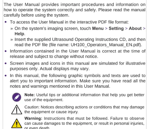
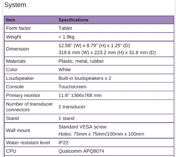
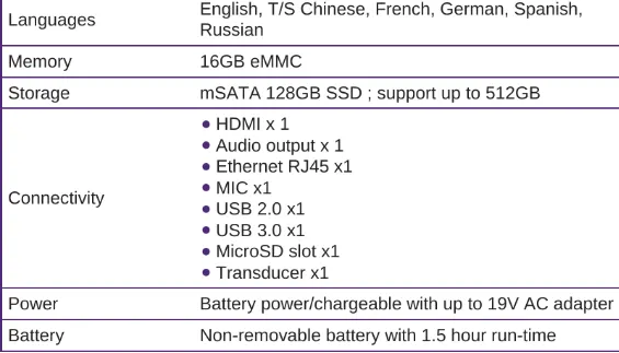
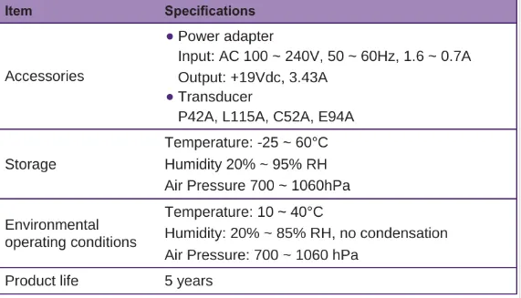
user manual