Project 2
1.
urinary system:
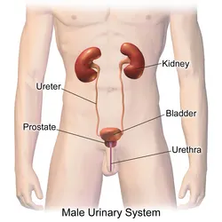
The structures that create and carry urine to the point of excretion are referred to as the urinary system. On both the left and right sides of the human urinary system, two kidneys are positioned between the dorsal body wall and the parietal peritoneum. The kidneys get a large amount of blood via the renal arteries, which exit the kidneys via the renal vein.Each kidney is made up of functioning components known as nephrons. Wastes (in the form of urine) depart the kidney via the ureters, which are tubes comprised of smooth muscle fibres that transport urine towards the urinary bladder, where it is stored and then released from the body by urination (voiding). The female and male urinary systems are essentially similar, with the urethra being the only difference. Urine is produced in the kidneys by blood filtration. The urine is subsequently transported through the ureters and stored in the bladder. Urine is transferred from the bladder to the exterior of the body via the urethra during urination. 800–2,000 milliliters (mL) of urine are normally produced every day in a healthy human. This amount varies according to fluid intake and kidney function.
Normal function of the organ
1. Kidney
This pair of organs, placed under the rib cage, works as a filtration system to remove waste from the blood and form urine. The filtration units called nephrons contain a renal tube and a ball wrapped in small blood capillaries. It also helps maintain the required levels of salts and important nutrients in the blood, and secretes hormones that help generate new red blood cells.
2. Ureter
The ureters are the thin passageways through which urine travels from kidneys to the bladder. The ureter walls are made of muscles that constantly contract and expand to push down the urine formed in the kidneys. A few millilitres of urine are passed through the ureters every 15 seconds. Its blockage can result in a urinary infection, affecting the function of urinary system.
3. Bladder
This works as a reservoir to store urine. It is held in the lower abdomen, by ligaments that connect it to organs and bones in the vicinity. The bladder generally has the ability to hold around 2 cups of urine for a time period ranging from 2 to 5 hours. When a person urinates, the muscular walls of this organ contract to push the urine into the urethra.
4. Urethra
This tube starts from the bladder and allows urine to escape the body. When the bladder’s walls contract, the sphincter muscles relax and urine is pushed through the urethra. Normal function of urinary system needs the brain to send signals to both the bladder muscles and the sphincter muscles in the right order. In males, urine is released through the penis, and in female placental mammals through the vulva.
5. Prostrate
The prostrate is only present in males. It is placed in front of the rectum, between the penis and the bladder. The tube connecting the bladder to the penis runs right through it. The secretions from this gland provide nourishment for sperm. When a male ejaculates, the fluid is secreted into the urethra and combines with sperm to form semen.
6. Others
- Nerves running through the bladder, which signal the brain when the bladder is full.
- Two sphincter muscles surround the exit of the bladder like a rubber band and prevent the leakage of urine from it.
Reason for the pathological condition
Kidney Stones Form from Substances in Urine
The kidneys produce urine to eliminate waste. Kidney stones can form when mineral and acid salts in the urine crystallize and stick together. If the stone is small, it can pass easily through the urinary system and out of the body. A larger stone can get stuck in the urinary tract, however. A stuck kidney stone causes pain and can block the flow of urine.
All 3D models in the page have loaded
Kidney Stones
Kidney stones form within the kidney when the urine becomes concentrated. This causes the debris to crystallize and form hard deposits.
Urinary Incontinence Is the Loss of Bladder Control
Most bladder control issues arise when the sphincter muscles of the urethra are too weak or too active. If the sphincter muscles are too weak, a cough or sneeze can cause urination. Sphincter muscles that are too active can trigger a sudden, strong urge to urinate with little urine in the bladder. These issues are diagnosed as urinary incontinence (UI). Women experience UI twice as often as men. It becomes more common with age.
Fluid-filled Cysts Can Develop in the Kidneys
A simple kidney cyst is a rounded pouch or a closed pocket that is usually filled with fluid. In polycystic kidney disease (PKD), clusters of cysts form inside the kidneys and take the place of the normal tissue. The affected kidneys become enlarged and work poorly. PKD is an inherited condition that often leads to kidney failure, requiring dialysis or kidney transplantation. Acquired cystic kidney disease (ACKD) typically affects people already on dialysis from chronic kidney disease. In ACKD the kidneys do not enlarge and no other symptoms occur.
Chronic Kidney Disease Can Lead to Kidney Failure
In chronic kidney disease (CKD), the kidneys are damaged and unable to filter blood properly. This damage can lead to a build-up of waste substances in the body and to other problems, including kidney failure. The most common causes of CKD include diabetes, heart disease, and high blood pressure. A diseased kidney may look smaller and have a granular surface.
Diagnostic methods selected by the surgeon for assessing the condition.
1. Blood urea nitrogen
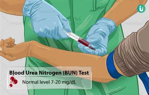
A blood urea nitrogen test is a vital test to know how well the kidneys function. It is a serum test that measures the amount of nitrogenous urea, a byproduct of protein metabolism in urea. BUN level indicates the extent of renal clearance of urea and nitrogenous waste. It can be due to many factors, such as poor renal function, dehydration, and so on. The normal blood urea nitrogen level is 10 mg/dL to 20 mg/dL. If there is any disturbance in these levels, it could be due to renal failure.
The test can be used to diagnose, screen for, and help treat patients. Blood urea nitrogen alone is often less helpful for these purposes than blood urea nitrogen combined with other renal function tests. A BUN test is usually analyzed in conjunction with a creatinine test to help determine how effectively the kidneys are operating.
The quantity of urea nitrogen in the blood is measured by a blood urea nitrogen (BUN) test. Urea nitrogen levels are an indicator of how effectively the kidneys are operating. Urea nitrogen is a typical byproduct produced by the body when eating. The liver breaks down the proteins in the daily diet, and as it does so, it produces blood urea nitrogen or BUN. The products are released into the bloodstream by the liver and finally end up in the kidneys.
Healthy kidneys efficiently eliminate BUN, leaving only a trace in the blood. However, for the most part, the kidneys eliminate it by filtering it all out of the body through urine. When the kidneys are not functioning properly, they have difficulty eliminating BUN and thus retaining it in the bloodstream. Therefore the amount of waste product is detected in the blood. If the levels are beyond the usual range, it might indicate that the kidneys or liver are not functioning correctly. A blood urea nitrogen test is also called urea nitrogen, BUN, or serum urea nitrogen.
2. cystoscopy
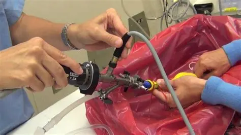
A cystoscope is a thin tube with a camera and light on the end. During a cystoscopy, a doctor inserts this tube through your urethra (the tube that carries urine out of your bladder) and into your bladder so they can visualize the inside of your bladder. Magnified images from the camera are displayed on a screen where your doctor can see them.
The procedure might be performed in a hospital or doctor’s office. You will need some form of anesthesia, so talk to your doctor about your options before the procedure. These include:
Local anesthesia: Outpatient procedures generally involve local anesthesia. This means you’ll be awake. You can drink and eat normally on your appointment day and go home immediately after the procedure.
General anesthesia: General anesthesia means you’ll be unconscious during the cystoscopy. With general anesthesia, you may need to fast for several hours ahead of time.
Regional anesthesia: Regional anesthesia involves an injection in your back. This will numb you below the waist. You might feel a sting from the shot.
With either regional or general anesthesia, you will probably need to stay in the hospital for a few hours after the procedure.
Just before the cystoscopy, you need go to the bathroom to empty your bladder. Then, you change into a surgical gown and lie down on your back on a treatment table. Your feet may be positioned in stirrups. The nurse may provide you with antibiotics to help prevent a bladder infection.
At this point, you’ll be given anesthesia. If you get general anesthesia, this will be all that you are conscious of until you wake up. If you get a local or regional anesthetic, you may also be given a sedative to relax you. Your urethra will be numbed with an anesthetic spray or gel. You’ll still feel some sensations, but the gel makes the procedure less painful. The doctor will lubricate the scope with gel and carefully insert it into the urethra. This may burn slightly, and it may feel like urinating.
If the procedure is investigatory, your doctor will use a flexible scope. biopsy or other surgical procedures require a slightly thicker, rigid scope. The bigger scope allows surgical instruments to pass through it.
Your doctor looks through a lens as the scope enters your bladder. A sterile solution also flows through to flood your bladder. This makes it easier for your doctor to see what’s going on. The fluid might give you an uncomfortable feeling of needing to urinate.
With local anesthesia, your cystoscopy may take less than five minutes. If you’re sedated or given general anesthesia, the entire procedure may take 15 to 30 minutes.
How the doctor identifies the problem
Most people have been asked to submit the urine sample for testing when they visit a health institution. The sample is used to analyze various urine test types that can help the doctors diagnose disease or monitor a patient’s progress. Urine elimination in the body is important as it helps maintain the body water balance as well as remove toxic substances produced during the metabolism process.
What Does A Urine Test Show?
The smell and the color of the urine sample can be an indication of something wrong. Adults should the urinalysis periodically to ensure that their health is intact. A urine test might result in different scenarios. Even before the test, there are some factors that indicate the patient is not okay.
- Having dark urine indicates there is less water intake or a problem with the kidneys while flaky and cloudy urine indicates the presence of a urinary tract infection.
- In some cases, the urine can be reddish indicating that there might be blood. To avoid speculations and to know further, it is essential to take a urine test strip which shows either a person is suffering from diabetes or urinary tract infection.
- Persistent occurrence of protein in urine tests is an early indicator of a chronic kidney disease while the glucose elements indicate signs of potential diabetes.
- The white blood cells in the urine indicate the presence of urinary tract infection. Undertaking a urine test is important as it helps the doctors to know exactly what they are dealing with and offer the suitable solutions.
The urine test types for people who are already suffering are done in intervals to monitor their progress especially those suffering from chronic kidney disease. The urine test show if the given treatment is reducing the protein in the urine or not.
Urine Test Types
There are various urine test types that are performed for various reasons in the quest to determine what is wrong with a patient. It may be part of a regular physical routine check
- Urinalysis
A complete urinalysis is done in the laboratory and performed on people who are about to go for surgery, having been admitted to the hospital or whose different urine results are abnormal. It is used to either determine or monitor bleeding in the urinary system, urinary tract infection, diabetes, kidney disease, bladder stones and blood disease.
- Rapid Urine Test
It is the easiest way to test urine and involves dipping the urine test strips in the urine samples. Diabetic people use this test to check their sugar levels. It can also be performed ion people with acute lower abdomen pains, backaches, blood in the urine, stomachache and persistent painful urination. The rapid urine test detects the following
- Glucose, Nitrate, Protein, Ketone in Urine
- White Blood Cells in Urine
- Red Blood Cells in Urine
- Bilirubin in Urine
- Pregnancy Test
Urine for pregnancy test determines whether a woman has conceived or not. The test is done like a rapid test, eight to ten days after a late menstrual date for accurate results. The test should not be done too soon when a woman is on medication or having taken a lot of fluid just before the test to avoid altering the results.
- Urine Culture Test
The urine test is done in the laboratory to determine the presence of fungi or bacteria. If they are found, the right kind of antibiotics is then administered for treatment.
- 24 Hour Urine Collection Test
This test is used to determine the presence of proteins, salt, metabolic products, hormones among other substances are excreted in the body. The urine is collected in 24-hour duration and stored in the refrigerator during that period.
How to Prepare for A Urine Test?
It is important to take normal meals when going for a urine test. It is also essential to take plenty of water. Let your physician know about any type of medicine, supplements or substance you have been using as some o them might alter the urine test results. Also, for women, let the doctor know if menstruating.
What is The Procedure of The Urine Test?
The health institution issues a container where the urine sample is collected. One can use the clean-catch collection method which involves washing up the urinary opening area first. One can then urinate in the toilet first then a few ounces in the container and finishing up in the toilet. The sample is handed over to the doctor for testing.
In some cases, the doctor might use a catheter which is inserted into the bladder through the urethra. The procedure can be discomforting causing mild pains. For the testing, the doctor can use either a visual exam, dipstick test or microscopic exam when conducting the urine test.
Urine Test Results and Interpretation
When the results are abnormal, the doctor determines the next step based on various factors. Previous diagnosis with urinary tract problems, kidney issues or any other related condition may result in further tests. It is vital to identify the occurrence of the abnormal contents of the urine.
Having high level proteins in urine indicates there are underlying issues that can result in kidney disease. Such conditions include leukemia, lupus, heart conditions, hypertension, sickle cell anemia and rheumatoid arthritis. Also, a high level of glucose in the urine can indicate the presence of diabetes.
Urine test pus cells indicate that the urinary tract has major issues. Most of the times it can either be a bladder infection or kidney infection. If there is the presence of too much pus cells in the urine test, then it means the patient has an abscess near the kidney, sexually transmitted disease, kidney infection or urinary tract infection. The condition can also be an indication of stones in the urinary passage.
It is important to have a regular checkup where the doctor takes different urine test types to determine your general health. Some of the diagnosed conditions don’t show the normal signs and symptoms thus the importance of carrying out the tests
Surgical solution
kidney transplant:
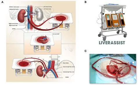
In most case kidney transplant is required for Kidney Failure – A condition in which the patient loses his/her ability to balance their fluids and remove waste from their body.
Kidney Transplant – is a surgical procedure performed to replace the damaged kidney with a donor’s healthy kidney. The new healthy kidney can come from an alive or deceased donor or a family member. People who choose to provide their kidney’s transplant can lead a healthy life with the one kidney, provided it remains healthy.
Kidney Transplant Procedure Step By Step
Step 1: Before proceeding with the surgery; first the doctors ensures to search for a kidney that can be donated by the donor or a family member.
Step 2: once the kidney is found, specific tests are run on the patient to match the new kidney with the body. If everything seems fine, the patient is given a surgery appointment.
Step 3: The patient is prepared for the surgery, before which his/her blood, mental stability, and concerned organs are closely monitored. An IV will be inserted in your arm with a few catheters in your wrist and arm for monitoring the heart rate of the patient.
Step 4: Now the patient will be put to sleep using general anesthesia. During the procedure, the patient is put on a ventilator allow him/her to breathe.
Step 5: A long incision is made on the lower abdomen, and the kidney is visually inspected by the doctor before replacing it with a new kidney.
Note: the left kidney of the donor is implanted on the right side of the patient, and the right kidney of the donor is placed on the patients left side. This allows the doctors to access the ureter and connect it to the bladder easily.
Step 6: Now the artery and veins of the donor’s kidneys are attached to the patient’s body, allowing the blood to flow in the vessels.
Step 7: Next the donor’s ureter is connected to the patient's bladder.
Step 8: Now that all the organs are connected to the new kidney, the incision is closed using surgical staples or stitches, and a sterile bandage is applied to the wound.
LIFESTYLE CHANGES TO MANAGE A URINARY TRACT INFECTION (UTI)
Urinary tract infections (UTIs) are very common, particularly in women. Around one in two women and one in 20 men will get a UTI in their lifetime. The urinary system is designed to minimise the risk of serious infection in the kidneys. It does this by preventing the urine from flowing back up into the kidneys from the bladder. Most urinary infections are confined to the bladder and, while causing symptoms, are not serious or life threatening.
Symptoms of UTIs
Some of the symptoms of UTIs include:
- wanting to urinate more often and urgently, if only a few drops
- burning pain or a ‘scalding’ sensation when urinating
- a feeling that the bladder is still full after urinating
- pain above the pubic bone
- Cloudy, bloody or very smelly urine.
Causes of UTIs
When bacteria enter the urinary tract and multiply, they can cause a UTI. To infect the urinary system, a micro-organism usually has to enter through the urethra or, rarely, through the bloodstream. The most common bacterium to cause UTIs is Escherichia coli (E. coli). It is usually spread to the urethra from the anus. Other micro-organisms, such as mycoplasma and chlamydia, can cause urethritis in both men and women. These micro-organisms are sexually transmitted so, when these infections are detected, both partners need medical treatment to avoid re-infection.
Prevention of UTIs
Although not always backed up by clinical research, some women have found some suggestions useful in reducing their risk of developing urinary tract infections, including:
- Drink plenty of water and other fluids to flush the urinary system.
- Treat vaginal infections such as thrush or trichomonas quickly.
- Avoid using spermicide-containing products, particularly with a diaphragm contraceptive device.
- Go to the toilet as soon as you feel the urge to urinate, rather than holding on.
- Wipe yourself from front to back (urethra to anus) after going to the toilet.
- Empty your bladder after sex.
- Avoid constipation.
Cranberries (usually as cranberry juice) have been used to prevent UTIs. Cranberries contain a substance that can prevent the E. coli bacteria from sticking to the urinary tract lining cells.
Doppelherz Urocalm: Contains Natural plant extracts, Cranberry extract rich in proanthocyanidins that prevent bacteria from sticking to the wall of the urinary tract and bladder and Dandelion extract to stimulate diuresis that helps to flush out bacteria. Doppelherz Urocalm combines the properties of cranberry and dandelion for a more efficient fight against recurrent urinary tract infections. Urocalm can be used as an adjunct with antibiotics for treating severe urinary tract infections.
Urocalm is very safe for prolong use (due to its natural source) in the treatment of urinary tract infection to prevent relapse in treatment.
Post surgical care required for the patients
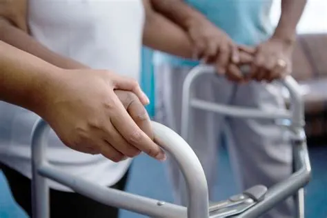
Postoperative infections are fatal infections that are completely preventable. Infection after surgery is a common problem in health care. The process of infection at the surgical site is complex and involves the interaction of several biological pathways at the molecular level. These infections cause pain, delayed recovery from surgery, long hospital stays, increased care costs, the need for long-term use of antibiotics, failure of surgery, and in acute cases, organ failure and even death.
Minimizing the number of infections depends on the patient's condition, the use of appropriate disinfectant solutions at the incision site, the sterile surgical environment, the use of sterilized instruments during surgery, and the appropriate and disinfected coverage of operating room staff. after surgery, what their symptoms are and how they are treated in this article.
Women have a 20% risk of developing a urinary tract infection (UTI) following urogynecologic surgery. This study assessed the association of post-operative UTI with bacteria in pre-operative samples of catheterized urine.
Immediately before surgery, vaginal swabs, perineal swabs and catheterized urine samples were collected and the V4 region of the 16S rRNA gene was sequenced. The cohort was dichotomized in two ways: 1) standard day-of-surgery urine culture result (positive/negative) and 2) occurrence of post-operative UTI (positive/negative). Characteristics of the bladder, vaginal and perineal microbiomes were assessed to identify factors associated with post-operative UTI.
87% of the 104 POP/UI surgical patients were white; the mean age was 57 years. The most common genus was Lactobacillus with a mean relative abundance of 39.91% in catheterized urine, 53.88% in vaginal swabs, and 30.28% in perineal swabs. Two distinct clusters, based on dispersion of catheterized urine (i.e., bladder) microbiomes, had highly significant (p<2.2e-16) differences in age, microbes and post-operative UTI risk. Post-operative UTI was most associated with the bladder microbiome; microbes in adjacent pelvic floor niches also contributed to UTI risk. UTI risk was associated with depletion of Lactobacillus iners and enrichment of a diverse mixture of uropathogens.
2.
Cardiovascular system
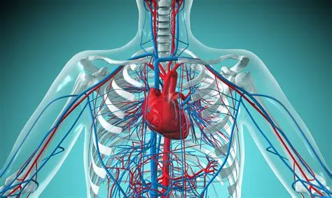
The cardiovascular system is sometimes called the blood-vascular, or simply the circulatory, system. It consists of the heart, which is a muscular pumping device, and a closed system of vessels called arteries, veins, and capillaries. As the name implies, blood contained in the circulatory system is pumped by the heart around a closed circle or circuit of vessels as it passes again and again through the various "circulations" of the body.
As in the adult, survival of the developing embryo depends on the circulation of blood to maintain homeostasis and a favorable cellular environment. In response to this need, the cardiovascular system makes its appearance early in development and reaches a functional state long before any other major organ system. Incredible as it seems, the primitive heart begins to beat regularly early in the fourth week following fertilization.
The vital role of the cardiovascular system in maintaining homeostasis depends on the continuous and controlled movement of blood through the thousands of miles of capillaries that permeate every tissue and reach every cell in the body. It is in the microscopic capillaries that blood performs its ultimate transport function. Nutrients and other essential materials pass from capillary blood into fluids surrounding the cells as waste products are removed.
Numerous control mechanisms help to regulate and integrate the diverse functions and component parts of the cardiovascular system in order to supply blood to specific body areas according to need. These mechanisms ensure a constant internal environment surrounding each body cell regardless of differing demands for nutrients or production of waste products.
Heart Attack:
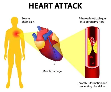
A heart attack occurs when the blood flow to the heart is severely restricted or blocked. The obstruction is often brought on by the build up of fat, cholesterol, and other substances in the heart's (coronary) arteries. The fatty, cholesterol-containing formations are known as plaques. The process of plaque accumulation is known as atherosclerosis. On rare occasions, a plaque may rupture, causing a clot to form and obstruct blood flow. A reduction in blood flow can damage or even destroy a portion of the heart muscle. A heart attack is also known as a myocardial infarction. When the blood flow that supplies oxygen to the heart muscle is significantly lowered or entirely stopped, a heart attack happens. These words are frequently used interchangeably, however they refer to distinct events. When the heart's blood supply is cut off, a heart attack occurs. It has to do with circulation. In cases of sudden cardiac arrest (SCA), the heart experiences a malfunction and unpredictably stops beating. An electrical issue causes sudden cardiac arrest.
A cardiac arrest may result from a heart attack. Death occurs in cardiac arrest, also known as sudden cardiac death (SCD), when the heart abruptly ceases functioning properly. Arrhythmias, or abnormal cardiac rhythms, are the root cause of this.
Ventricular fibrillation is the most typical arrhythmia in cardiac arrest. The bottom chambers of the heart suddenly begin to pulse erratically and stop pumping blood at this time. Within minutes of the heart stopping, death happens.If CPR (cardiopulmonary resuscitation) is carried out and a defibrillator is utilised within minutes to shock the heart and re establish a normal heart rhythm, cardiac arrest may be reversed.
Causes of a Heart Attack
Plaque accumulation in the arteries is the root cause of the blockage (atherosclerosis). Deposits, cholesterol, and other materials make up plaque. Immediately after a plaque ruptures, a blood clot forms. The heart attack is actually brought on by a blood clot.
Heart muscle cells start to incur damage and begin to die if the blood and oxygen supply is cut off. Within 30 minutes of obstruction, irreversible harm starts. The upshot is that oxygen-deprived cardiac muscle ceases to function properly.0
The heart's four primary functions include:
- Pumping oxygen-rich blood to other bodily components.
- delivering hormones and other essential nutrients to various bodily areas.
- receiving and transporting metabolic waste products from the body to the lungs where it is oxygenated.
- keeping blood pressure constant.
What is the heart's homeostasis?
The regulation of blood flow to satisfy the needs of the various organ and tissue systems is referred to as hemodynamic homeostasis.Throughout our lifetime, the heart's output is kept in a homeostatic balance to ensure that it always meets the needs of the body's tissues. The heart may not be able to sustain an adequate flow when it suffers damage, like after a heart attack. Blood pressure drops as a result, setting off homeostatic feedback loops to try to restore normal blood pressure (and cardiac output).
A heart attack is one method in which the heart may be harmed. This happens when coronary blood arteries, which provide oxygen and nutrients to the heart's cells, dramatically narrow or are entirely stopped by a buildup of plaque and thrombus (clot). The lack of oxygen during a heart attack causes the cells to perish. The heart weakens so that it can no longer pump as much blood, which causes the various side effects linked to this condition if enough heart muscle cells perish.
An additional prevalent ailment that affects cardiac output is congestive heart failure. It could appear as a result of a heart attack or asa result of numerous other heart ailments. The issue with congestive heart failure is that the heart muscle is so weak that it struggles to effectively pump out the blood that is entering it (the blood entering the heart is referred to as venous return). In actuality, one of the primary feedback loops that controls blood pressure alters the body's blood supply.
This is due to the fact that, given a healthy heart, the more blood we have, the higher our venous return and cardiac output will be. More venous return may actually result in reduced cardiac output due to the weakened heart caused by congestive heart failure. The body responds to a drop in blood pressure by increasing blood volume, which raises venous return and lowers cardiac output even further. It is a positive feedback loop, and I don't want to be in one where the issue with cardiac output and blood pressure keeps getting worse.
Diagnostic methods selected by the surgeon for assessing the condition.
Angiography:
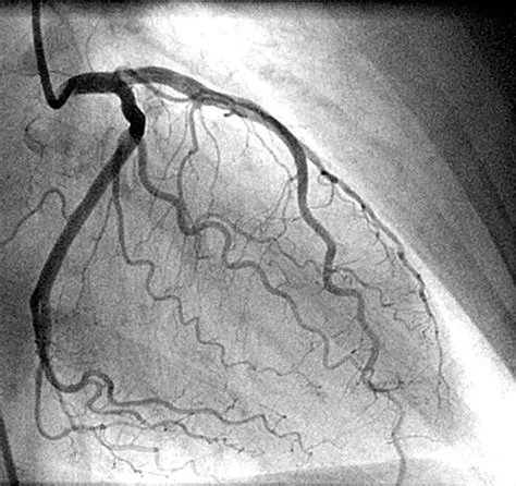
With a focus on the arteries, veins, and heart chambers, angiograms are a type of medical imaging treatment used to observe the interior, or lumen, of the body's blood channels and organs. Angiography is an X-ray method used to evaluate blood vessels. A particular dye must first be injected into your blood because a regular X-ray does not properly show blood vessels. Your blood vessels will be easier to see, which can help your doctor identify any problems. The X-ray images created during an angiography are known as angiograms. Your blood vessels' health and how blood flows through them are both assessed. It can help with the investigation or diagnosis of several conditions involving blood vessels, such as heart attack, peripheral arterial disease, blood clots, etc.
The angiography procedure
While having angiography you'll be asked to wear a hospital gown and lie on an X-ray table
To be tested:
Although you'll typically be awake, general anaesthesia, which puts you to sleep, may be used on very young infants
Local anaesthetic is applied to numb the area where a small cut is made in the skin over one of your arteries, generally near your groyne or wrist, so it won't hurt.
The artery is carefully guided to the area being inspected as a long, thin, flexible tube (catheter) is inserted. When this is done, you could feel some pulling and pushing, but it shouldn't hurt.
The artery is carefully guided to the area being inspected as a long, thin, flexible tube (catheter) is inserted. When this is done, you could feel some pulling and pushing, but it shouldn't hurt.
Through the catheter, a specific dye (contrast agent) is administered. After this is completed, a series of X-rays are taken as the dye travels through your blood vessels, which may cause you to feel warm, flushed, and as though you need to urinate for a short while.
Treatment can occasionally be administered simultaneously, such as when a balloon or tiny tube is inserted to widen a constricted artery. Angioplasty is the term used for this.
The catheter is taken out after the procedure, and pressure is applied to the wound to halt any bleeding. There's no need for stitches.
Magnetic Resonance Imaging:
Can cardiac issues be found via an MRI?
Because MRI gives medical professionals a more detailed view of the heart, they can provide patients with more precise diagnosis and customised treatments. Dr. Brian Shapiro: Since you can clearly see the location and severity of the heart attack.
What exactly is a heart MRI?
A huge magnet, radio signals, and a computer are used in the test known as magnetic resonance imaging (MRI) to create images of the body's organs and tissue. The heart is visualised in this instance. The MRI machine is a sizable, cylindrical device. A powerful magnetic field is produced around the body. Some MRI units are more transparent.Within your body, the hydrogen protons are aligned by the magnetic field. The protons are then moved out of place by the radio waves. They transmit radio signals as they reposition themselves in their right location. The signals are gathered and transformed into photographs of the body by a computer. This picture can be seen on a monitor.When examining soft tissues or organs, MRI may be used instead of a CT scan.
An MRI of the heart may be performed to examine any indications or symptoms that could mean:
- Atherosclerosis. This is the result of fatty compounds and other substances in the blood stream gradually blocking the arteries. It takes many years to develop.
- Cardiomyopathy. This occurs as a result of the heart muscle becoming weak and thick.
- congenital cardiomyopathy These are cardiac abnormalities that develop as the foetus grows. An illustration is a hole in the heart's two bottom chambers' wall (ventricular septal defect).
- a heart attack. Because to this illness, the heart's muscle is frail and unable to adequately supply the body with blood.
- Aneurysm. This results in the aorta or a portion of the heart muscle expanding and weakening.
- heart valve illness Heart blood flow can be obstructed when heart valves are damaged.
- heart tumour. Heart tumours can develop either inside or outside of the organ.
Echocardiogram
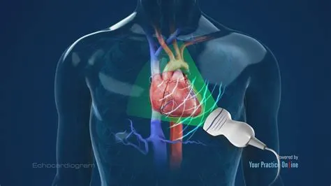
An echocardiography, often known as an ultrasound examination of the heart, is performed to look at the composition and functionality of the organ. An echo can be used to diagnose a number of conditions, including cardiomyopathy and valve failure. An echocardiogram (echo) is a visual representation of your heartbeat. You will undergo an echo test so that the physician can use ultrasonography (high-frequency sound waves) from a hand-held wand implanted on your chest to capture images of the chambers and valves of your heart. This helps the doctor determine how efficiently your heart is pumping. Medical experts routinely combine echo, Doppler ultrasonography, and colour Doppler treatments to evaluate blood flow across your heart's valves. Echocardiography uses no radiation. This sets an echo apart from other tests, such X-rays and CT scans, which use almost any radiation.
How come I could require an echocardiogram?
An echocardiography may be performed for a more thorough assessment of indications or symptoms that could indicate:
Atherosclerosis: a condition when fatty molecules and other substances in the bloodstream gradually block the arteries. It may result in issues with your heart's pumping or wall movements.
Cardiomyopathy: a cardiac enlargement brought on by thick or frail heart muscle congenital cardiomyopathy defects that appear in one or more cardiac components while the foetus is still developing, like a ventricular septal defect (hole in the wall between the 2 lower chambers of the heart.
a heart attack: a condition when blood cannot be pumped effectively because the heart muscle has grown weak or tight due to cardiac relaxation. Edema (swelling) in the feet, ankles, and other areas of the body may result from this, as well as fluid accumulation (congestion) in the blood vessels and lungs.
Aneurysm: an enlargement and weakening of the aorta or a portion of the heart muscle (the large artery that carries oxygenated blood out of the heart to the rest of the body). There's a chance the aneurysm will burst.
heart valve illness failure of one or more heart valves, which could result in irregular blood flow within the heart. Blood cannot flow through the heart or out to the lungs and body when the valves are constricted. Blood might seep backwards through the defective valves as well. The tissue of the heart valves can be examined for infection using an echocardiography.
heart tumour: a cardiac tumour that can develop on the heart's exterior, inside one or more of its chambers, or within the muscular tissue (myocardium) of the heart.
Pericarditis: the sac that surrounds the heart being inflamed or infected.
Pericardial tamponade or effusion. Blood, fluid, or infection can fill the sac that surrounds the heart. The heart muscle may get compressed as a result, failing to beat and pump blood normally. This may result in symptoms such as feeling faint or dizzy or a potentially dangerous drop in blood pressure.
flaws in the septum or atrial wall. Uneven pathways between the right and left sides of the heart might exist from birth, result from trauma, or develop during a heart attack. These flaws might be found in the lower pumping chambers or the top filling chambers (atria) (ventricles). This could result in poor blood flow, cardiac failure, or an elevated risk of stroke.
Shunts: Shunts are a common finding in atrial and ventricular septal abnormalities, as well as when the liver and lungs drive erratic blood flow through the circulatory system.
MEDICATIONS
Medications to treat a heart attack might include:
Aspirin reduces blood clotting. It helps keep blood moving through a narrowed artery. If you called 911 or your local emergency number, you may be told to chew aspirin. Emergency medical providers may give you aspirin immediately.
Clot busters (thrombolytics or fibrinolytics).These drugs help break up any blood clots that are blocking blood flow to the heart. The earlier a thrombolytic drug is given after a heart attack, the less the heart is damaged and the greater the chance of survival.
Other blood-thinning medications.A medicine called heparin may be given by IV or injection. Heparin makes the blood less sticky and less likely to form clots.
This medication widens the blood vessels. It helps improve blood flow to the heart. Nitroglycerin is used to treat sudden chest pain (angina). It's given as a pill under the tongue, as a pill to swallow or as an injection.
This medicine is given to relieve chest pain that doesn't go away with nitroglycerin.
Beta blockers.These medications slow the heartbeat and decrease blood pressure. Beta blockers can limit the amount of heart muscle damage and prevent future heart attacks. They are given to most people who are having a heart attack.
ACEThese drugs lower blood pressure and reduce stress on the heart.
These drugs help lower unhealthy cholesterol levels. Too much bad (low-density lipoprotein, or LDL) cholesterol can clog arteries.
Surgical and other procedures
If you've had a heart attack, a surgery or procedure may be done to open a blocked artery. Surgeries and procedures to treat a heart attack include:
Coronary angioplasty and stenting.This procedure is done to open clogged heart arteries. It may also be called percutaneous coronary intervention (PCI). If you've had a heart attack, this procedure is often done during a procedure to find blockages (cardiac catheterization).
During angioplasty, a heart doctor (cardiologist) guides a thin, flexible tube (catheter) to the narrowed part of the heart artery. A tiny balloon is inflated to help widen the blocked artery and improve blood flow.
A small wire mesh tube (stent) may be placed in the artery during angioplasty. The stent helps keep the artery open. It lowers the risk of the artery narrowing again. Some stents are coated with a medication that helps keep the arteries open.
Coronary artery bypass surgery (CABG). This is open-heart surgery. A surgeon takes a healthy blood vessel from another part of the body to create a new path for blood in the heart. The blood then goes around the blocked or narrowed coronary artery. CABGmay be done as an emergency surgery at the time of a heart attack. Sometimes it's done a few days later, after the heart has recovered a bit.
Cardiac rehabilitation
Cardiac rehabilitation is a personalized exercise and education program that teaches ways to improve heart health after heart surgery. It focuses on exercise, a heart-healthy diet, stress management and a gradual return to usual activities. Most hospitals offer cardiac rehabilitation starting in the hospital. The program typically continues for a few weeks or months after you return home.
People who attend cardiac rehab after a heart attack generally live longer and are less likely to have another heart attack or complications from the heart attack. If cardiac rehab is not recommended during your hospital stay, ask your provider about it.
Surgical solution in detail
Heart Procedures and Surgeries
If you've had a heart attack, you may have already had certain procedures to help you survive your heart attack and diagnose your condition. For example, many heart attack patients have undergone thrombolysis, a procedure that involves injecting a clot-dissolving agent to restore blood flow in a coronary artery. This procedure is administered within a few (usually three) hours of a heart attack. If this treatment isn't done immediately after a heart attack, many patients will need to undergo coronary angioplasty or coronary artery bypass graft surgery (CABG) later to improve blood supply to the heart muscle.
Heart Procedures and Surgeries
Angioplasty
Also known as Percutaneous Coronary Interventions [PCI], Balloon Angioplasty and Coronary Artery Balloon Dilation.
What the Procedure Does
Special tubing with an attached deflated balloon is threaded up to the coronary arteries. The balloon is inflated to widen blocked areas where blood flow to the heart muscle has been reduced or cutoff. Often combined with implantation of a stent (see below) to help prop the artery open and decrease the chance of another blockage. Considered less invasive because the body is not cut open. Lasts from 30 minutes to several hours. May require an overnight hospital stay.
Reason for the Procedure
Greatly increases blood flow through the blocked artery.
Decreases chest pain (angina).
Increases ability for physical activity that has been limited by angina or ischemia.
Can also be used to open neck and brain arteries to help prevent stroke.
Medications That Your Doctor May Prescribe Post-Procedure
Learn more about cardiac medications, including dual antiplatelet therapy, that you may need to take after your procedure to prevent complications and to put you on the path for the best recovery.
Angioplasty, Laser
What the Procedure Does
Similar to angioplasty except that the catheter has a laser tip that opens the blocked artery. Pulsating beams of light vaporize the plaque buildup.
Reason for the Procedure
Increases blood flow through blocked arteries.
Artificial Heart Valve Surgery
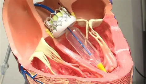
(Also known as Heart Valve Replacement Surgery)
What the Procedure Does
Replaces an abnormal or diseased heart valve with a healthy one.
Reason for the Procedure
Restores function of the heart valves.
Atherectomy
What the Procedure Does
Similar to angioplasty except that the catheter has a rotating shaver on its tip to cut away plaque from the artery.
Reason for the Procedure
Increases blood flow through the blocked artery by removing plaque buildup.
May also be used in carotid arteries (major arteries of the neck leading to the brain) to remove plaque and reduce risk for stroke.
Bypass Surgery
(Also known as CABG, pronounced "cabbage," Coronary Artery Bypass Graft done via Open-Heart Surgery)
What the Procedure Does
Treats blocked heart arteries by taking arteries or veins from other parts of your body — called grafts — and using them to reroute the blood around the clogged artery to supply blood flow to your heart muscle. A patient may undergo one, two, three or more bypass grafts, depending on how many coronary arteries are narrowed. Requires several days in the hospital.
Reason for the Procedure
One of the most common and effective procedures to manage blockage of blood to the heart muscle.
Improves the supply of blood and oxygen to the heart.
Relieves (angina).
Reduces risk of heart attack.
Improves ability for physical activity that has been limited by angina or ischemia.
Medications That Your Doctor May Prescribe Post-Procedure
Cardiomyoplasty
What the Procedure Does
An experimental procedure in which skeletal muscles are taken from a patient's back or abdomen. Then they're wrapped around an ailing heart. This added muscle, aided by ongoing stimulation from a device similar to a pacemaker, may boost the heart's pumping motion.
Reason for the Procedure
Increases the pumping motion of the heart.
Heart Transplant
What the Procedure Does
Removes a diseased heart and replaces it with a healthy human heart when a heart is irreversibly damaged. Uses hearts from organ donation.
Reason for the Procedure
Recognized as a proven procedure to restore heart health in appropriately selected patients.
Minimally Invasive Heart Surgery
(Also known as Limited Access Coronary Artery Surgery and includes Port-Access Coronary Artery Bypass (PACAB or PortCAB) and Minimally Invasive Coronary Artery Bypass Graft (MIDCAB or minimally invasive CABG)
What the Procedure Does
An alternative to standard bypass surgery (CABG). Small incisions ("ports") are made in the chest. Chest arteries or veins from your leg are attached to the heart to "bypass" the clogged coronary artery or arteries. The instruments are passed through the ports to perform the bypasses. The surgeon views these operations on video monitors rather than directly. In PACAB, the heart is stopped and blood is pumped through an oxygenator or "heart-lung" machine. MIDCAB is used to avoid the heart-lung machine. It's done while the heart is still beating. Requires several days in the hospital.
Reason for the Procedure
Manages blockage of blood flow to the heart and improves the supply of blood and oxygen to the heart.
Relieves
Reduces risk of heart attack.
Improves ability for physical activity.
Radiofrequency Ablation
(Also known as Catheter Ablation)
What the Procedure Does
A catheter with an electrode at its tip is guided through the veins to the heart muscle with real-time, moving X-rays (fluoroscopy) displayed on a video screen. The catheter is placed at the exact site inside the heart where cells give off the electrical signals that stimulate the abnormal heart rhythm. Then a mild, painless radiofrequency energy (similar to microwave heat) is transmitted to the pathway. This destroys carefully selected heart muscle cells in a very small area (about 1/5 of an inch).
Reason for the Procedure
Preferred treatment for many types of rapid heartbeats (arrhythmias) especially supraventricular tachyarrhythmias.
What precautions should be taken after heart attack?
follow the tips below for a healthy lifestyle.
-Stop smoking Tobacco use is a major risk factor for heart disease.
-Control your blood pressure.
-Control your cholesterol levels.
-Check for diabetes.
-Regular cardio exercise can make your heart stronger.
-Eat a heart-healthy diet.
-Control your stress level
Surgical solution in detail
Despite the advantages, surgery can trigger cardiac events including heart attacks, heart failure, heart rhythm disturbances, and death. Previous research has shown that nearly three-quarters of patients who die after surgery were never admitted to critical care,3 suggesting that their risk was unrecognised.
Each year, cardiac complications occur within 30 days after major noncardiac surgery in more than 10 million people worldwide; postoperative mortality is 1.5%. Enhanced patient monitoring and measurement of natriuretic hormone and troponin levels may improve outcomes.
INTRODUCTION
Cardiac complications occurring in the postanesthetic care unit (PACU) are typically due to hypotension, hypertension, and dysrhythmias. Patients with known coronary artery disease or congestive heart failure are more prone to these complications after surgical procedure.
HYPOTENSION
Decreased intravascular volume, or hypovolemia is due to inadequate intravenous fluid administration or blood loss. Patients can be resuscitated with crystalloids, colloids, and various blood products. If fluid resuscitation is inadequate to perfuse end organs, then vasopressors and inotropes should be added.
Myocardial ischemia with acute heart failure and ventricular or valvular dysfunction can also lead to hypotension. This may be associated with tachycardia and ST segment changes on electrocardiogram. A history of coronary artery disease predisposes patients to these complications and should be noted on preoperative evaluation. Drug-eluting stents typically require antiplatelet therapy for surgical procedures; if antiplatelet therapy is halted, patients may be at increased risk for acute coronary events. Suspected coronary thrombosis requires immediate evaluation for cardiac catheterization.
Decreased systemic vascular resistance in the PACU setting is usually iatrogenic and leads to hypotension. Disease states that cause decreased SVR include sepsis, spinal shock from spinal cord injury, and histamine release during anaphylactic reactions. While supportive measures are instituted, the underlying cause should be identified and treated. Residual effects of anesthetics, including inhalational, intravenous, and neuraxial agents, also produce hypotension. Treatment is indicated if mean arterial pressure is 20% less than baseline.
HYPERTENSION
Pain is a common cause of hypertension in the PACU. Surgical trauma and pain cause increased sympathetic tone leading to hypertension and tachycardia. Multimodal pain management strategies are preferable.
Hypercarbia from respiratory failure also leads to hypertension. Treatment includes promoting effective gas exchange via invasive or noninvasive, positive pressure ventilation.
Urinary retention and bladder distention are a common cause of hypertension in the PACU. It is more common after inguinal hernia repair, neuraxial anesthesia, and in elderly men with prostatic obstruction. Patients may require bladder catheterization. Patients who remain intubated in the PACU, if not adequately sedated, may become hypertensive from irritation of the endotracheal tube.
ARRHYTHMIAS
Arrhythmias occur often in the PACU and some can be life threatening. If cardiac arrest should occur, PACU treatment may have to be tailored to accommodate surgical incisions. Thorough review of current Advance Cardiac Life Support (ACLS) algorithms should be reviewed.
Bradycardia in the PACU can be due to vasovagal reflexes, residual effects of anticholinesterases, β-blockers, or opioids. Bradycardia may also result from severe myocardial infarction with complete heart block. The ACLS algorithm should be consulted for unstable bradycardia. Anticholinergic medications and pacing options must be readily available.
Sinus tachycardia can be due to pain, hypovolemia, fever, sepsis, or certain drugs such as albuterol or anticholinergics.
Atrial.
BYPASS SURGERY OVERVIEW
Coronary artery bypass graft surgery, also known as CABG or bypass surgery, can help to restore blood flow to an area of the heart. However, surgery does not stop the progression of atherosclerosis (coronary heart disease), which deposits fatty material into artery walls, narrowing them and eventually limiting blood flow at other sites in the bypassed arteries or in previously normal coronary arteries.
Patients and healthcare providers must work together after surgery to treat the underlying atherosclerosis and the factors that can cause progression of heart disease.
This topic review discusses treatments that are recommended after coronary artery bypass graft surgery. These treatments can help to:
Reduce the risk of developing complications of coronary heart disease, including having a subsequent heart attack or dying.
Help a person to feel better and have more energy.
An overview of coronary artery bypass graft surgery is discussed in detail separately.
CARE AT HOME AFTER BYPASS SURGERY
Care after bypass surgery aims to reduce the risk factors for heart disease and includes strategies to help patients and family members stop smoking, control high blood pressure, improve cholesterol levels, begin exercising regularly, reduce weight if necessary, and reduce stress. Some of these changes can be made by adjusting lifestyle habits through diet and exercise. However, lifestyle changes alone may not be adequate and medications are often needed.
Discharge from the hospital — Patients with an uncomplicated CABG usually go home after about five days in the hospital. In some cases, the hospital stay is longer. If complications have occurred, discharge is delayed until the person's condition is stable.
Before leaving the hospital, it is important for the patient and family to participate in and understand the discharge plan. Make sure all questions are answered and obtain written directions for how to take all medications (new and old). After bypass surgery, it is common to start new medications and stop or adjust the doses of previous medications.
Medications — Most people who have had bypass surgery are sent home with prescriptions for several medications, most of which are taken every day. Some of these drugs improve survival, decrease the risk of complications, and help to prevent or treat recurrent chest pain.
Antiplatelet therapy– Aspirin is an antiplatelet medication that is given to help prevent the formation of blood clots that can block either your coronary arteries or coronary bypass graft. It is usually recommended indefinitely.
Beta blockers– Beta blockers slow the heart rate, lower blood pressure, and decrease the heart's demand for oxygen. They are given to some patients with high blood pressure, heart failure, some rhythm changes or a heart attack, and to some patients in whom bypass surgery is not expected to relieve all symptoms of angina. If a person cannot tolerate a beta blocker, a calcium channel blocker may be substituted.
Nitrates– A nitrate, either as short-acting nitro glycerin, or as a long-acting preparation (isosorbide mononitrate or dinitrate). These drugs dilate coronary blood vessels, bringing more blood to the heart muscle. Nitrates also reduce the amount of blood returning to the heart, which decreases the heart's demand for oxygen. Nitrates are often given to treat or prevent further episodes of chest pain. Nitrates may be given to patients after bypass surgery if some of the coronary blood vessels could not be bypassed.
ACE inhibitor– Angiotensin converting enzyme (ACE) inhibitors are often used to treat high blood pressure.
Examples of ACE inhibitors include captopril (brand name: Capoten), enalapril (brand name: Vasotec), lisinopril (sample brand names: Zestril or Prinivil), and ramipril (brand name: Altace).
Some patients who cannot tolerate an ACE inhibitor (often because of a chronic cough) may be prescribed an angiotensin II receptor blocker (ARB). These related drugs are satisfactory replacements.
Examples of ARBs include losartan (brand name: Cozaar), valsartan (brand name: Diovan), and irbesartan (brand name: Avapro).
Lipid-lowering therapy– Almost all patients are given a medication to lower lipids after CABG. Cholesterol lowering can be beneficial both before and after CABG because it can reduce the progression of atherosclerosis in both native and graft vessels.
Other medications– Other medications may be given on a short-term basis to prevent the development of an irregular heart, to manage discomfort associated with healing incisions, or to allow for regular bowel movements.
Lipid therapies are recommended even for patients who have values that are in the "normal" range. The goal level for "bad" cholesterol (called LDL or low density lipoprotein) is less than 70 mg/dL (1.8 mmol/L).
Statins are the most common medications used to lower cholesterol levels. Other drugs may be used as well
Wound care — After discharge from the hospital, the patient is usually given instructions about how to care for their chest and/or leg wounds. It is important to follow these instructions closely and to notify a healthcare provider immediately if there are questions or concerns.
Avoid heavy lifting and extremes of shoulder movement (eg, as in tennis, baseball, and golf) for six to eight weeks after surgery to allow for complete healing of the breast bone (sternum)
When to seek help — If the patient develops any of the following signs or symptoms of wound infection, a healthcare provider should be contacted immediately. Most wound infections develop within 14 days of the surgery.
Fever greater than 100.4º F (38º C)
New or worsened pain in the chest or around the incision
A rapid heart rate
Reddened skin, bleeding or pus-like drainage from the incision