Project 1
-
Part A - Detailed study of anatomical basis of surgical procedures or Clinical management in association with any of the organ system
-Kidney stone
-Chronic Obstructive Pulmonary Disease
-Hernia
1.Kidney stone
Kidney stones (also called renal calculi, nephrolithiasis or urolithiasis) are hard deposits made of minerals and salts that form inside kidneys.
urinary system:
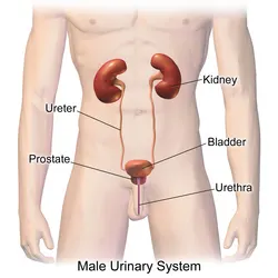
The structures that create and carry urine to the point of excretion are referred to as the urinary system. On both the left and right sides of the human urinary system, two kidneys are positioned between the dorsal body wall and the parietal peritoneum. The kidneys get a large amount of blood via the renal arteries, which exit the kidneys via the renal vein.Each kidney is made up of functioning components known as nephrons. Wastes (in the form of urine) depart the kidney via the ureters, which are tubes comprised of smooth muscle fibres that transport urine towards the urinary bladder, where it is stored and then released from the body by urination (voiding). The female and male urinary systems are essentially similar, with the urethra being the only difference. Urine is produced in the kidneys by blood filtration. The urine is subsequently transported through the ureters and stored in the bladder. Urine is transferred from the bladder to the exterior of the body via the urethra during urination. 800–2,000 milliliters (mL) of urine are normally produced every day in a healthy human. This amount varies according to fluid intake and kidney function.
Normal function of the organ
1. Kidney
This pair of organs, placed under the rib cage, works as a filtration system to remove waste from the blood and form urine. The filtration units called nephrons contain a renal tube and a ball wrapped in small blood capillaries. It also helps maintain the required levels of salts and important nutrients in the blood, and secretes hormones that help generate new red blood cells.
2. Ureter
The ureters are the thin passageways through which urine travels from kidneys to the bladder. The ureter walls are made of muscles that constantly contract and expand to push down the urine formed in the kidneys. A few millilitres of urine are passed through the ureters every 15 seconds. Its blockage can result in a urinary infection, affecting the function of urinary system.
3. Bladder
This works as a reservoir to store urine. It is held in the lower abdomen, by ligaments that connect it to organs and bones in the vicinity. The bladder generally has the ability to hold around 2 cups of urine for a time period ranging from 2 to 5 hours. When a person urinates, the muscular walls of this organ contract to push the urine into the urethra.
4. Urethra
This tube starts from the bladder and allows urine to escape the body. When the bladder’s walls contract, the sphincter muscles relax and urine is pushed through the urethra. Normal function of urinary system needs the brain to send signals to both the bladder muscles and the sphincter muscles in the right order. In males, urine is released through the penis, and in female placental mammals through the vulva.
5. Prostrate
The prostrate is only present in males. It is placed in front of the rectum, between the penis and the bladder. The tube connecting the bladder to the penis runs right through it. The secretions from this gland provide nourishment for sperm. When a male ejaculates, the fluid is secreted into the urethra and combines with sperm to form semen.
6. Others
- Nerves running through the bladder, which signal the brain when the bladder is full.
- Two sphincter muscles surround the exit of the bladder like a rubber band and prevent the leakage of urine from it.
Kidney Stones Form from Substances in Urine
The kidneys produce urine to eliminate waste. Kidney stones can form when mineral and acid salts in the urine crystallize and stick together. If the stone is small, it can pass easily through the urinary system and out of the body. A larger stone can get stuck in the urinary tract, however. A stuck kidney stone causes pain and can block the flow of urine.
Kidney Stones
Kidney stones form within the kidney when the urine becomes concentrated. This causes the debris to crystallize and form hard deposits.
Diet, excess body weight, some medical conditions, and certain supplements and medications are among the many causes of kidney stones. Kidney stones can affect any part of your urinary tract — from your kidneys to your bladder. Often, stones form when the urine becomes concentrated, allowing minerals to crystallize and stick together.
Passing kidney stones can be quite painful, but the stones usually cause no permanent damage if they're recognized in a timely fashion. Depending on your situation, you may need nothing more than to take pain medication and drink lots of water to pass a kidney stone. In other instances — for example, if stones become lodged in the urinary tract, are associated with a urinary infection or cause complications — surgery may be needed.
Your doctor may recommend preventive treatment to reduce your risk of recurrent kidney stones if you're at increased risk of developing them again.
Symptoms
A kidney stone usually will not cause symptoms until it moves around within the kidney or passes into one of the ureters. The ureters are the tubes that connect the kidneys and bladder.If a kidney stone becomes lodged in the ureters, it may block the flow of urine and cause the kidney to swell and the ureter to spasm, which can be very painful. At that point, you may experience these symptoms:
- Severe, sharp pain in the side and back, below the ribs
- Pain that radiates to the lower abdomen and groin
- Pain that comes in waves and fluctuates in intensity
- Pain or burning sensation while urinating
Other signs and symptoms may include:
- Pink, red or brown urine
- Cloudy or foul-smelling urine
- A persistent need to urinate, urinating more often than usual or urinating in small amounts
- Nausea and vomiting
- Fever and chills if an infection is present
Pain caused by a kidney stone may change — for instance, shifting to a different location or increasing in intensity — as the stone moves through your urinary tract.
When to see a doctor
Make an appointment with your doctor if you have any signs and symptoms that worry you.
Seek immediate medical attention if you experience:
- Pain so severe that you can't sit still or find a comfortable position
- Pain accompanied by nausea and vomiting
- Pain accompanied by fever and chills
- Blood in your urine
- Difficulty passing urin
Kidney stone removal
A urologist can remove the kidney stone or break it into small pieces with the following treatments:
Shock wave lithotripsy
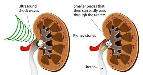
The doctor can use shock wave lithotripsy NIH external link to blast the kidney stone into small pieces. The smaller pieces of the kidney stone then pass through your urinary tract. A doctor can give you anesthesia NIH external link during this outpatient procedure.
Cystoscopy and ureteroscopy
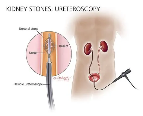
During cystoscopy, the doctor uses a cystoscope to look inside the urethra and bladder to find a stone in your urethra or bladder. During ureteroscopy, the doctor uses a ureteroscope, which is longer and thinner than a cystoscope, to see detailed images of the lining of the ureters and kidneys. The doctor inserts the cystoscope or ureteroscope through the urethra to see the rest of the urinary tract. Once the stone is found, the doctor can remove it or break it into smaller pieces. The doctor performs these procedures in the hospital with anesthesia. You can typically go home the same day.
Percutaneous nephrolithotomy
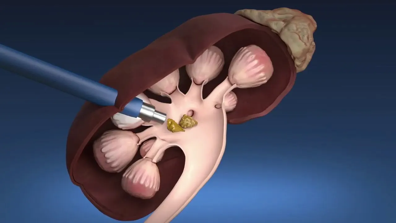
The doctor uses a thin viewing tool, called a nephroscope, to locate and remove the kidney stone. The doctor inserts the tool directly into your kidney through a small cut made in your back. For larger kidney stones, the doctor also may use a laser to break the kidney stones into smaller pieces. The doctor performs percutaneous nephrolithotomy NIH external link in a hospital with anesthesia. You may have to stay in the hospital for several days after the procedure.
After these procedures, sometimes the urologist may leave a thin flexible tube, called a ureteral stent, in your urinary tract to help urine flow or a stone to pass. Once the kidney stone is removed, your doctor sends the kidney stone or its pieces to a lab to find out what type it is.
The health care professional also may ask you to collect your urine for 24 hours after the kidney stone has passed or been removed. The health care professional can then measure how much urine you produce in a day, along with mineral levels in your urine. You are more likely to form stones if you don’t make enough urine each day or have a problem with high mineral levels.
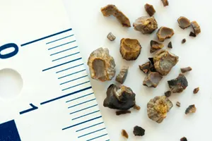
Health care professionals usually treat kidney stones based on their size and what they are made of. How can I prevent kidney stones?
To help prevent future kidney stones, you also need to know what caused your previous kidney stones. Once you know what type of kidney stone you had, a health care professional can help you make changes to your eating, diet, and nutrition to prevent future kidney stones.
Drinking liquids
In most cases, drinking enough liquids each day is the best way to help prevent most types of kidney stones. Drinking enough liquids keeps your urine diluted and helps flush away minerals that might form stones.
Though water is best, other liquids such as citrus drinks may also help prevent kidney stones. Some studies show that citrus drinks, such as lemonade and orange juice, protect against kidney stones because they contain citrate, which stops crystals from turning into stones.
Unless you have kidney failure, you should drink six to eight, 8-ounce glasses a day. If you previously had cystine stones, you may need to drink even more. Talk with a health care professional if you can’t drink the recommended amount due to other health problems, such as urinary incontinence, urinary frequency, or kidney failure.
The amount of liquid you need to drink depends on the weather and your activity level. If you live, work, or exercise in hot weather, you may need more liquid to replace the fluid you lose through sweat. A health care professional may ask you to collect your urine for 24 hours to determine the amount of urine you produce a day. If the amount of urine is too low, the health care professional may advise you to increase your liquid intake.
After surgery care
Postoperative infections are fatal infections that are completely preventable. Infection after surgery is a common problem in health care. The process of infection at the surgical site is complex and involves the interaction of several biological pathways at the molecular level. These infections cause pain, delayed recovery from surgery, long hospital stays, increased care costs, the need for long-term use of antibiotics, failure of surgery, and in acute cases, organ failure and even death.
Minimizing the number of infections depends on the patient's condition, the use of appropriate disinfectant solutions at the incision site, the sterile surgical environment, the use of sterilized instruments during surgery, and the appropriate and disinfected coverage of operating room staff. after surgery, what their symptoms are and how they are treated in this article.
Women have a 20% risk of developing a urinary tract infection (UTI) following urogynecologic surgery. This study assessed the association of post-operative UTI with bacteria in pre-operative samples of catheterized urine.
Immediately before surgery, vaginal swabs, perineal swabs and catheterized urine samples were collected and the V4 region of the 16S rRNA gene was sequenced. The cohort was dichotomized in two ways: 1) standard day-of-surgery urine culture result (positive/negative) and 2) occurrence of post-operative UTI (positive/negative). Characteristics of the bladder, vaginal and perineal microbiomes were assessed to identify factors associated with post-operative UTI.
87% of the 104 POP/UI surgical patients were white; the mean age was 57 years. The most common genus was Lactobacillus with a mean relative abundance of 39.91% in catheterized urine, 53.88% in vaginal swabs, and 30.28% in perineal swabs. Two distinct clusters, based on dispersion of catheterized urine (i.e., bladder) microbiomes, had highly significant (p<2.2e-16) differences in age, microbes and post-operative UTI risk. Post-operative UTI was most associated with the bladder microbiome; microbes in adjacent pelvic floor niches also contributed to UTI risk. UTI risk was associated with depletion of Lactobacillus iners and enrichment of a diverse mixture of uropathogens.
2.Chronic Obstructive Pulmonary Disease-
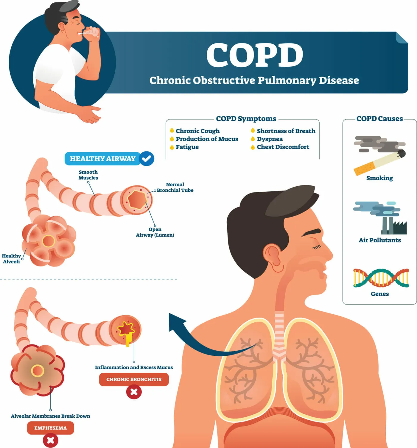
Chronic Obstructive Pulmonary Disease, or COPD. Airways get clogged as a result of lung inflammation, which is its defining feature.
COPD is a catch-all term for a number of lung disorders that worsen with time. Emphysema and chronic bronchitis can potentially lead to COPD. A COPD diagnosis indicates that you may have signs of both of these lung-damaging conditions or just one of them. As COPD worsens over time, breathing becomes more difficult.
If you’re having any of the signs or symptoms of COPD, don't wait for your next appointment to call your provider. Report these symptoms promptly, even if you don't feel sick. Don't wait for symptoms to become so severe that you need to seek emergency care. If you notice your symptoms early, your provider might change your treatment or medications to relieve your symptoms.
COPD results in persistent and progressive respiratory symptoms, such as coughing up phlegm and having trouble breathing.
Vital points
- 3.23 million people died from chronic obstructive pulmonary disease (COPD), the third most common cause of mortality in the world in 2019.
- In low- and middle-income nations, COPD fatalities in people under 70 years of age account for about 90% of all mortality (LMIC).
- To stop the progression of symptoms and lessen flare-ups, early diagnosis and treatment are necessary, including help for quitting smoking.
- Important COPD risk factors include occupational exposure to dust, gases, and chemicals, indoor air pollution, and tobacco smoke exposure.
- Long-term exposure to toxic chemicals and particles mixed with individual factors, such as childhood lung development-influencing events and genetics, cause COPD.
Causes:
- Prolonged exposure to particulate matter
- Smoke from cigarettes
- persistent bronchitis
- Emphysema
Symptoms:
- Having trouble breathing
- Cough
- mucus formation
- Wheezing
- Not enough energy
- Shortness of breath, especially during physical activities
- Wheezing
- chest constriction
- a persistent cough that may discharge clear, white, yellow, or greenish mucus (sputum)
- Frequent respiratory infections
- Unwanted loss of weight (in later stages)
- ankles, feet, or legs swelling
Therapy:
- Oxygen administration
- lung surgery
- Transplanting lungs
- Lifestyle adjustments, such as giving up smoking
- Bronchodilators to be inhaled
- Corticosteroids
Prevention:
- One should refrain from smoking.
- You should stay away from polluted locations.
- Frequent exercise
- Consume healthily
- Vaccine
How your lungs are affected?
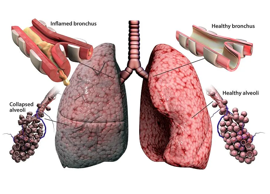
Two substantial tubes carry air into your lungs from your windpipe (trachea) (bronchi). Like the branches of a tree, these tubes within your lungs repeatedly split into smaller tubes called bronchioles, which then terminate in collections of tiny air sacs (alveoli).
The walls of the air sacs are extremely thin and filled with microscopic blood veins (capillaries). These blood arteries allow the oxygen in the air you breathe to enter your bloodstream. Carbon dioxide, a gas that is a waste product of metabolism, is also exhaled at the same time.
To force air out of your body, your lungs rely on the bronchial tubes' and air sacs' inherent suppleness.
Describe the common disorders in association with the organ
- Pleural Effusion
- Emphysema
- Lung Cancer
- Pulmonary Embolism
- Pulmonary Edema
- Chronic Obstructive Pulmonary Disease
- Bronchitis
- Pneumothorax
- Pneumonia
- Asthma
- COVID-19
Enumerate the various diagnostic techniques and investigations used in diagnosis of the disorder
- Spirometry
- Bronchodilator reversibility test
- Blood tests
- Genetic testing
- Chest X-ray or CT scan
- Sputum examination
- Electrocardiogram (ECG or EKG)
Spirometry:
Spirometry is the most used and reliable approach for identifying COPD. The pulmonary function test, or PFT, is another name for it. This quick, painless test evaluates lung capacity and function.
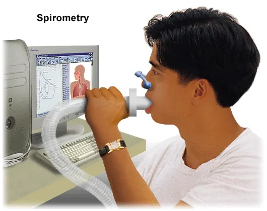
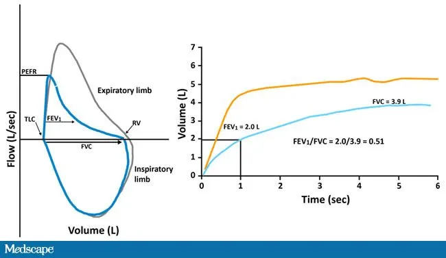
You will exhale as forcefully as you can into a tube that is attached to the spirometer, a little machine, to conduct this test. The forced vital capacity refers to the total amount of air that leaves your lungs each breath
The FEV1 is the proportion of the FVC that is driven out in the first second. Forced expiratory volume is referred to as FEV. Peak expiratory flow rate refers to the fastest rate at which you can empty your lungs (PEFR).
The results of a spirometry test assist in identifying the type and severity of your lung disease. The outcomes are immediately interpretable.
The most accurate test is one that can identify COPD even before serious symptoms manifest. It can also help your doctor track the progression of COPD and monitor the effectiveness of treatment.
Precautions
- Spirometry is not advised for someone who has recently undergone heart surgery or had a heart attack because it needs you to exhale strongly.
- Prior to testing, it's crucial to be fully healed from any severe illnesses or ailments. You might have a little breathlessness and lightheadedness just after the test, even if you generally have good health.
Risk factors for COPD include:
- Exposure to tobacco smoke.The most significant risk factor for COPD is long-term cigarette smoking. The more years you smoke and the more packs you smoke, the greater your risk. Pipe smokers, cigar smokers and marijuana smokers also may be at risk, as well as people exposed to large amounts of secondhand smoke.
- People with asthma.Asthma, a chronic inflammatory airway disease, may be a risk factor for developing COPD. The combination of asthma and smoking increases the risk of COPD even more.
- Occupational exposure to dusts and chemicals.Long-term exposure to chemical fumes, vapors and dusts in the workplace can irritate and inflame your lungs.
- Exposure to fumes from burning fuel.In the developing world, people exposed to fumes from burning fuel for cooking and heating in poorly ventilated homes are at higher risk of developing COPD.
- The uncommon genetic disorder alpha-1-antitrypsin deficiency is the cause of some cases of COPD. Other genetic factors likely make certain smokers more susceptible to the disease.
Prevention/ Management
- Unlike some diseases, COPD typically has a clear cause and a clear path of prevention, and there are ways to slow the progression of the disease. The majority of cases are directly related to cigarette smoking, and the best way to prevent COPD is to never smoke — or to stop smoking now.
- If you're a longtime smoker, these simple statements may not seem so simple, especially if you've tried quitting — once, twice or many times before. But keep trying to quit. It's critical to find a tobacco cessation program that can help you quit for good. It's your best chance for reducing damage to your lungs.
- Occupational exposure to chemical fumes and dusts is another risk factor for COPD. If you work with these types of lung irritants, talk to your supervisor about the best ways to protect yourself, such as using respiratory protective equipment.
- Here are some steps you can take to help prevent complications associated with COPD:
- Quit smoking to help reduce your risk of heart disease and lung cancer.
- Get an annual flu vaccination and regular vaccination against pneumococcal pneumonia to reduce your risk of or prevent some infections.
- Talk to your doctor if you feel sad or helpless or think that you may be experiencing depression.
- Surgical procedures for the treatment of COPD
- Only very advanced cases of COPD are thought to demand surgical interventions. The volume of the lungs can be decreased via a variety of methods to facilitate breathing. Lung transplantation may be an option in really severe COPD cases.
Lung volume reduction
The hyperinflated areas of the lungs are removed or have their size decreased in this sort of treatment. As a result, the initial volume of the lungs is smaller overall. However, the healthy parts of the lungs have more room, which makes it simpler for them to oxygenate the blood. The purpose of this treatment is to improve overall breathing, lessen shortness of breath, and lessen stress on the breathing muscles.
There are two types of lung volume reduction treatment:
- Surgery: Either a major cut in the chest is made to expose the rib cage or multiple minor apertures are used (keyhole surgery). In order to improve breathing, the surgeon eliminates inflated lung tissue.
- Surgery: Either a major cut in the chest is made to expose the rib cage or multiple minor apertures are used (keyhole surgery). In order to improve breathing, the surgeon eliminates inflated lung tissue.
Treatment
COPD patients may benefit from medication that reduces lung volume, but there are also certain dangers involved. Every process has advantages and disadvantages. Nowadays, bronchoscopy is a more common operation than surgery, although less study has been done on them.
Surgery The likelihood of surviving longer may be increased by lung volume reduction surgery. After five years, the following results were observed in the largest trial on this therapy:
Additionally, those who underwent the procedure had improved quality of life, were physically fitter, and experienced fewer episodes of breathing problems. Additionally, they were less likely to require hospitalisation if their symptoms suddenly got severe (exacerbation).
However, there were drawbacks to the surgery as well: more patients in the surgery group passed away in the first year following the procedure (due to consequences of the operation). The surgery carries dangers like inflammations and issues with wound healing, much like any operations. These negative effects, however, weren't sufficiently examined in the studies.
Bronchoscopic procedures:
Bronchoscopic procedures using valves or coils can improve your physical fitness and quality of life. Coils were also found to reduce the likelihood of breathing difficulties. It’s not clear whether this treatment can help people to live longer. The studies so far have lasted one year at most. That's too short to be able to answer this question.
One disadvantage of this treatment is that people's breathing sometimes suddenly gets worse afterwards. This can be caused by the endoscope that is inserted into the bronchi (air tubes in the lungs). It can lead to a collapsed lung (pneumothorax) too. People often cough up a bit of blood after the procedure, and the treatment is associated with the usual surgery-related risks. If coils are used, there is a greater risk of pneumonia.
Less is known about the pros and cons of procedures that use steam, foam and stents because there’s a lack of research on these approaches.
Bullectomy
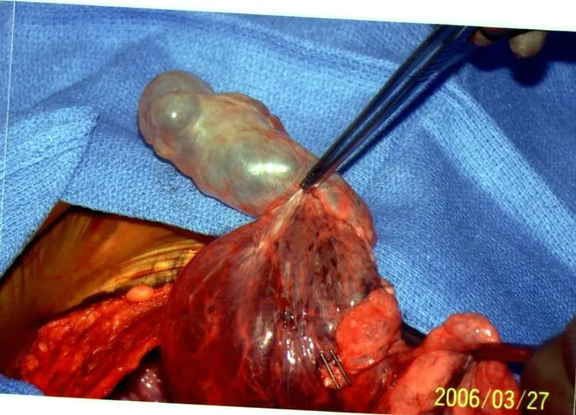 This treatment is similar to lung volume reduction. It specifically gets rid of large air-filled spaces (known as bullae). Bullectomy is considered as a treatment option if individual air pockets take up more than a third of a lung and put pressure on nearby tissue. Like other treatments mentioned above, bullectomy is a minimally invasive procedure. In other words, it is done through small cuts in the chest area (keyhole surgery).
Transplants
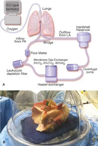 Lung transplants are only considered for people who have advanced pulmonary emphysema, and only if they have already tried out all other treatment options. Due to a lack of donor lungs, this treatment isn't always possible. People who receive new lungs can usually breathe a lot better and their quality of life improves.
Transplants are associated with special risks because they involve major surgery and the body’s immune system might attack the new organ afterwards. To lower the risk of the body rejecting the organ, people have to take medication to suppress their immune system for the rest of their lives. But this medication sometimes has serious side effects.
Lung transplants are only considered as a treatment option if someone is very likely to die of COPD in the next few years – and if they don’t have any other serious illnesses. They have to stop smoking quite a long time before having the transplant, and they have to be prepared to actively take part in rehabilitation activities and exercises. Because people over the age of 65 are more likely to have other serious illnesses, most patients who have a lung transplant are younger.
Hernia
The male reproductive system includes a group of organs that make up a man’s reproductive and urinary system. These organs do the following jobs within your body:
- They produce, maintain and transport sperm (the male reproductive cells) and semen (the protective fluid around sperm).
- They discharge sperm into the female reproductive tract.
- They produce and secrete male sex hormones.
The male reproductive system is made up of internal (inside your body) and external (outside your body) parts. Together, these organs help you urinate (rid your body of liquid waste materials), have sexual intercourse and make children.
FUNCTION
The entire male reproductive system is dependent on hormones. These are chemicals that stimulate or regulate the activity of your cells or organs. The primary hormones involved in the functioning of the male reproductive system are follicle-stimulating hormone (FSH), luteinizing hormone (LH) and testosterone.
FSH and LH are produced by the pituitary gland. It’s located at the base of your brain and it’s responsible for many functions in your body. FSH is necessary for sperm production (spermatogenesis). LH stimulates the production of testosterone, which is necessary to continue the process of spermatogenesis. Testosterone is also important in the development of male characteristics, including muscle mass and strength, fat distribution, bone mass and sex drive.
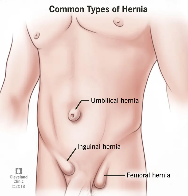
The most common forms of hernia are:
- Inguinal hernia: In men, the inguinal canal is a passageway for the spermatic cord and blood vessels leading to the testicles. In women, the inguinal canal contains the round ligament that gives support for the uterus. In an inguinal hernia, fatty tissue or a part of the intestine pokes into the groin at the top of the inner thigh. This is the most common type of hernia, and affects men more often than women.
- Femoral hernia: Fatty tissue or part of the intestine protrudes into the groin at the top of the inner thigh. Femoral hernias are much less common than inguinal hernias and mainly affect older women.
- Umbilical hernia: Fatty tissue or part of the intestine pushes through the abdomen near the navel (belly button).
- Hiatal (hiatus) hernia: Part of the stomach pushes up into the chest cavity through an opening in the diaphragm (the horizontal sheet of muscle that separates the chest from the abdomen).
Other types of hernias include:
- Incisional hernia: Tissue protrudes through the site of an abdominal scar from a remote abdominal or pelvic operation.
- Epigastric hernia: Fatty tissue protrudes through the abdominal area between the navel and lower part of the sternum (breastbone).
- Spigelian hernia: The intestine pushes through the abdomen at the side of the abdominal muscle, below the navel.
- Diaphragmatic hernia: Organs in the abdomen move into the chest through an opening in the diaphragm.
How common are hernias?
Of all hernias that occur:
- 75 to 80% are inguinal or femoral.
- 2% are incisional or ventral.
- 3 to 10% are umbilical, affecting 10 to 20% of newborns; most close by themselves by 5 years of age.
- 1 to 3% are other types.
What causes a hernia?
Inguinal and femoral hernias are due to weakened muscles that may have been present since birth, or are associated with aging and repeated strains on the abdominal and groin areas. Such strain may come from physical exertion, obesity, pregnancy, frequent coughing, or straining on the toilet due to constipation.
Adults may get an umbilical hernia by straining the abdominal area, being overweight, having a long-lasting heavy cough or after giving birth.
The cause of hiatal hernias is not fully understood, but a weakening of the diaphragm with age or pressure on the abdomen could play a part.
What are the symptoms of a hernia
A hernia in the abdomen or groin can produce a noticeable lump or bulge that can be pushed back in, or that can disappear when lying down. Laughing, crying, coughing, straining during a bowel movement, or physical activity may make the lump reappear after it has been pushed in. More symptoms of a hernia include:
- Swelling or bulge in the groin or scrotum (the pouch that contains the testicles).
- Increased pain at the site of the bulge.
- Pain while lifting.
- Increase in the bulge size over time.
- A dull aching sensation.
- A sense of feeling full or signs of bowel obstruction.
In the case of hiatal hernias there are no bulges on the outside of the body. Instead, symptoms may include heartburn, indigestion, difficulty swallowing, frequent regurgitation (bringing food back up) and chest
hernia diagnosed
It is usually possible to see or feel a bulge in the area where a hernia has occurred by physical exam. As part of a male’s typical physical exam for inguinal hernias, the doctor feels the area around the testicles and groin while the patient is asked to cough. In some cases, soft-tissue imaging like a CT scan will accurately diagnose the condition.
Management
When you have a hernia, treatment will start with your primary care provider. If you need surgery to repair the hernia, you’ll be referred to a general surgeon. In fact, ventral hernia repairs are one of the most common operations U.S. general surgeons perform.
If you think you have a hernia, don’t wait to seek help. A neglected hernia can grow larger and more painful — this can lead to complications and possibly emergency surgery. Early repair is more successful, less risky and offers a better recovery and outcome.
Hernia treatment
Hernias usually do not get better on their own, and surgery may be the only way to repair them. However, your doctor will recommend the best therapy to address your hernia, and may refer you to a surgeon. If the surgeon thinks it is necessary to repair your hernia, then the surgeon will tailor the method of repair that best meets your needs.
In the case of an umbilical hernia in a child, surgery may be recommended if the hernia is large or if it has not healed by the age of 4 to 5 years old. By this age, a child can usually avoid surgical complications.
If an adult has an umbilical hernia, surgery is usually recommended because the condition will not likely improve on its own and the risk of complications is higher.
One of three types of hernia surgery can be performed:
- Open surgery
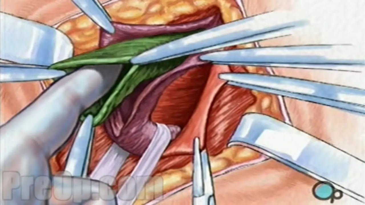 In which a cut is made into the body at the location of the hernia. The protruding tissue is set back in place and the weakened muscle wall is stitched back together. Sometimes a type of mesh is implanted in the area to provide extra support.
- Laparoscopic surgery
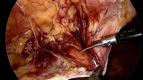
involves the same type of repairs. However, instead of a cut to the outside of the abdomen or groin, tiny incisions are made to allow for the insertion of surgical tools to complete the procedure.
- Robotic hernia repair
laparoscopic surgery, uses a laparoscope, and is performed with small incisions. With robotic surgery, the surgeon is seated at a console in the operating room, and handles the surgical instruments from the console. While robotic surgery can be used for some smaller hernias, or weak areas, it can now also be used to reconstruct the abdominal wall.
Each type of surgery has its advantages and disadvantages. The best approach will be decided by the patient’s surgeon.
What can happen if a hernia is not treated?
Other than umbilical hernias in babies, hernias will not disappear on their own. Over time, a hernia can grow larger and more painful or can develop complications.
Complications of an untreated inguinal or femoral hernia may include:
- Obstruction (incarceration): Part of the intestine becomes stuck in the inguinal canal, causing nausea, vomiting, stomach pain, and a painful lump in the groin.
- Strangulation: Part of the intestine is trapped in a way that cuts off its blood supply. In such cases, emergency surgery (within hours of occurring) is necessary to prevent tissue death.
PREVENTION
- Maintain ideal body weight by eating a healthy diet and exercising.
- Eat enough fruits, vegetables and whole grains to avoid constipation.
- Use correct form when lifting weights or heavy objects. Avoid lifting anything that is beyond your ability.
- See a doctor when you are ill with persistent coughs or sneezing.
- Don’t smoke, as the habit can lead to coughing that triggers a hernia.
After surgery, you will be given instructions. These include what diet to follow, how to care for the incision site, and how to take care to avoid physical strain. Hernias may recur regardless of the repair operations. This is sometimes caused by inherent tissue weakness or protracted healing. Smoking and obesity are also major risk factors for hernia recurrence.
Part B – Case Study
Bone matrix, which includes both organic and inorganic components, makes up bones. Osteoblasts lay down the collagen, commonly referred to as osteoid, that makes up bone matrix. Inorganic salts like calcium and phosphate and the compounds that the osteoblasts release during the mineralization process harden osteoid.
An osteon is the basic microscopic component of bone (or Haversian system). Osteons are essentially cylinder-shaped structures that can be several millimetres long and have a diameter of 0.2 mm.
A central canal is surrounded by lamellae of compact bone tissue that make up each osteon. The bone's blood vessels are located in the Haversian canal. The cement line designates an osteon's edge. Osteons can be organised into lamellar or woven bone
Osteon: A photo taken through a microscope that shows the anatomy of compact bone with a detailed view of an osteon.
Blood supply to the long bones comes from these three main sources:
Nutrient artery system
Metaphyseal-epiphyseal system
Periosteal system
The external carotid, internal carotid, and vertebrobasilar circulations all supply tributaries of a complicated artery network that supplies the cranial nerves with blood. Many times, each of these areas contributes to the circulatory supply of a specific cranial nerve along its path.
The major systemic arteries branch into the high-pressure nutrition artery system. It moves into the medullary canal after entering the cortex via the nutrient foramen. The inner 2/3 of the bone in the Haversian system are supplied by arterioles, which further branch out into ascending and descending branches.
The periarticular plexus, which is located surrounding the joint region of a long bone, is the source of the metaphyseal-epiphyseal arteries. The outer third of the bone is supplied by the low-pressure periosteal artery system, which is connected by the Haversian and Volkmann canals. The osteon structure of the cortex includes these channels.
Our bones become weaker as we age. A variety of calcium-rich foods, including dairy products, broccoli, and specific seafood, are advised to maintain them robust. A lifetime of movement is supported by your skeletal system with the aid of weight-bearing exercise.
The outer third of the bone is supplied by the low-pressure periosteal artery system, which is connected by the Haversian and Volkmann canals. The osteon structure of the cortex includes these channels.
Elaborate on bone remodeling and new bone formation
Osteoclast cells in cell remodelling.
steoblast cells helps in bone formation.
To sustain mechanical forces, growth, and reparative processes, bones are formed. Bone is continuously resorbed by osteoclasts and replaced by osteoblasts during the process of bone remodelling.
- As mechanosensors, osteoocytes guide osteoclasts in where to remove bone and osteoblasts in when and where to add new bone.
-There are four stages to the remodelling of bones. First comes the activation phase, which entails the osteoclast detecting a remodelling signal as a result of changes in physical stresses sensed by the osteocytes or damage to the bone matrix as a result of osteocyte apoptosis.
-The bone matrix is broken down by osteoclasts during the second phase, which is called resorption. The undigested, demineralized collagen matrix in the lacunae is removed by mononuclear cells during the reverse phase, which also prepares the bone surface for new bone formation.
- Osteoblastic bone development is the final stage. Bone resorption is prevented and stimulated by mechanical stimulation. Conditions of rest prevent bone growth. When an equal amount of resorbed bone has been rebuilt, the remodelling cycle is complete. The majority of the time, remodelling happens at random, although it can sometimes happen in damaged places.
Growing cartilage is gradually replaced by bone to generate long bones during the process known as endochondral ossification. The physis and epiphysis of the bone are the two primary places where this process takes place. Additionally, the cuboidal carpal and tarsal bones are undergoing the same process. Mesenchymal progenitor cells that differentiate into chondrocytes play a role in the development of endochondral bone. The cartilaginous template is laid down by chondrocytes, which is later mineralized and replaced by bone. The physis contains chondrocytes that can be separated into several zones or layers. The resting or reserve zone is located in the distalmost zone (furthest from the metaphysis). The proliferative zone, where chondrocyte division takes place, is located just next to this zone. The development of the flat bones of the skull, mandible, maxilla, and clavicles is followed by the differentiation of mesenchymal progenitor cells directly into osteoblasts.
The steps in intramembranous ossification are:
Ossification center formation
Calcification
Trabeculae formation
Periosteum development
Bone implant – an overview
- Biological principles and biomechanical considerations of implants, miniplates and miniscrew implants
- Distance and Contact Osteogenesis
- Bone Density
- Biological compatibility of materials
- Effect of surface topography on osseointegration
- Occlusal Considerations for Implant-Supported Prostheses
- Biocompatibility of Mesoporous Materials
Biocompatibility, Surface Engineering, and Delivery of Drugs, Genetic
The correct material choice for an orthopaedic implant depends on its intended use. Orthopedic implants frequently use ceramics, polymers, and metallic alloys. Different physical, chemical, and biological characteristics of these materials allow them to be used in various applications.
Postoperative Complications
Mobile Implant.
The absence of bone-implant integration requires the urgent removal of the implant because infection and more bone loss could follow. The implant may occasionally migrate within tissue cavities, or it may be eaten or aspirated.
With the counter-torque ratchet approach, an abutment or an engaging extraction tool is inserted into the implant and reverse torque is applied to the implant in a counterclockwise direction. This method is typically fairly non-traumatic.
To encourage ectopic bone growth, demineralized bone matrix can be injected into muscle or soft tissue. When transplanted into rats with normal vitamin D levels, demineralized allogeneic bone matrix from vitamin D-deficient rats is less successful in inducing bone formation than matrix from control animals. It was speculated that the lack of a mitogen in the vitamin D-deficient matrix may be the reason for the decreased induction of osteoblasts.
Conventional Extraction Methods: To avoid the possibility of breaking the buccal or lingual plate, this technique should only be used with minimal luxation and conventional forceps and elevators. Before grafting or reimplantation, all soft tissue around the implant site should be removed after removal.
Enumerate the indications and contraindications for the bone implant
Indications and contraindications for bone surgery in periodontology and implant dentistry
The extraction of teeth following trauma results in a loss of substance, creating functional and esthetic consequences that are significant during oral rehabilitations.
bone resorption may have four main causes:
Pathologic (eg, periodontal diseases, cysts)
Surgical (eg, extraction of impacted cuspids, apicoectomy)
Congenital (eg, micrognathy, oligodontia, palatal clefts)
Physiologic (eg, tooth loss, age, pneumatization of the maxillary sinus)
The most common causes of bone material loss are aggressive periodontitis and post-extraction injuries brought on by using a detachable prosthesis that is not properly fitted. The effects of these bone diseases are illustrated by the subsequent clinical case.
In this radiographic example, a 40-year-old woman with severe chronic periodontitis who was otherwise healthy and whose condition had been stabilised for two years had undergone traditional periodontal therapy (i.e., initial nonsurgical treatment, periodontal surgery, and replacement of tooth 16 with a conventional fixed bridge).
She has received routine maintenance every four months for the previous 15 years. After 15 years, the periodontal condition was acceptable. But on the distal of tooth 15, where occlusal stress was also evident, bone loss developed.
The maintenance care was strengthened after an occlusal adjustment was finished. After two years (1997), the patient went back for follow-up care with a fixed prosthesis that was noticeably mobile. The radiograph revealed irreversible bone loss, necessitating tooth extraction number 15. After healing, there will unavoidably be a three-dimensional vertical and horizontal loss of material.
Conditions that can be healed depend on three things:
Defect size: The critical size of the bone defect, which is unique to the kind of bone, determines the healing quality. A defect that is too large to mend correctly will result in substance loss.
Number of walls: The behaviour of the soft tissues above is influenced by the number of bony walls. If there are only one or two bone walls left, the unsupported soft tissues will fall into the defect. Defect type: A close infrabony defect will mend more quickly than an open defect because it can be filled mechanically. The presence of an open lesion implies the requirement for bone surgery. A bone block graft, a bone filler, or a membrane with or without bone chips must be used to restore the lost substance. It is now possible to execute osteotomies, osteoplasties, and the separation of the soft and hard tissues to rectify these substance losses with the use of potent ultrasonic devices and their wide range of tips mountable on a handpiece.
As with any oral surgery, bone surgical techniques (eg, manual, mechanical, or ultrasonic) present indications and contraindications.