Project 2
- Project(a):
1.PATOLOGICAL CONDITION : CANCER IN LIVER (GASTRO INTENSTIAL SYSTEM)
A.LIVER FUNCTION:
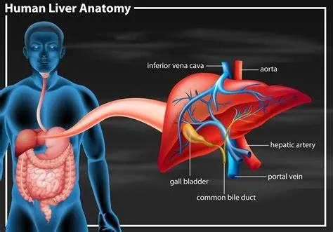
- Bile production and excretion.
- Excretion of bilirubin, cholesterol, hormones, and drugs.
- Metabolism of fats, proteins, and carbohydrates.
- Enzyme activation.
- Storage of glycogen, vitamins, and minerals.
- Synthesis of plasma proteins, such as albumin, and clotting factors.
B. PATHOLOGY STAGE: LIVER CANCER
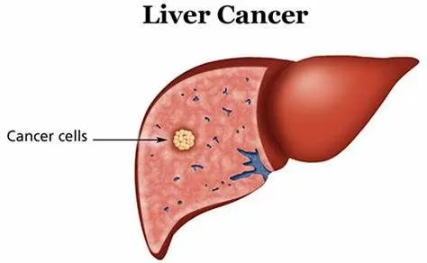
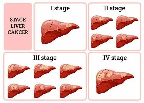
- Cancer is a disease in which cells in the body grow out of control.
- When cancer starts in the liver, it is called liver cancer.
- liver cancer affect function of liver.
- when cancer cell grow very high they can also damage the liver.
C. RESON FOR THE PATHOLOGICAL CONDITION:
- Hepatitis B virus (HBV) infection: HBV can be transmitted in blood, semen or other body fluids. The infection can be passed from mother to child during childbirth, through sexual contact, or by sharing needles that are used to inject drugs. It can cause inflammation (swelling) of the liver that leads to cancer.
- Hepatitis C virus (HCV) infection: HCV can be transmitted in the blood. The infection can be spread by sharing needles that are used to inject drugs or, less often, through sexual contact. In the past, it was also spread during blood transfusion or organ transplants. Today, blood banks test all donated blood for HCV, which greatly lowers the risk of getting the virus from blood transfusions. It can cause cirrhosis that may lead to liver cancer
- Cirrhosis: The risk of developing liver cancer is increased for people who have cirrhosis, a disease
- Heavy alcohol use: Heavy alcohol use can cause cirrhosis, which is a risk factor for liver cancer.
- Aflatoxin B1: The risk of developing liver cancer may be increased by eating foods that contain aflatoxin B1 . It is most common in sub-Saharan Africa, Southeast Asia, and China.
- Nonalcoholic steatohepatitis (NASH): NASH is a condition that can cause cirrhosis that may lead to liver cancer.
- Cigarette smoking: Cigarette smoking has been linked to a higher risk of liver cancer.
D. EFFECT HOMEOSTASIS IN HUMAN BODY:
- Losing weight without trying
- Loss of appetite
- Yellow discoloration of your skin and the whites of your eyes (jaundice)
- White, chalky stools
2. Description of the diagnostic methods selected by the surgeon for assessing the condition.
A. DIAGNOSTIC METHOD:
- Blood test
- imaging test like ultrasound,ct scan,mri
- biopsy
B. Data : By recive the images from ct-scan, ultrasound and blood tests and tissue test which aquried through biopsy.
C. Doctor identify the liver cancer to saw size of the tumor and area of liver affected in the image of various imaging technique. and chek the result of blood test and liver tissue laboratory test
3. CLINICAL SOLUTION:
- Localized treatments
- Localized treatments for liver cancer are those that are administered directly to the cancer cells or the area surrounding the cancer cells. Localized treatment options for liver cancer include:
- Heating cancer cells.
- Freezing cancer cells.
- Injecting alcohol into the tumor..
- Injecting chemotherapy drugs into the liver.
- Placing beads filled with radiation in the liver.
- Radiation therapy
- This treatment uses high-powered energy from sources such as X-rays and protons to destroy cancer cells and shrink tumors.
- Targeted drug therapy
- Targeted drug treatments focus on specific abnormalities present within cancer cells. By blocking these abnormalities, targeted drug treatments can cause cancer cells to die.
- Immunotherapy
- Immunotherapy uses your immune system to fight cancer.
- Chemotherapy
- Chemotherapy uses drugs to kill rapidly growing cells, including cancer cells.
- Surgery
- Operations used to treat liver cancer include:
-
Surgery to remove the tumor: In certain situations, your doctor may recommend an operation to remove the liver cancer and a small portion of healthy liver tissue that surrounds it if your tumor is small and your liver function is good.
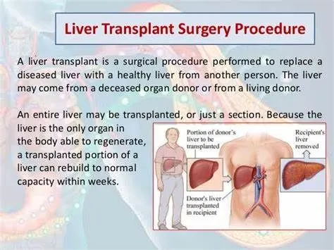
- Liver transplant surgery. During liver transplant surgery, your diseased liver is removed and replaced with a healthy liver from a donor. Liver transplant surgery is only an option for a small percentage of people with early-stage liver cancer.
4.SURGICAL SOLUTION:.
A. LIVER TRANSPLANT
B.SUPINE POSITION
C. PROCEDURE:
- If the liver comes from a deceased donor, the surgical procedure begins when the liver arrives at the hospital.
- f the liver comes from a living donor, the medical team schedules the procedures in advance and the recipient and donor have surgery at the same time.
- Before the surgery, the transplant recipient will receive a genral anesthetic. This means they will be asleep during surgery.
- To replace the liver, a surgeon cuts across the recipient’s abdomen and toward their chest.
- They then remove the damaged liver and insert the new one, connecting it to the recipient’s blood vessels and bile ducts, which are the tubes connecting the liver to the small intestine.
- The surgeon then closes the wound with clips or stitches.
D:instruments required during the surgery .
- surgical blade
- Tissue Forcep
- neddle hoder
- various claps
- sutures
- surgical staplar
5.Brief description about - Post surgical care required for the patients
- After your liver transplant, you can expect to:
- Possibly stay in the intensive care unit for a few days. Doctors and nurses will monitor your condition to watch for signs of complications. They'll also test your liver function frequently for signs that your new liver is working.
- Spend 5 to 10 days in the hospital. Once you're stable, you're taken to a transplant recovery area to continue recuperating.
- Have frequent checkups as you continue recovering at home.
- Your transplant team designs a checkup schedule for you. You may undergo blood tests a few times each week at first and then less often over time.
- Take medications for the rest of your life. You'll take a number of medications after your liver transplant. You'll need to continue taking many of them for the rest of your life
- Drugs called immunosuppressants help keep your immune system from attacking your new liver. Other drugs help reduce the risk of other complications after your transplant.
- Expect six months or more of recovery time before you'll feel fully healed after your liver transplant surgery. You may be able to resume normal activities or go back to work a few months after surgery.
- How long it takes you to recover may depend on how ill you were before your liver transplant.
6.List all the medical devices used in this case study
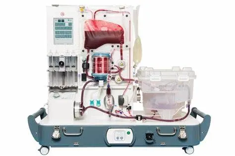
- Liver perfusion device: preserve donor livers prior to transplantation. This system includes a portable enclosure designed to warm the donor liver, provide it with oxygen, nutrients, and blood, and collect bile from the donor liver
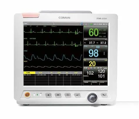
- Patient monitor : devices used to measure, record, and display various patient parameters such as heart rate and rhythm, SPO2, blood pressure, temperature, respiratory rate, blood pressure, blood oxygen saturation, et cetera to keep a track of the patient’s health and provide them with high-quality health care.
- Project(b):
1.PATOLOGICAL CONDITION : BRAIN ANEURYSM (NERVOUS SYSYTEM)
A.BRAIN FUNCTION:
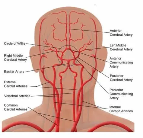
- The human brain is magnificent and complex.
- The brain is made up of many parts, each with a specific and important function.
- It controls our ability to balance, walk, talk, and eat.
- It coordinates and regulates our breathing, blood circulation, and heart rate.
- It is responsible for our ability to speak, to process and remember information, make decisions, and feel emotions.
- Every brain is unique, ever-changing, and extremely sensitive to its environment.
B. PATHOLOGY STAGE:BRAIN ANEURYSM
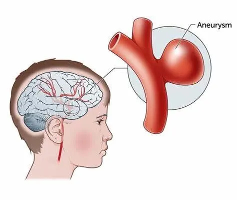
- When a brain aneurysm ruptures, the bleeding usually lasts only a few seconds.
- However, the blood can cause direct damage to surrounding cells, and the bleeding can damage or kill other cells.
- It also increases pressure inside the skull.If the pressure becomes too high, it may disrupt the blood and oxygen supply to the brain and loss of consciousness or even death may occur.
C. RESON FOR THE PATHOLOGICAL CONDITION:
- Older age
- Cigarette smoking
- High blood pressure
- Drug abuse, particularly the use of cocaine
- Heavy alcohol consumption
- Risk factors present at birth:
- Inherited connective tissue disorders, such as Ehlers-Danlos syndrome, that weaken blood vessels
- Polycystic kidney disease, an inherited disorder that results in fluid-filled sacs in the kidneys and usually increases blood pressure
- Narrow aorta (coarctation of the aorta), the large blood vessel that delivers oxygen-rich blood from the heart to the body
- Brain arteriovenous malformation (AVM), in which the arteries and veins in the brain are tangled, interrupting blood flow
- Family history of brain aneurysm, particularly a first-degree relative, such as a parent, brother, sister or child
D.EFFECT HOMEOSTASIS IN HUMAN BODY:
- The type of symptoms you have from a brain aneurysm depend on whether it ruptures or not.
- Ruptured brain aneyrysum symptoms
- Intense headache that comes on suddenly
- Loss of consciousness
- vomating
- Drowsiness
- Loss of balance in things like walking and normal coordination
- stiff neck
- Dilated pupils
- Sensitivity to light
- Sudden blurred or double vision
- Drooping eyelid
- Confusion or trouble with mental awareness
- seizure
- Unruptured brain aneurysm symptom
- Dilated pupils
- Blurred or double vision
- Drooping eyelid
- A hard time speaking
- Weakness and numbness in one side of your face
2. Description of the diagnostic methods selected by the surgeon for assessing the condition.
A. DIAGNOSTIC METHOD:
- ct scan: This exam creates images of your brain. You will lie on a table that slides into a CT scanner. A technician will inject contrast dye into one of your veins to make it easier to see the blood flow and spot aneurysms in your brain.
- MRI: This exam is similar in that you lie on a table that slides into a scanner. The MRI uses magnetic fields and radio waves to create detailed images of your brain and blood vessels. MRIs and CT scans can detect aneurysms larger than 3 to 5 millimeters.
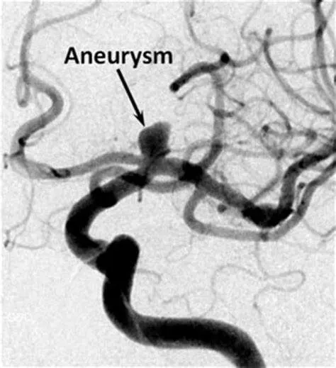
- Angiogram: This test, considered the most reliable way to detect aneurysms, shows the weak spots in your blood vessels. During the test, you lie on an X-ray table, and you will be given something to numb any pain. Your doctor will insert a small flexible tube through a blood vessel in the leg. They’ll guide that tube, called a catheter, into the blood vessels in your neck that reach the brain. Then they’ll inject a contrast dye into you, and X-rays will be taken that show all the blood vessels in the brain. This gives your doctor a map of your blood vessels, pinpointing the aneurysm.
- Cerebrospinal fluid test: Your doctor may order this test if they think an aneurysm may have ruptured.
B. Data : By recive the images from ct-scan, mri,angiogram and test of cerebrospinal fluid test
C. Doctor detect aneurysm from the image of various imaging technique. and chek the result of cerebrospinal fluid test.
3. CLINICAL SOLUTION:
- Ruptured brain aneurysm treatment
- You need treatment as soon as possible if you have a ruptured brain aneurysm, because it’s likely that it will bleed again. Treatment involves stopping the blood flow into the aneurysm.
- Surgical clipping: A section of your skull is removed to locate the aneurysm. A metal clip is placed on the opening of the aneurysm to cut off the blood flow. Your skull is then sealed shut.
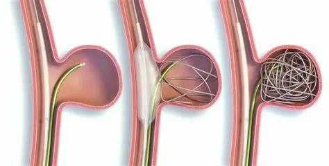
- Endovascular coiling: This doesn’t require surgery that opens the skull. Your doctor will insert a catheter into your groin to reach the affected blood vessel where the aneurysm is located.The doctor will send tiny platinum coils through the tube and place them inside the aneurysm. The coils conform to the shape of the aneurysm, stopping the blood flow there. This may be safer than surgical clipping, but it has a higher chance of the aneurysm bleeding again.
- Flow diverter surgery: This option is for larger brain aneurysms in which neither clipping nor coiling would work. In this procedure, your doctor inserts a stent, usually made of metal mesh, inside the artery. It becomes a wall inside the vessel to divert blood away from the aneurysm.
- To help manage symptoms and prevent complications of an aneurysm, your doctor may suggest:
- Treatments to prevent a stroke, such as drugs that widen the blood vessels so blood will flow through narrowed blood vessels, or a procedure called an angioplasty that uses a small balloon to widen blood vesselsAnti-seizure medication
- Ventricular or lumbar draining catheters to reduce pressure on the brain
- Shunt surgery
- Rehabilitative therapy to help you relearndication skills you may have lost from damage to your brain
- Unruptured brain aneurysm treatment
- Small aneurysms that haven’t ruptured and aren’t causing symptoms may not need treatment. But this depends on your health and the aneurysm. You can talk this all over with your doctors.
4.SURGICAL SOLUTION:
A Endovascular coiling
B.SUPINE POSITION
C. PROCEDURE:
- A microcatheter is inserted through the initial catheter. The coil is attached to the microcatheter.
- When the microcatheter has reached the aneurysm and has been inserted into the aneurysm, an electrical current is used to separate the coil from the catheter.
- The coil seals off the opening of the aneurysm.
- The coil is left in place permanently in the aneurysm.
- Depending on the size of the aneurysm, more than one coil may be needed to completely seal off the aneurysm.
- the coils used in this procedure are made of soft platinum metal, and are shaped like a spring.
- These coils are very small and thin, ranging in size from about twice the width of a human hair (largest) to less than one hair's width (smallest).
- Fluoroscopy (a special type of x-ray, similar to an x-ray "movie") aids in this procedure. The catheter, which is inserted into an artery in the groin, is guided by a small wire inside of the catheter along the length of the blood vessel to reach the area of the aneurysm. The physician uses fluoroscopy to guide the catheter to the aneurysm's location in the brain.
D:instruments required during the surgery .
- cathetar
- surgical blade
- gude wire
- patient monitor
- fluroscope
5.Brief description about - Post surgical care required for the patients
- After surgery, a patient might expect to return home after spending one night in the Neuro Intensive Care Unit, and may expect to return to normal activities within 2 days.
- Your physician will provide specific details regarding your post-surgical care prior to your discharge from the hospital.
6.List all the medical devices used in this case study
- fluroscope:Fluoroscope is a medical device` that makes a real-time video of the movements inside a part of the body by passing x-rays through the body over a period of time
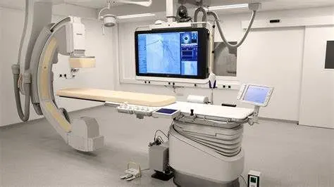
- patient monitor :Patient monitors are devices used to measure, record, and display various patient parameters such as heart rate and rhythm, SPO2, blood pressure, temperature, respiratory rate, blood pressure, blood oxygen saturation, et cetera to keep a track of the patient’s health and provide them with high-quality health care.