Project 1
- PART A
- RESPIRATORY SYSTEM - LUNG CANCER
1. Describe one common clinical condition/ disorder associated with the system involved
- LUNG:
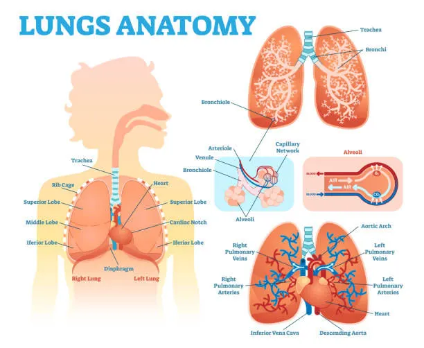
- One of a pair of organs in the chest that supplies the body with oxygen, and removes carbon dioxide from the body.
- The lungs are two spongy organs located in the thorax.
- They consist of elastic tissue, filled with an interlacing network of tubes and sacs that carry air and blood vessels that carry blood.
- Each lung is divided into lobes.
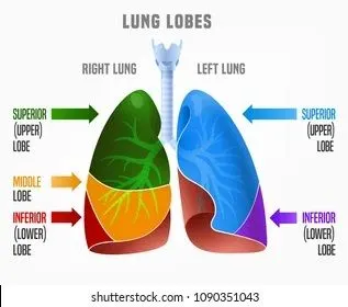
- The left lung is divided into two lobes, upper and lower, by the oblique (major) fissure.
- The right lung has two fissures, oblique fissure and horizontal fissure, which separate the lung into three lobes - upper, middle, and lower.
- The left lung has an indentation called the cardiac depression or notch for placement of the heart.
- At the end of each bronchiole are the alveoli.
- The lungs contain about 300 million alveoli sacs, which are the air cells where the exchange of oxygen and carbon dioxide takes place with the capillaries
- The base of the lungs rest on the Diaphram, a muscular wall separating the thorax from the abdominal cavity.
- It is involved in respiration, drawing downward in the chest during inhalation, and pushing upward during exhalation.
- Pleural fluid is a liquid that is located between the layers of the pleura.
- The pleura is a two-layer membrane that covers the lungs and lines the chest cavity.
- Pleural fluid keeps the pleura moist and reduces friction between the membranes when you breathe.
2. Describe in common disorders in association with the organ
- LUNG CANCER:
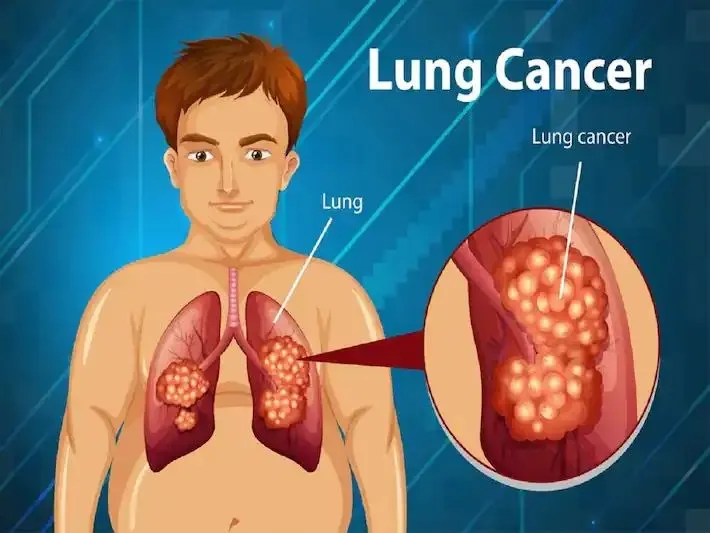
- Cancer starts when cells in the body begin to grow out of control.
- Lung cancer is cancer that forms in tissues of the lung, usually in the cells that line the air passages.
- It is the leading cause of cancer death in both men and women.
- There are two main types: small cell lung cancer and non-small cell lung cancer.
- These two types grow differently and are treated differently.
- Non small cell lung cancer(NSCLC):
- About 80% to 85% of lung cancers are NSCLC.
- The main subtypes of NSCLC are adenocarcinoma, squamous cell carcinoma, and large cell carcinoma.
- These subtypes, which start from different types of lung cells are grouped together as NSCLC because their treatment and prognoses (outlook) are often similar.
- Adenocarcinoma: Adenocarcinomas start in the cells that would normally secrete substances such as mucus.This type of lung cancer occurs mainly in people who currently smoke or formerly smoked, but it is also the most common type of lung cancer seen in people who don't smoke. It is more common in women than in men, and it is more likely to occur in younger people than other types of lung cancer.Adenocarcinoma is usually found in the outer parts of the lung and is more likely to be found before it has spread.
- Squamous cell carcinoma: Squamous cell carcinomas start in squamous cells, which are flat cells that line the inside of the airways in the lungs. They are often linked to a history of smoking and tend to be found in the central part of the lungs, near a main airway (bronchus).
- Large cell carcinoma: Large cell carcinoma can appear in any part of the lung. It tends to grow and spread quickly, which can make it harder to treat. A subtype of large cell carcinoma, known as large cell neuroendocrine carcinoma, is a fast-growing cancer that is very similar to small cell lung cancer.
- Small cell lung cancer (SCLC):
- 10% to 15% of all lung cancers are SCLC and it is sometimes called oat cell cancer.
- This type of lung cancer tends to grow and spread faster than NSCLC.
- About 70% of people with SCLC will have cancer that has already spread at the time they are diagnosed.
- Since this cancer grows quickly, it tends to respond well to chemotherapy and radiation therapy.
- SYMPTOMS:
- Chest pain or discomfort
- A cough that doesn't go away or gets worse over time
- troble breathing
- Wheezing
- Blood in sputum (mucus coughed up from the lungs)
- Hoarseness
- Loss of appetite
- Weight loss for no known reason
- fatigue
- Swelling in the face and/or veins in the neck
- Diagnostic Techniques:
- A medical history, which includes asking about your symptoms
- A family history
- A physical exam
- Imaging test, such as a chest x-ray or chest Ct-scan.
- Lab tests, including tests of your blood and sputum
- A biopsy of the lung
- A biopsy of the lung:
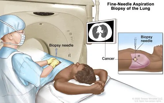
- A biopsy is a procedure performed to remove tissue or cells from the body for examination under a microscope.
- A lung biopsy is a procedure in which samples of lung tissue are removed to determine if lung disease or cancer is present.
- The various biopsy procedures include:
- Needle biopsy: After a local anesthetic is given, the doctor uses a needle that is guided through the chest wall into a suspicious area with Ct scan and fluroscopy to obtain a tissue sample.
- Transbronchial biopsy: This type of biopsy is performed through a fiberoptic bronchoscope.through the main airways of the lungs (bronchoscopy).
- Thoracoscopic biopsy:After a general anesthetic is given, an endoscope is inserted through the chest wall into the chest cavity. Various types of biopsy tools can be inserted through the endoscope to obtain lung tissue for examination. This procedure may be referred to as video-assisted thoracic surgery (VATS) biopsy.
- Open biopsy: After a general anesthetic is given, the doctor makes an incision in the skin on the chest and surgically removes a piece of lung tissue.
3. Describe the management and treatment plan utilized in the disorder
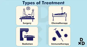
- Medical Management
- Radiation therapy
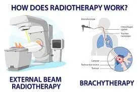
- Radiation therapy uses high-powered energy beams from sources such as X-rays and protons to kill cancer cells.
- During radiation therapy, you lie on a table while a machine moves around you, directing radiation to precise points on your body.
- For people with locally advanced lung cancer, radiation may be used before surgery or after surgery.
- It's often combined with chemotherapy treatments. If surgery isn't an option, combined chemotherapy and radiation therapy may be your primary treatment.
- Chemotherapy
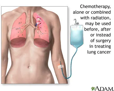
- Chemotherapy uses drugs to kill cancer cells.
- One or more chemotherapy drugs may be given through a vein in your arm (intravenously) or taken orally.
- A combination of drugs usually is given in a series of treatments over a period of weeks or months, with breaks in between so that you can recover.
- Chemotherapy is often used after surgery to kill any cancer cells that may remain. It can be used alone or co
- mbined with radiation therapy.
- Chemotherapy may also be used before surgery to shrink cancers and make them easier to remove
- Stereotactic body radiotherapy
- Stereotactic body radiotherapy
- also known as radiosurgery, is an intense radiation treatment that aims many beams of radiation from many angles at the cancer.
- Stereotactic body radiotherapy treatment is typically completed in one or a few treatments.
- Stereotactic body radiotherapy may be an option for people with small lung cancers who can't undergo surgery.
- Targeted drug therapy
- Targeted drug treatments focus on specific abnormalities present within cancer cells.
- By blocking these abnormalities, targeted drug treatments can cause cancer cells to die.
- Many targeted therapy drugs are used to treat lung cancer, though most are reserved for people with advanced or recurrent cancer.
- Some targeted therapies only work in people whose cancer cells have certain genetic mutations. Your cancer cells may be tested in a laboratory to see if these drugs might help you.
- Immunotherapy
- Immunotherapy uses your immune system to fight cancer.
- Your body's disease-fighting immune system may not attack your cancer because the cancer cells produce proteins that help them hide from the immune system cells.
- Immunotherapy works by interfering with that process.
- Immunotherapy treatments are generally reserved for people with locally advanced lung cancers and cancers that have spread to other parts of the body.
- SURGICAL PROCEDURE FOR LUNG CANCER
- Wedge resection : to remove a small section of lung that contains the tumor along with a margin of healthy tissue
- Segmental resection : to remove a larger portion of lung, but not an entire lobe
- Lobectomy : to remove the entire lobe of one lung
- Pneumonectomy : to remove an entire lung
4. Describe the surgical management of the disorder
- LOBECTOMY:
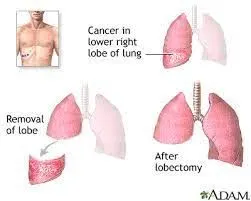
- A lobectomy is a surgery to remove one of the lobes of the lungs.
- The lungs have sections called lobes. The right lung has 3 lobes.
- The left lung has 2 lobes. A lobectomy may be done when a problem is found in just part of a lung.
- The affected lobe is removed, and the remaining healthy lung tissue can work as normal.
- A lobectomy is most often done during a surgery called a thoracotomy. During this type of surgery, the chest is opened.
- PROCDURE:
- In most cases, during a lobectomy the cut (incision) is made at the level of the affected lobe.
- The cut is most often made on the front of the chest under the nipple and wraps around the back under the shoulder blade.
- The surgeon gets access to the chest cavity through the exposed ribs to remove the lobe.
- The cut will be closed with stitches (sutures) or staples. A bandage or dressing will be put on the area.
- A thin tube (epidural catheter) may be put in the area of the lower spine. This is done to send pain medicine into your back. It may be done in the operating room or in the recovery room.
- POST OPRATION CARE:
- After the procedure, Patient may be sleepy and confused.
- healthcare team will watch patient's vital signs, such as your heart rate and breathing.
- patient will be given pain medicine if you need it.
- A chest X-ray may be done right after the surgery. This is to make sure lungs are OK.
- patient will stay in the hospital for several days.
- patient will be taught deep-breathing exercises and coughing methods to help lungs re-expand after surgery.
- This is to help breathing and prevent pneumonia. patient may need oxygen for a period of time after surgery.
- In most cases, the oxygen will be stopped before patient go home.
- INSTRUMENT USED FOR SURGERY:
- Lobectomy and Segmental Lung Resection Instrument Set is needed to treat lobectomy and segmental lung resection.
- This set includes tissue forceps, needle holders, rib spreaders, lung retractors, and many more.
- All instruments come in multiple variations. So, surgeons can pick the best one of their choice.
- This amazing set is manufactured from premium-grade German surgical stainless material.
- Sterilization is essential before and after using instruments.
- The stainless body renders the surface inert to many chemical reactions.
- Therefore, it gives an exceptional built-in corrosion resistance.
- They are reusable tools that can be sterilized easily.
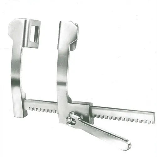
( rib spreaders) ( tissue forceps)
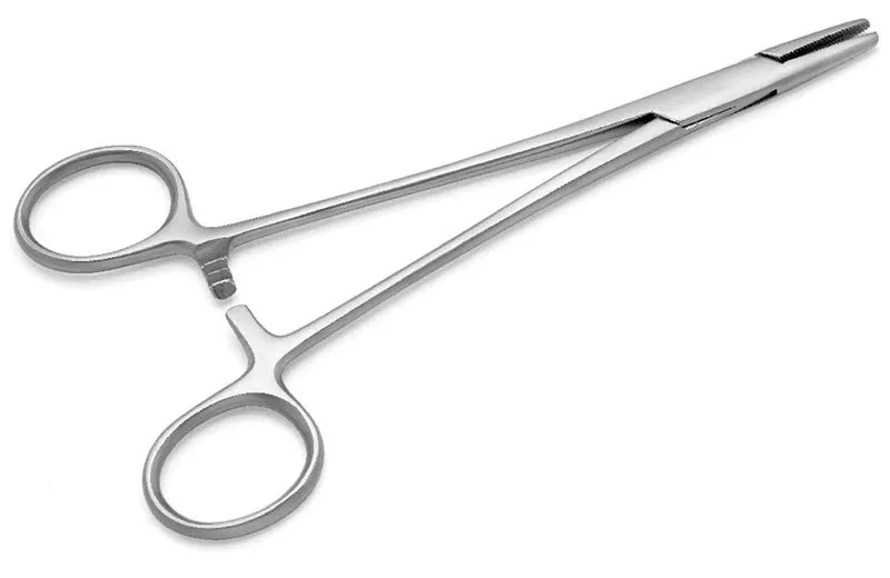
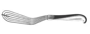
(needle holder) (lung resection)
5. Self study on the recent advances in the field
- ROBOTIC LOBECTOMY:
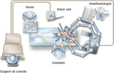
- During a robotic-assisted lobectomy, a surgeon removes a diseased or cancerous lobe of the lung using robotic technology.
- The robot is actually a system of three to four mechanical arms that hold and manipulate the surgical instruments.
- It acts as a helper and a tool that allows the surgeon to work delicately inside the body.
- Because robotic tools enable surgeons to perform complex procedures in small spaces, robotic-assisted surgery is conducted through a number of small incisions, rather than a single large one.
- This means that patients who receive robotic-assisted lobectomies also tend to leave the hospital earlier, recover more quickly, and experience less pain than patients treated with traditional, open procedures.
- DIGESTIVE SYSTEM - APPENDICITIS
1. Describe one common clinical condition/ disorder associated with the system involved
- LARGE INTENSTINE:
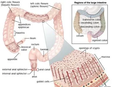
- The large intestine is the last part of the gastrointestinal (GI) tract, the long, tube-like pathway that food travels through your digestive system.
- It follows from the small intenstine and ends at the anal canal, where food waste leaves your body.
- The large intestine, also called the large bowel, is where food waste is formed into poop, stored, and finally excreted.
- The large intestine is one long tube, but slightly different things happen in different parts of it.
- Its three parts are the colon, the rectum and the anus.
- The colon can also be divided into parts. The entry point, about six inches long, is called the cecum. The rest of the colon is divided into segments: the ascending colon (traveling up), the transverse colon (traveling across to the left), the descending colon (traveling down) and the sigmoid colon (headed back across to the right).
- The large intestine is in your lower abdominal cavity from your waist down.
- It surrounds the small intestine in a sort of a square question mark shape, with the tail of the question mark ending at the anal canal.
- The large intestine looks like a semi-flat, segmented tube that lays loosely around the edges of the abdominal cavity. A seam runs vertically down the middle of the tube, making the segments bulge on either side of it.
- The large intestine is about six feet long — much shorter than the small intestine, which is 22 feet. It’s called the large intestine because it's wider — about three inches, while the small intestine is only one inch in diameter.
- Layers of muscles and tissues make up the intestinal walls. Separate layers of circular muscles and longitudinal muscles allow the intestine to contract in different ways. The mucous lining provides blood supply, nerve endings and glands that secrete and absorb.
- The appendix is a small, thin pouch about 5 to 10cm (2 to 4 inches) long. It's connected to the large intestine.
- FUNCTION
- When the large intestine receives food from the small intestine, the food has been liquified by the digestive process and most of the nutrients have been absorbed.
- The colon’s job is to dehydrate what’s left of the food and form it into stool.
- It does this by slowly absorbing water and electrolytes as its muscle system moves the waste along.
- Meanwhile, bacteria living in your colon feed on the waste and break it down further, completing the chemical part of the digestive process
2. Describe in common disorders in association with the organ
- Appendicitis
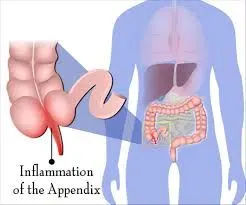
- appendix is a finger-sized tube located where the large and small intestines connect.
- It has no known function, but if it gets inflamed or infected (appendicitis), you’ll need immediate treatment.
- An inflamed appendix may cause pain off and on.
- Or it may burst open (rupture), causing sudden, severe pain.
- A ruptured appendix can spread bacteria through the abdominal cavity. These bacteria trigger a serious, sometimes-fatal infection called peritonisis.
- CAUSES OF APPENDIX:
- Abdominal injury or trauma.
- Blockage at the opening where the appendix connects to the intestines.
- Digestive tract infection.
- inflammatory bowel disease.
- Growths inside the appendix.
- SYMPTOMS:
- Abdominal pain or tenderness that hurts more when you cough, sneeze, inhale or move.
- Swollen belly.
- Diarrhea.
- Inability to pass gas.
- Loss of appetite (not feeling hungry when you usually would).
- Low-grade fever (below 100 degrees F).
- vometing
- DIAGNOSTIC METHOD:
- Physical exam
- blood test for infection
- imaging like abdominal ultrasound,ct scan,mri
- Abdominial ultrasound:
- ultrasound, also called sonography or diagnostic medical sonography, is an imaging method that uses sound waves to produce images of structures within your body.
- The images can provide valuable information for diagnosing and directing treatment for a variety of diseases and conditions.
- Most ultrasound examinations are done using an ultrasound device outside your body, though some involve placing a small device inside your body
- Abdominal ultrasound is a type of imaging exam that uses sound waves to create pictures of the inside of the abdomen and/or pelvis.
3. Describe the management and treatment plan utilized in the disorder
- medical managment:
- There are two type of medical mangement for appendicitis
- Antibiotic therapy
- antibiotic to treat infection.
- Some cases of mild appendicitis get better with antibiotics alone.
2. Surgery
-
doctor will watch you closely to determine if you need surgery.
-
Surgery is the only way to treat abdominal infection when the appendix ruptures.
- there are two type of surgery:
- appendectomy
- laproscopic appendectomy
4. Describe the surgical management of the disorder
- LAPROSCOPIC APPENDECTOMY
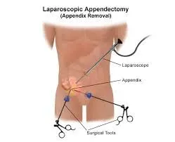
- This method is less invasive. That means it’s done without a large incision.
- Instead, from 1 to 3 tiny cuts are made. A long, thin tube called a laparoscope is put into one of the incisions.
- It has a tiny video camera and surgical tools.
- The surgeon looks at a TV monitor to see inside your abdomen and guide the tools.
- PROCDURE:
- A tiny incision will be made for the tube (laparoscope). More cuts may be made so that other tools can be used during surgery.
- Carbon dioxide gas will be used to swell up your abdomen so that your appendix and other organs can be easily seen.
- The laparoscope will be put in and your appendix will be found.
- Your appendix will be tied off with stitches and removed through an incision.
- When the surgery is done, the laparoscope and tools will be removed. The carbon dioxide will be let out through the cuts. A small tube may be placed in the cut to drain out fluids.
- POST OPRATION CARE:
- After surgery, patient will be taken to the recovery room.
- healthcare team will watch patient vital signs, such as your heart rate and breathing.
- patient recovery will depend on the type of surgery that was done and the type of anesthesia you had. Once blood pressure, pulse, and breathing are stable and patient are awake and alert.
- patient will have pain medicine as needed. patient may have a thin plastic tube that goes through your nose into your stomach.
- This is used to remove stomach fluids and air that you swallow. The tube will be taken out when patient bowels are working normally. patient not be able to eat or drink until the tube is removed.
- patient will be asked to get out of bed a few hours after a laparoscopic surgery or by the next day after an open surgery.
- patient may be allowed to drink liquids a few hours after surgery.
- instrument used for surgery:
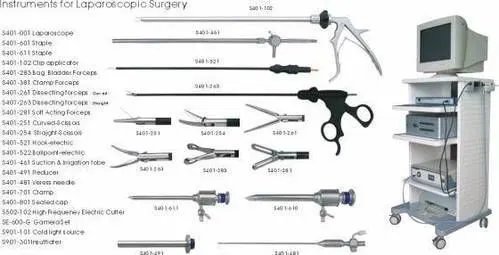
- Laproscope
- trocar
- light cable
- light source
- co2 insufflator
- monitor
- surgical innstrument like forceps, scissors, probes, dissectors, hooks, and retractors etc.
- PART B - CASE STUDY
- A 25 year old biker indulged in illegal racing met with a road traffic accident and severed head injury and skull fracture. After resuscitation and operation done for the hematoma it was resorted to do skull implant for his fracture by the orthopaedician
- Describe the type of bone the skull belongs to
- There are flat bones in the skull (occipital, parietal, frontal, nasal, lacrimal, and vomer),
- Flat bones are somewhat flattened, and can provide protection, like a shield; flat bones can also provide large areas of attachment for muscles.
- Flat bones are bones whose principal function is either extensive protection or the provision of broad surfaces for muscular attachment.
- These bones are expanded into broad, flat plates as in the cranium , the ilium .
- These bones are composed of two thin layers of compact bone enclosing between them a variable quantity of cancellous bone which is the location of red bone marrow.
- In an adult, most red blood cell are formed in flat bones.
- In the cranial , the layers of compact tissue are familiarly known as the tables of the skull; the outer one is thick and tough; the inner is thin, dense, and brittle, and hence is termed the vitreous (glass-like) table.
- Describe the microscopic anatomy of bone
-
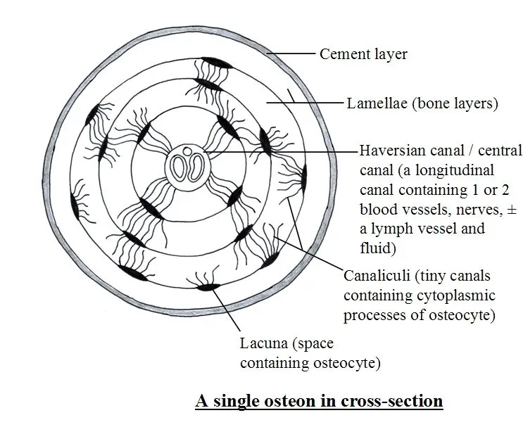
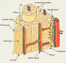
- the basic unit of bone is osteon which is also called system they contain four part
- Centarl canal:- A cental tube is called centarl canal which carries blood vessle and nerve
- Performing canal:- located perpendicular to central canal and carries blood vessle and nerve
- Lacunae :- Lacunae is cavities containing bone cell
- Canalicui- canaliculi is the tiny canal radiate from the central canal to the lacunae and form a transport system
- harversion system provide strength and support of bone
- Explain the blood supply of bone
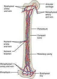
- In a typical long bone, blood is supplied by three separate systems: a nutrient artery, periosteal vessels, and epiphyseal vessels.
- The diaphysis and metaphysis are nourished primarily by the nutrient artery,
- which passes through the cortex into the medullary cavity and then ramifies outward through haversian and volkmens canal to supply the cortex.
- extensive vessels in the peristium, the membrane surrounding the bone, supply the superficial layers of the cortex and connect with the nutrient-artery system.
- The epiphysis are supplied by a separate system that consists of a ring of arteries entering the bone along a circular band between the growth plate and the joint capsule.
- In the adult these vessels become connected to the other two systems at the metaphyseal-epiphyseal junction,
- Drainage of blood is by a system of veins that runs parallel with the arterial supply and by veins leaving the cortical periosteum through muscle insertions.
- Elaborate on bone remodeling and new bone formation
- Bone remodeling involves the removal of mineralized bone by osteoclasts followed by the formation of bone matrix through the osteoblasts that subsequently become mineralized.
- The remodeling cycle consists of three consecutive phases: resorption, during which osteoclasts digest old bone; reversal, when mononuclear cells appear on the bone surface; and formation, when osteoblasts lay down new bone until the resorbed bone is completely replaced.
- Bone remodeling serves to adjust bone architecture to meet changing mechanical needs and it helps to repair microdamages in bone matrix preventing the accumulation of old bone.
- It also plays an important role in maintaining plasma calcium homeostasis. The regulation of bone remodeling is both systemic and local.
- SKULL IMPLANT:
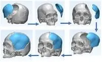
- cranioplasty is the surgical repair of a bone defect in the skull resulting from a previous operation or injury.
- There are different kinds of cranioplasties, but most involve lifting the scalp and restoring the contour of the skull with the original skull piece or a custom contoured graft made from material such as:
- Titanium (plate or mesh).
- Synthetic bone substitute (in liquid form).
- Solid biomaterial (prefabricated customized implant to match the exact contour and shape of the skull).
- Conventional cranioplasty methods, which have been used by neurosurgeons for more than 100 years, involve peeling back all five layers of the scalp to place the bone remnant or custom implant into the proper cranial location.
- For the pericranial-onlay cranioplasty, a newer technique developed here at Johns Hopkins by Chad Gordon and his team, the surgeon gently pulls back only the three uppermost layers of the scalp and inserts the bone or implant in between the bottom layers of the scalp protecting the brain. This type of cranioplasty procedure is safer and less invasive.