Project 1
Part A - Detailed study of anatomical basis of surgical procedures or Clinical management in association with any of the organ system (select 3 system from Module 1 to module 6))
UROLITHIASIS
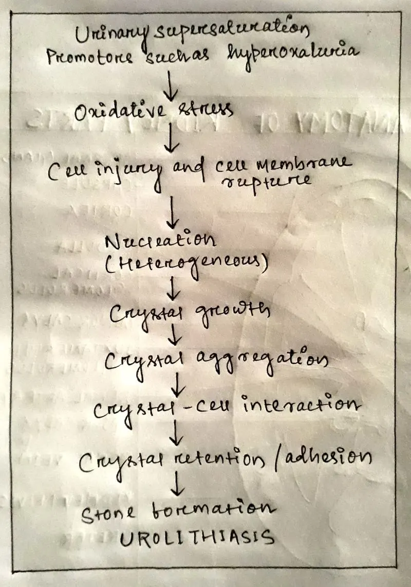
Urolithiasis; generally known as Kidney stones also called renal calculi / nephrolithiasis. A small, hard deposit like crystals that forms in the kidneys and it is painful when passing through the urinary tract. Kidney stones are hard deposits of minerals and acid salts that stick together in concentrate urine. It can be occurred anywhere in the urinary system including the kidneys & bladder.
CAUSES
- Sometimes oxalate enrichment foods; beets, spinach, tea, nuts etc. form kidney stones
- Excess body weight
- Diet
- Taking of alcohol, dark cola beverages, artificial fruit punch
- Lack of drinking water
- Taking salt in excess amount etc.
SYMPTOMS
- Severe pain in the side & back, below the ribs and pain radiates to the lower abdomen & groin
- Pain or burning sensation while urinating
- Cloudy or foul-smelling urine
- Nausea & Vomiting
- Fever
- Blood in urine
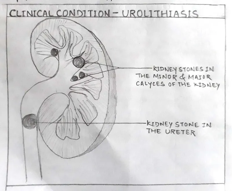
Organ involved - Kidney
KIDNEY
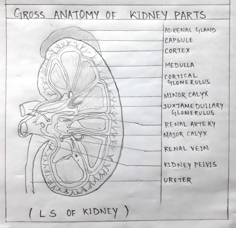
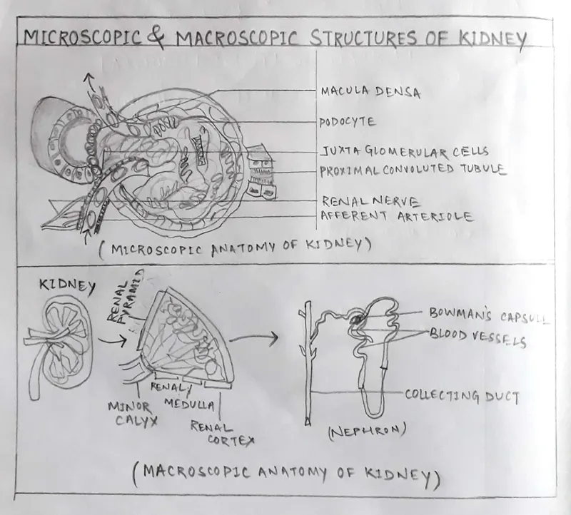
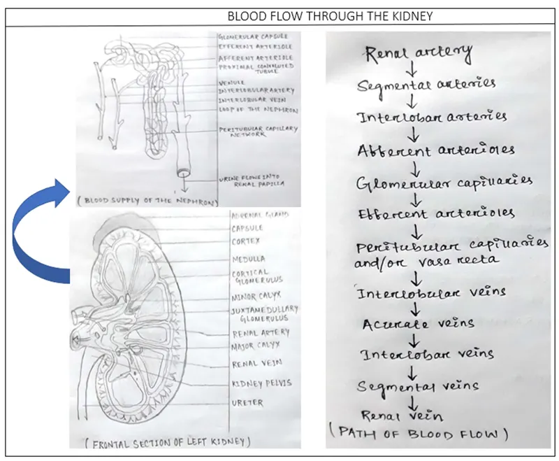
- Kidneys are bean shaped having a convex boarder and a concave boarder hilum and Hilum transmits the renal vessels, nerves & renal pelvis.
- Kidneys lie obliquely on the posterior abdominal wall behind the peritoneum on either side of the vertebral column under the cover of the costal margin.
- The right and left kidneys weighed 108.7 +/- 22.6g and 111.8 +/- 23.3 g (P>0.05) respectively in males corresponding weights in females were 96.6 +/- 19.3g and 99.4 +/- 25.3 g (P>0.05) for the right and left kidneys respectively.
- Renal pelvis: The expanded proximal end of the ureter and the renal pelvis receives urine from the calyces.
- Medulla: The middle portion of the internal structure of the kidney.
- Cortex: The outer portion of the internal structure of the kidney.
- Calyx: The cup like extension of the renal pelvis that urine empties into.
- Adrenal gland: An endocrine gland that sits on the top of the kidney.
- Renal capsule: The tough white fibrous membrane surrounding the kidney.
- Renal artery: Carries oxygenated blood to the kidney.
- Renal vein: carries deoxygenated blood away from the kidney.
- Abdominal aorta: The renal arteries that branch off from the large blood vessel.
- Interior vena cava: The main large blood vessel that drains blood from the lower part of the body.
- Ureter: Tubule that drains urine from the kidney into the bladder. The ureter is a 25-30cm long that begins as a continuation of the renal pelvis.
DIFFERENT METHODS OF DIAGNOSIS
- Blood testing: Blood testing used to check calcium and uric acid level in blood. It helps to monitor the health of kidneys, also helps to check other medical conditions.
- Urine testing: The 24 hr urine collection test may show that you’re excreting too many stone forming minerals or too few preventing substances. For this test doctor may request to perform two urine collections over two consecutive days.
- Imaging: Imaging techniques like ultrasound, CT scan may show stones in kidneys and urinary tracts. High speed or dual energy computerized tomography may reveal even tiny stones. Simple abdominal X-rays are used less frequently because this imaging test can miss small kidney stones. Ultrasound is another imaging option to diagnose kidney stones, it’s a noninvasive test that is quick and easy to perform
- Cystoscopy: A cystoscopy allows doctors to examine the inside of the bladder and urethra. A cystoscope is a long, thin instrument with an eyepiece on the external end and a tiny lens and a light on the end that is inserted into the bladder. The cystoscope is inserted into the patient’s urethra and the small lens magnifies the inner lining of the urethra and bladder allowing to see inside the hollow bladder.
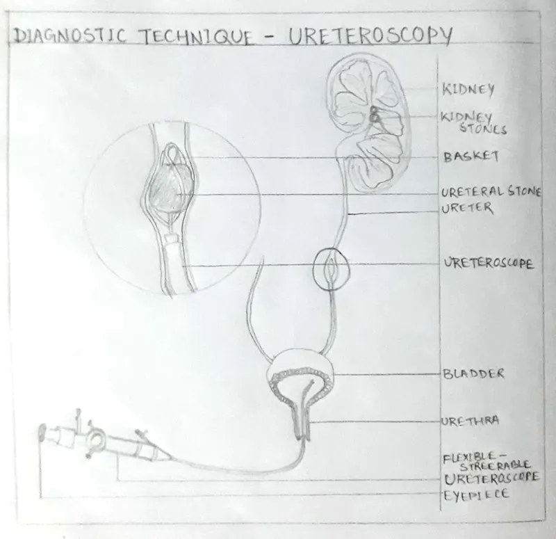
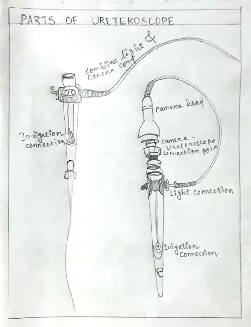
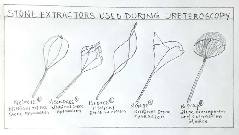
- Ureteroscopy: It is a procedure to address kidney stones as well as stone present in ureter and involves the passage of a small telescope called ureteroscope through the urethra & bladder and up the ureter to the point where the sone is located. It is performed in the operating room with general or spinal anesthesia and is typically an out patient procedure. Small stones can be removed with the use of a tiny wire basket. Larger stones can be fragmented with the use of a holmium laser that creates tiny shock waves that break the stones into small fragments that can be passed out the ureter.
POST-OPERATIVE CARE
A care that the patient receives after a surgical procedure and it depends upon the patient’s past clinical history & types of surgery. It often depends upon pain management and wound care. So a complete rest is required.
You may return to your normal diet and light activity as tolerated after surgery but avoid driving while on narcotic pain medications. Drink water as much as you can take.
Try to avoid any heavy lifting more than 10lbs or strenuous physical activities altogether for several days and then ease back into your regular activity.
Avoid nonsteroidal anti-inflammatory drugs or any other blood thinners if your urine is very bloody until the bleeding has improved.
Staying well hydrated is recommended to help dilute the urine and flush away any potential debris or residual bleeding.
MEDICATIONS
Medications can control the number of salts and minerals in the urine, also helpful in people who form certain kind of kidney stones.
Calcium stones: To prevent from calcium stones forming, doctor may prescribe a thiazide diuretic or a phosphate containing preparation.
Uric acid stones: Doctor prescribes Zyloprm, Aloprim to reduce uric acid levels in blood.
Drinking water: Drinking 3-4 L water per day will keep urine dilute and prevent in stone forming.
Pain relievers: Passing a small stone can cause some discomfort with severe pain. To relieve pain doctor recommends pain relievers such as Ibuprofen, Advil, Motrin, Naproxen sodium and others.
Urinary biomarkers are neutrophil gelatinase-associated lipocalin (NGAL), kidney injury molecule-1 (KIM-1) and N-acetyl-B-D-glucosamindase (NAG), recognised as being useful for the detection of kidney tubular damage but their role in the diagnosis and prognosis of kidney stone disease.
Part B- Case study
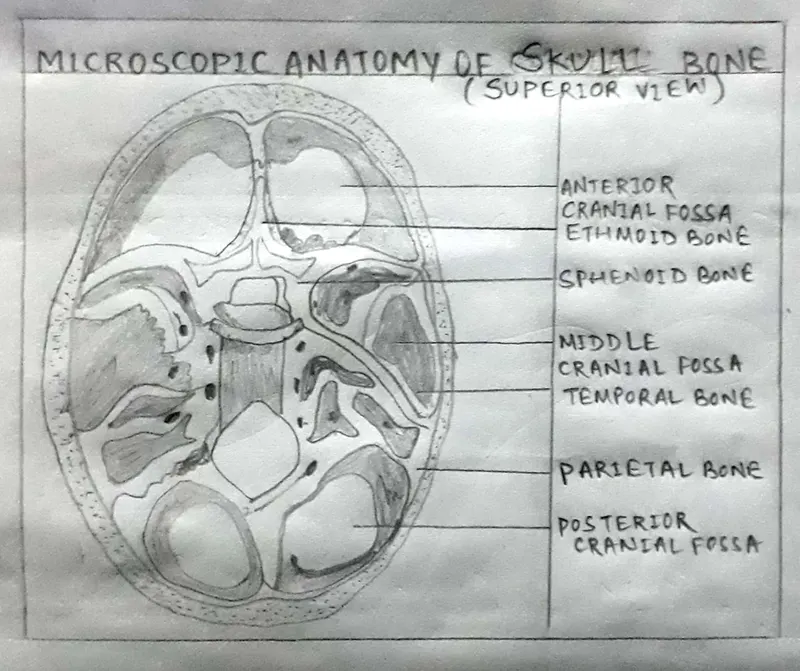
- The human skull is generally consisting of twenty-two bones including eight cranial bones and fourteen facial skeleton bones. In neurocranium these are the occipital bone, two temporal bones, two parietal bones, the sphenoid bones, ethmoid and frontal bones. The skull provides structure to head and face while also protecting our brain. It can be divided into the cranial bones which form the cranium and facial bones which make up our face.
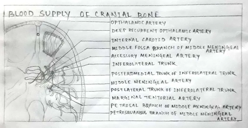
- Bone remodeling involves the removal of mineralized bone by osteoclasts followed by the formation of bone matrix through the osteoblasts that subsequently become mineralized.
- Bone remodeling also known as bone metabolism. This process also controls the reshaping and replacement of bone injuries like fractures.
- Bone remodeling has five steps; activation, resorption, reversal, formation, and quiescence. The total process takes about 4 to 8 months and occurs continually throughout our lives.
- New bone formation is known as Osteogenesis or bone ossification. This process begins the sixth and seventh weeks of embryonic development and continues until about age 25, although this varies slightly based on the individual and his functional activities.
- Bone implant depends upon physical and chemical factors, methodologies, locations, size and shape , proper biomaterials etc.