Project 1
PART A
1 - organ - Kidney
clinical condition - kidney stones form in your kidney . as stones move into your ureters - the thin tubes that allow urine to pass from your kidneys to your bladder- signs and symptoms can result.
signs and symptoms of kidney stones can include severe pain, nausea, vomiting, fever, chills and blood in your urine.
gross anatomy - the kidney are bean - shaped structures and weigh about 150g in the male and about 135g in the female. they are typically 10-12 cm in lenght, 5-7 cm in width, and 2-3 cm in thickness
kidney is covered by 3 layers of fascioa from inside out
- renal capsule
- perirenal fat
- renal fascia
microscopic unit of kidney - the uriniferous tubule is the basic functional unit of the kidney. each tubule consists of two distinct morphological entities
1 - the nephron, which produces the filtrate
2 - the collecting tubuke which serves to concentrate the filtrate by the further removal of water
macroscopic - kidney consists of two distinct regions, both an outer cortex and an inner medulla
blood supply - The renal arteries carry a large volume of blood from the heart to the kidneys. The kidneys filter waste and excess fluid from the blood. You have two renal arteries. The right renal artery supplies blood to the right kidney, while the left artery sends blood to the left kidney.
nerve supply - Kidney innervation consists of both afferent and efferent nerves, of which the efferent is strictly sympathetic. These nerves make up the renal plexus, and receive inputs from the celiac and aorticorenal plexuses as well as the least splanchnic nerves.
2 - the most common form of kidney disease is chronic kidney disease. chronic kidney disease is along term condition that does not improve over time. its commonly caused by high blood pressure.
clincal - chronic kidney disease is a condition in which the kidneys are damaged and cannot filter blood as well as they should. Because of this, excess fluid and waste from blood remain in the body and may cause other health problems, such as heart disease and stroke.
diagnostic technique - ultrasound scan , MRI Scan or CT scan
ultrsound scan in detail - A kidney ultrasound is a noninvasive diagnostic exam that produces images, which are used to assess the size, shape, and location of the kidneys. Ultrasound may also be used to assess blood flow to the kidneys. Ultrasound uses a transducer that sends out ultrasound waves at a frequency too high to be heard.
3 - Your doctor may give you a medication to help pass your kidney stone. This type of medication, known as an alpha blocker, relaxes the muscles in your ureter, helping you pass the kidney stone more quickly and with less pain
Depending on your situation, you may need nothing more than to take pain medication and drink lots of water to pass a kidney stone. In other instances — for example, if stones become lodged in the urinary tract, are associated with a urinary infection or cause complications — surgery may be needed.
medical management -
1. A clinician should perform a screening evaluation consisting of a detailed medical and dietary history, serum chemistries and urinalysis on a patient newly diagnosed with kidney or ureteral stones.
2. Clinicians should obtain serum intact parathyroid hormone (PTH) level as part of the screening evaluation if primary hyperparathyroidism is suspected.
3. When a stone is available, clinicians should obtain a stone analysis at least once.
4. Clinicians should obtain or review available imaging studies to quantify stone burden.
5. Clinicians should perform additional metabolic testing in high-risk or interested first-time stone formers and recurrent stone formers.
6. Metabolic testing should consist of one or two 24-hour urine collections obtained on a random diet and analyzed at minimum for total volume, pH, calcium, oxalate, uric acid, citrate, sodium, potassium and creatinine.
7. Clinicians should not routinely perform "fast and calcium load" testing to distinguish among types of hypercalciuria.
surgical procedure - The doctor uses a thin viewing tool, called a nephroscope, to locate and remove the kidney stone. The doctor inserts the tool directly into your kidney through a small cut made in your back. For larger kidney stones, the doctor also may use a laser to break the kidney stones into smaller pieces.
4 - SURGICAL OPTIONS The most commonly used surgical procedures for stone removal are minimally invasive techniques such as shock wave lithotripsy (SWL), ureteroscopy (URS), and percutaneous nephrolithotomy (PNL). Other, less commonly performed, procedures include laparoscopic, robot-assisted, and open surgery
anatomical - Kidney stones often go unnoticed until they block the renal pelvis or travel through the ureter. The main symptom is pain, which can range from feeling just a little unwell to waves of severe pain. The pain may affect your lower abdomen, belly or back "flank pain," depending on which part of the ureter the stone is in.
instruments -
Cystoscopy and ureteroscopy are common procedures performed by a urologist to look inside the urinary tract.
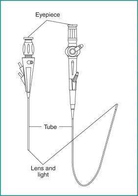
Cystoscopy is a procedure that uses a cystoscope to look inside the urethra and bladder. A cystoscope is a long, thin optical instrument with an eyepiece at one end, a rigid or flexible tube in the middle, and a tiny lens and light at the other end of the tube. A urologist fills the bladder with fluid and looks at detailed images of the urethra and bladder linings on a computer monitor.
Ureteroscopy is a procedure that uses a ureteroscope to look inside the ureter and kidney Like a cystoscope, a ureteroscope has an eyepiece at one end, a rigid or flexible tube in the middle, and a tiny lens and light at the other end of the tube. However, a ureteroscope is longer and thinner than a cystoscope so the urologist can see detailed images of the lining of the ureters and kidneys.
operative care -Most people can go back to their regular daily activities 1 or 2 days after this procedure. Drink a lot of water in the weeks after treatment. This helps pass any pieces of stone that still remain. Your health care provider may give you a medicine called an alpha blocker to make it easier to pass the pieces of stone.
5 - recent advances -Right now, many kidney stones can be treated with a procedure called shock wave lithotripsy. It delivers high-energy sound waves through the skin to break the stone into tiny fragments that can then be passed.
therapeutic -For certain kidney stones — depending on size and location — your doctor may recommend a procedure called extracorporeal shock wave lithotripsy (ESWL). ESWL uses sound waves to create strong vibrations (shock waves) that break the stones into tiny pieces that can be passed in your urine.
diagnostic -Imaging tests may show kidney stones in your urinary tract. High-speed or dual energy computerized tomography (CT) may reveal even tiny stones. Simple abdominal X-rays are used less frequently because this kind of imaging test can miss small kidney stones.
PART B
Type of bone - flat bone
microscopic anatomy of bone - Flat bones are made up of a layer of spongy bone between two thin layers of compact bone. They have a flat shape, not rounded. Examples include the skull and rib bones. Flat bones have marrow, but they do not have a bone marrow cavity.
blood supply of bone -The blood supply to bone is delivered to the endosteal cavity by nutrient arteries, then flows through marrow sinusoids before exiting via numerous small vessels that ramify through the cortex.
bone remodeling - Bone remodeling serves to adjust bone architecture to meet changing mechanical needs and it helps to repair microdamages in bone matrix preventing the accumulation of old bone. It also plays an important role in maintaining plasma calcium homeostasis. The regulation of bone remodeling is both systemic and local.
implant -Titanium is a common metal used for implantation in orthopedic surgery. While titanium is a metallic element, the majority of orthopedic "titanium implants" are, in fact, alloys. These alloys are typically proprietary blends - differing from manufacturer to manufacturer.