Project 2
PROJECT (a)
- Clinical condition : Peptic ulcers
- Normal function is digesting the food (Organ involved : Stomach)
- Ulcers are sores which causes high abdominal pain, burning sensation, vomiting and loss of appetite
- Helicobacter pylori (bacterium) infection Or Use of anti-inflammatory drugs (NSAID), non steroidal drugs for long term.
- Low blood pressure due to bleeding ulcers, imbalance in the action of agents
- Diagnostic methods selected by the surgeon for assessing the condition
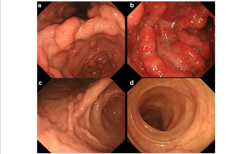
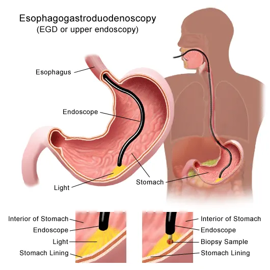
- Endoscopy e., Gastrointestinal endoscopy with biopsy
- Data can be acquired through endoscopy report
- Doctor identifies the state how brutal the walls of stomach was damaged
- Presribing medications Or if severe surgery can be done.
- Available clinical solutions to the pathology condition
- Medications that block stomach acid and supports healing, Antibiotics, Antacids, Antidiarrhoeal. Medications include Bismuth subsalicylate (Pepto-Bismol)
- Severe conditions includes surgery i.e., truncal vagotomy-antrectomy
- Surgical solution in detail
- Truncal vagotomy antrectomy
- Bookwalter style self retaining retractor
- Vagotomy is a surgery where part of your vagus nerve is removed. Until antacid medications and stomach bacteria treatments came along, this was one of the main treatments for peptic ulcer disease.
Entrance into the peritoneal cavity
- An upper midline incision is made just superior to the umbilicus and extends superiorly to the xiphoid process.
- The peritoneal cavity is opened and inspected.
- A self-retaining retractor is placed in the abdomen for exposure, and a liver retractor is placed to facilitate access to the lesser curvature and diaphragmatic hiatus.
Distal esophageal mobilization
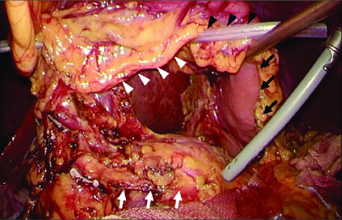
- The assistant retracts the greater curvature inferiorly to expose the GEJ.
- Bookwalter style self retaining retractor (commonly used retractor system for abdominal surgery, and is placed after draping the patient)
- The peritoneal covering of the GEJ is incised, and the esophagus is encircled with a Penrose drain.
- The esophagus should be mobilized for a distance of 4 to5 cm above the GEJ. If the left lobe of the liver is obstructing exposure, the left triangular ligament may be divided.
Identification and division of the anterior vagal trunk
- A single vagal trunk is identified, typically 2 to 4 cm superior to the GEJ.
- The nerve is mobilized for a minimum of 2 cm.
- The trunk is then clipped and divided proximally and distally with medium clips for a minimum resected specimen of 2 cm.
Identification and division of the posterior vagal trunk
- The esophagus is now more mobile, having taken the anterior vagus nerve and is retracted anteriorly and to the patient's left side.
- The posterior trunk is identified along the right edge of the esophagus.
- The nerve is mobilized for a minimum of 2 cm. The trunk is then clipped and divided proximally and distally with medium clips for a minimum resected specimen of 2 cm.
Pathologic confirmation via frozen section
- Drainage procedure or gastric resection with intestinal reconstruction
- Drainage procedures include gastroenterostomy, pyloroplasty and pyloromyotomy.
- Gastric resection can vary in location depending on the location of the ulcer but is typically distal and includes the antrum.
- Reconstruction after distal gastrectomy includes gastroduodenostomy (Billroth I), gastrojejunostomy (Billroth II), and the Roux-en-Y gastrojejunostomy.
Abdominal wall and skin closure
- Implants or instruments required during the surgery
- Penrose drain is a soft, flat, flexible tube made of latex. It lets blood and other fluids move out of the area of your surgery.
- Bookwalter style self retaining retractor (commonly used retractor system for abdominal surgery, and is placed after draping the patient)
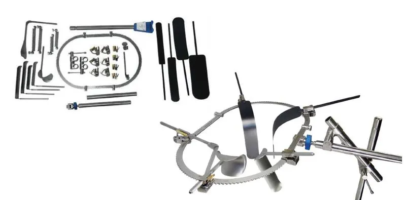
https://www.surgicalholdings.co.uk/bookwalter-retractor.html
- Post surgical care
- Wound care and dressing
- Vitamin suppliments
- Avoid fluids along with food
- Analyze cardiac status, respiratory and fluid balance
- Assesses for the return of peristalsis
6) Endoscopy i.e., Gastrointestinal endoscopy with biopsy
PROJECT (b)
Nervous system (Targeted organ : Brain)
1.
Clinical condition : Brain tumor
- The largest part of the brain, the cerebrum initiates and coordinates movement and regulates temperature.
- We are focusing on tumors that usually develop in cerebrum
- Symptoms include new or increasingly strong headaches, blurred vision, loss of balance, confusion and seizures. In some cases, there may be no symptoms.
- Mutations of brain cell DNA leads to tumors
- Primary brain tumors begin when normal cells develop changes (mutations) in their DNA. A cell's DNA contains the instructions that tell a cell what to do. The mutations tell the cells to grow and divide rapidly and to continue living when healthy cells would die. The result is a mass of abnormal cells, which forms a tumor.
- Effect of homoeostasis imbalance on human body include thermoregulation, dementia and depression
Description of the diagnostic methods selected by the surgeon for assessing the condition
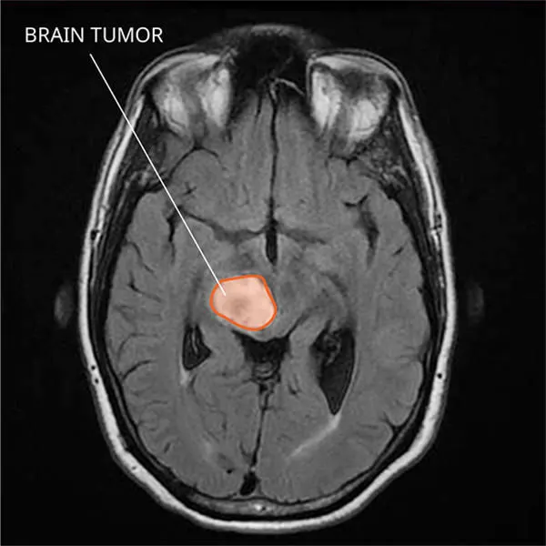
- MRI scan or CT scan
- Imaging includes dark or light spots (i.e., MR image)
- Analysis and observing MR image can detect the brain tumor
- Usually medications incase severe suggests surgery
https://www.cancer.gov/news-events/cancer-currents-blog/2018/liquid-biopsy-childhood-brain-tumors
3.
Available clinical solutions to the pathology condition
- Unexplained nausea and vomiting
- Unhealthy diets, no exercises and stress
- Surgery : Craniotomy
4.
Surgical solution
- Craniotomy
- Lateral position
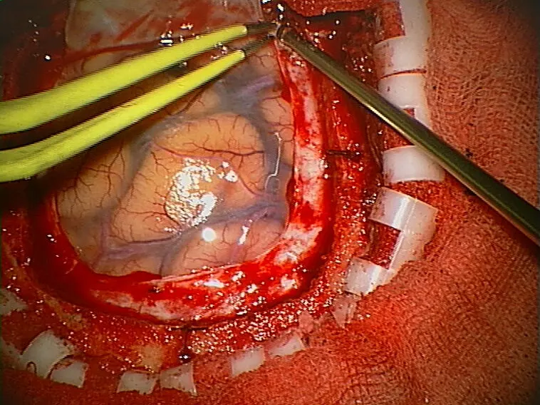
- A craniotomy is the surgical removal of part of the bone from the skull to expose the brain. Specialized tools are used to remove the section of bone called the bone flap. The bone flap is temporarily removed, then replaced after the brain surgery has been done.
- stereotactic biopsy of the brain (a needle is guided into an abnormal area so that a piece of tissue may be removed for exam under a microscope), stereotactic aspiration (removal of fluid from abscesses, hematomas, or cysts), and stereotactic radiosurgery (such as gamma knife radiosurgery).
https://en.wikipedia.org/wiki/Craniotomy
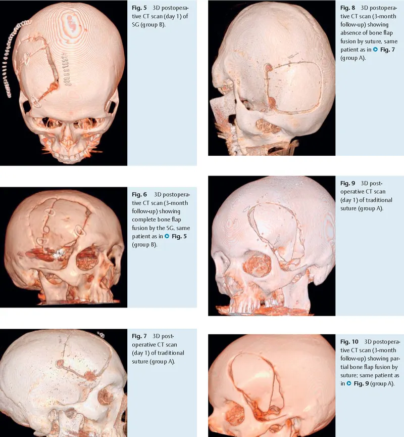
- Skull Grip bone fixation
- Nonabsorbable suture
5.
Post surgical care
- Keep the incision clean
- Watch the incision for signs of infection or complications
- Control Pain
- Gradually return to activity
https://www.curetoday.com/view/post-craniotomy-care-
6.
List all the medical devices used
http://www.abrahamblacksmith.co.uk/Basic-Craniotomy-Set.php
- frazier suction tube (used to suction away debris and liquid from a wound during surgical procedures)
- Scalpel handle (A Beaver blade handle is a scalpel blade handle used by Podiatrists to hold micro blades)
- Adson forceps (Adson Tissue (Thumb) Forceps are used for holding and manipulating delicate tissues. These forceps have a wide, flat thumb grasp area that is commonly serrated)
- Backhaus towel clamp (The Backhaus towel clamp is a perforating clamp used for grasping tissue, securing towels or drapes, and holding or reducing small bone fractures)