Project 1
PROJECT
Detailed study of anatomical basis of surgical procedures or Clinical management in association with any of the organ system
- Describe one common clinical condition or disorder associated with the system selected
Clinical condition : Bronchial asthma
Gross Anatomy of the Lower Respiratory Tract :
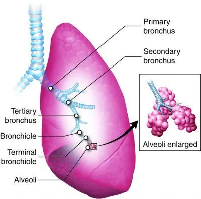
Air flow continues from the larynx to the windpipe i.e., Trachea
Trachea -> Primary bronchus -> Secondary bronchus -> Tertiary bronchus -> Bronchiole -> Terminal bronchiole -> Lungs
The human trachea is a cylinder, about 25 to 30 cm (9.8–11.8 in) long, which sits in front of the esophagus and extends from the pharynx into the thoracic cavity to the lungs.
- Trachea is made of C-shaped rings of cartilage and smooth muscle. The cartilage provides strength and support to the trachea to keep the passage open.
- The last cartilage in the trachea has a projection from the anterior surface that extends into the lumen.
- This projection, the carina, is very sensitive to particulate material and causes coughing when stimulated.
- The openings of the C-shaped cartilages face the posterior of the trachea and contain smooth muscle. This muscle constricts when someone coughs, increasing the force of the cough.
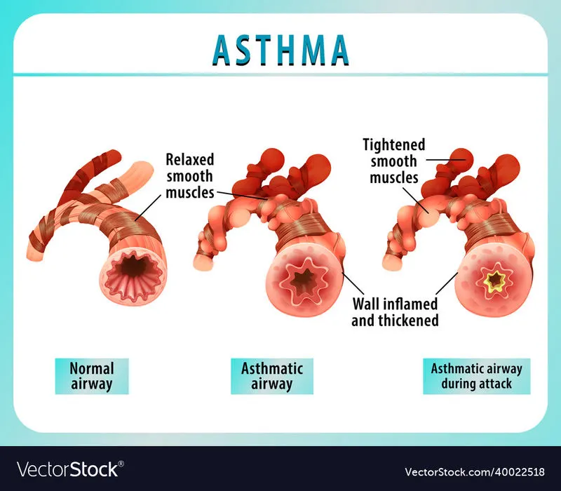
- The muscle also constricts during an asthmatic reaction, shrinking the airway and making it harder to breathe.
- The trachea is lined with cells that have cilia and secrete mucus. The mucus catches particles that have been inhaled, and the cilia move the particles toward the pharynx.
- The main function of the trachea is to funnel the inhaled air to the lungs and the exhaled air back out of the body.
Bronchi are the large tubes that connect to your trachea and direct the air you breathe to your right and left lungs. They are in your chest. Bronchi is the plural form of bronchus. The left bronchus carries air to your left lung. The right bronchus carries air to your right lung. Your bronchi are an essential part of your respiratory system. As you breathe and your lungs expand, your bronchi distribute the air within your lung.
- Bronchi carry air to and from your lungs. The bronchi also help moisturize the air you breathe and screen out foreign particles. Airways are lined with cells that create mucus.
- The mucus keeps your airways moist. It also traps bacteria, viruses, fungi and other particles to protect your lungs and prevent infection.
- The bronchi are lined with cilia, tiny hairlike structures. The cilia help move mucus (phlegm) and particles out of your lungs. When you cough or swallow, the particles trapped in the mucus move out of your body or into your digestive tract, where your body can dispose of them.
When the passages are under 1 mm in diameter when they are called bronchioles, and they split and spread through the lung.
- Bronchioles are some of the smallest airways in your lungs. Inhaled air passes through tiny ducts from the bronchioles into elastic air sacs (alveoli).
- The alveoli are surrounded by the alveolar-capillary membrane, which normally prevents liquid in the capillaries from entering the air sacs.
The lungs take up nearly all of the space in the thorax, superior to the diaphragm. As an organ, the lung is made up of airway tubes and alveoli, making it very light.
- Elastic connective tissues in the lungs allow them to expand with incoming air and recoil when expelling air.
- The lungs contain a large amount of surface area in order to efficiently support the exchange of oxygen and carbon dioxide.
Asthma is caused by inflammation of the small tubes, called bronchi, which carry air in and out of the lungs. If you have asthma, the bronchi will be inflamed and more sensitive than normal.
https://www.nhsinform.scot/illnesses-and-conditions/lungs-and-airways/asthma
Structure of bronchi :
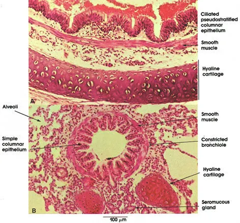
https://www.anatomyatlases.org/MicroscopicAnatomy/Section11/Plate11225.shtml
Bronchi are lined by pseudostratified columnar epithelium with goblet cells.
The thickness and layering of the epithelium decreases gradually with the decrease in size of bronchi.
A smooth muscle layer encircles a thin connective tissue lamina propria.
In contrast to the trachea, the smooth muscle of the bronchus is arranged in interlacing spirals around the bronchus.
Between the smooth muscle layer and the cartilage is the submucosa, which may contain seromucous glands.
The hyaline cartilage is arranged in discontinuous plates around the bronchus.
The connective tissue outside the muscle contains seromucous glands.
Solitary lymph nodules may be present in the mucosa and in the connective tissue around the cartilage.
As the bronchi undergo progressive reduction in size through dichotomous branching, the cartilage support is eventually lost, at which time the passageway is termed a bronchiole.
Blood supply and nerve supply of bronchi :
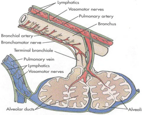
Bronchial arteries is responsible for blood supply of bronchi.
The lungs have a dual blood supply. The pulmonary artery brings oxygen-poor blood from the right ventricle of the heart. This blood passes through the pulmonary capillaries, where some carbon dioxide will leave the blood and a large amount of oxygen will be acquired.
The newly oxygenated blood enters the pulmonary veins and returns back to the left side of heart. The pulmonary circulation holds about 500 ml of blood, or about 10 percent of the body’s supply.
About 75 ml of blood is in the pulmonary capillaries for gas exchange at any one time.
The blood supply that nourishes the tissues of the lungs arrives through the bronchial artery, which branches off of the aorta and carries oxygen-rich blood to support the lung tissues.
Nerves from the pulmonary plexus enter the lungs at the hilus. These nerves contain a mixture of visceral sensory and autonomic nerve fibers that follow the bronchial tree and blood vessels.
Parasympathetic nerve stimulation results in bronchoconstriction, constriction of the bronchioles, while sympathetic nerve stimulation results in bronchodilation, dilation of the bronchioles.
https://www.sciencedirect.com/topics/agricultural-and-biological-sciences/pulmonary-circulation
Common disorders associated with bronchi :
The most common problem with the bronchi is bronchitis, an inflammation of the tubes. It can be acute or chronic.
Other problems include:
- Bronchiectasis - a condition in which damage to the airways causes them to widen and become flabby and scarred
- Exercise-induced bronchospasm - a breathing problem that happens when your airways shrink while you are exercising
- Bronchiolitis - an inflammation of the small airways that branch off from the bronchi
- Bronchopulmonary dysplasia - a chronic lung condition in infants, most often premature infants
Clinical presentation of bronchial asthma :
https://thoracickey.com/asthma-clinical-presentation-and-management/
https://www.japi.org/u2d4c494/clinical-presentations-and-investigations-in-asthma
Introduction :
Asthma is chronic inflammatory disorder of the airways characterized by marked variability in airflow obstruction.
Symptoms include wheezing, chest tightness, cough, and, occasionally, dyspnea and contributes to the heightened airway hyperresponsiveness to specific and nonspecific stimuli; a pathognomonic feature of asthma.
An estimated 300 million people worldwide suffer from asthma, with 250,000 annual deaths attributed to the disease.
Clinical presentation :
Asthma is heterogeneous in its presentation and severity, the clinical features of asthma show great variability both between individual asthmatics, and also within the same patient over time.
Asthma is often associated with different comorbidities including allergic rhinitis, atopic dermatitis, rhinosinusitis, gastroesophageal reflux disease, diabetes, depression, obesity, all of which may affect the clinical expression and severity of the disease.
Triger factors associated with asthma :
The common allergic trigger factors that may play a role in an asthmatic attack are house dust mite, pollen, fungal spore and pet. Cats are the most common household pets triggering asthma.
Non allergic triggering factors include exercise, food, drinks, drugs, viral infections, air pollution, smoking, psychological factors and climatic variations.
Association of atopy associated with asthma :
Atopy is the tendency of an individual to be hyper-responsive to certain allergens. It may be hereditary and occurs after an initial exposure with a particular allergen. It may present in various ways like eczema, allergic rhinitis, allergic conjunctivitis or allergic asthma.
Common presentations of asthma :
During childhood
- Transient infant wheezing
- Non atopic wheezing of toddler
- Late onset childhood asthma
During adulthood
- Intrinsic asthma or non-atopic asthma
Asthma is visualized after 40 years. No atopy is observed. Asthma is severe. This is characterised by female predominance and has more frequent association with nasal polyposis. The risk factors associated with this subtype are smoking and chronic rhinosinusitis.
- Asthma occurring after lower respiratory infection
Asthma followed by a severe respiratory infection. The two organisms commonly causing respiratory infection in such cases are chlamydia pneumoniae and mycoplasma pneumonia.
- Aspirin exacerbated respiratory disease (AERD)
Asthma due to allergic to aspirin called as aspirin exacerbated respiratory disease. It consists of symptom triad named Sampter's triad which include asthma, chronic rhinosinusitis with nasal polyposis and reaction to aspirin. Symptoms usually manifest 30 minutes to 3 hours after ingestion of aspirin. It is a type of pseudoallergy which is non IgE mediated and manifests as bronchospasm and nasal congestion.
- Occupational asthma
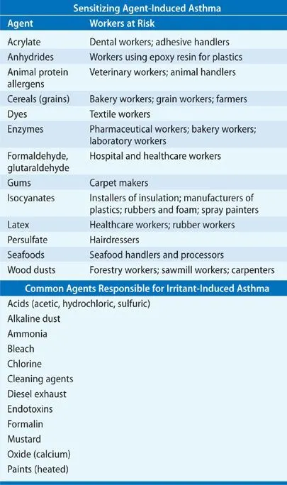
Asthma is triggered at workplace which is called occupational asthma and are immunologic and non immunologic stimuli. In immunologic type, reactive airway dysfunction syndrome (RADS) can be observed. The development of occupational asthma in workers depends on many factors such as intensity of exposure, the particular sensitising agent and host factors such as atopy, cigarette smoking and genetic predisposition
- Severe asthma
Asthma can also present in severe attacks. This type of asthma can occur at any age from childhood to adulthood and is called as severe asthma. It can be allergic or non-allergic in nature. Genetic factors found associated with these attacks are IL-12 promoter and RANTES-28L/G.
- Asthma with fixed airway obstruction
Approximately 35 to 50% of adult asthmatics suffer from fixed airflow obstruction instead of the normal variability seen in asthma. Sometimes this is seen even in children. It is due to airway inflammation and remodelling. Adult onset asthmatic males are generally more prone to develop fixed airflow obstruction. There is usually absence of atopy and persistent eosinophilia.
- Steroid resistant asthma
Glucocorticoids are the most effective agents in asthma treatment but some patients present with steroid resistant asthma. Steroid resistant asthma is defined as an FEV1 less than 75 percent of predicted, with failure to improve by 15 percent after an adequate dose and duration of glucocorticoid therapy (e.g., 40 mg/day of prednisolone for one to two weeks).
- Exercise induced asthma (EIA)
When an asthmatic patient complains of bronchoconstriction after exercise, it is called as exercise induced asthma. The magnitude of EIB is proportional to the degree of inflammation of airways. It presents as shortness of breath, chest tightness and cough. Exercise testing is usually done to confirm the diagnosis. A fall in FEV1 of more than 10% is considered to be positive.
Various diagnostic techniques and investigations used in diagnosis of the disorder :
Various laboratory investigations include
- Lung function tests (i.e., Spirometry; Bronchodilator)
- Body Plethysmography
- Bronchial challenge testing
- Blood tests
- Skin tests
- Chest Imaging (Chest radiography)
- Exhaled Nitric Oxide
- Sputum examination
One diagnostic technique in detail
Spirometry
Before going to discuss spirometry let us concentrate on Respiratory volume
Respiratory volume is an important aspect of pulmonary function testing because it can provide information about physical condition of lungs.
Respiratory volume is the volume of gas at a given time during the respiratory cycle.
Types include
- Tidal Volume (TV)
It is the amount of air inhaled or exhaled during a respiratory cycle which provides information about the mechanics of muscles, chest walls and lungs.
- Inspiratory reserve volume (IRV)
It is the amount of air that can be forcibly inhaled after a normal tidal volume. It is usually kept in reserve but is used during deep breathing. Adult can inhale between 1900 – 3300 ml
- Expiratory reserve volume (ERV)
It is the volume of air that can be exhaled forcibly after exhalation of normal tidal volume. ERV for a normal adult is between 700 – 1200 ml
- Residual volume (RV)
It is the volume of air remaining in the lungs after forced exhalation. RV for a normal adult value is averaged at 1200 ml
Total lung capacity is the maximum amount of air the lungs can accommodate.
- Total lung capacity = Sum of all volumes = TV+IRV+ERV+RV
- Vital capacity = TV+ERV+IRV (TV+ERV+IRV)
Respiratory volume is an important aspect of pulmonary function testing because it can provide information about physical condition of lungs.
It is measured by Spirometer and testing process called Spirometry
Spirometry is diagnostic procedure used to analyse proper functioning of lungs.
https://en.wikipedia.org/wiki/Spirometry
Spirometry analyse the breathing patterns in order to analyse health condition over time.
Spirometry generates pneumotachographs, which are charts that plot the volume and flow of air coming in and out of the lungs from one inhalation and one exhalation.
Spirometer is a device which is used to perform spirometry test which displays results in graphs called spirograms
Spirometers i.e., Volumetric spirometers & Flow measuring spirometers were used accordingly
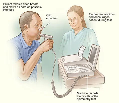
A spirometer is a diagnostic device that measures the amount of air you're able to breathe in and out. It also tracks the time it takes you to exhale completely after you take a deep breath. A spirometry test requires you to breathe into a tube attached to a machine called a spirometer.
Key spirometry measurements include the following:
Forced vital capacity (FVC) : This is the largest amount of air that you can forcefully exhale after breathing in as deeply as you can. A lower than normal FVC reading indicates restricted breathing.
Forced expiratory volume (FEV) : This is how much air you can force from your lungs in one second. This reading helps your doctor assess the severity of your breathing problems. Lower FEV-1 readings indicate more significant obstruction.
A spirometer measures ventilation, the movement of air into and out of the lungs. The spirogram will identify two different types of abnormal ventilation patterns, obstructive and restrictive. There are various types of spirometers that use a number of different methods for measurement (pressure transducers, ultrasonic, water gauge).
https://en.wikipedia.org/wiki/Spirometer
Whole body plethysmograph: This type of spirometer gives a more accurate measurement for the components of lung volumes as compared to other conventional spirometers. A person is enclosed in a small space when the measurement is taken.
Pneumotachometer : This spirometer measures the flow rate of gases by detecting pressure differences across fine mesh. One advantage of this spirometer is that the subject can breathe fresh air during the experiment.
Fully electronic spirometer : Electronic spirometers have been developed that compute airflow rates in a channel without the need for fine meshes or moving parts. They operate by measuring the speed of the airflow with techniques such as ultrasonic transducers, or by measuring pressure difference in the channel. These spirometers have greater accuracy by eliminating the momentum and resistance errors associated with moving parts such as windmills or flow valves for flow measurement. They also allow improved hygiene by allowing fully disposable air flow channels.
Incentive spirometer : This spirometer is specially designed to encourage improvement of one's lung function.
Peak flow meter : Peak expiratory flow device is useful for measuring how well a person's lungs expel air.
Windmill-type spirometer : This type of spirometer is used especially for measuring forced vital capacity without using water; it has broad measurements ranging from 1000 ml to 7000 ml. It is more portable and lighter than traditional water-tank type spirometers. This spirometer should be held horizontally while taking measurements because of the presence of a rotating disc.
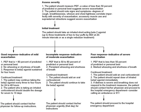
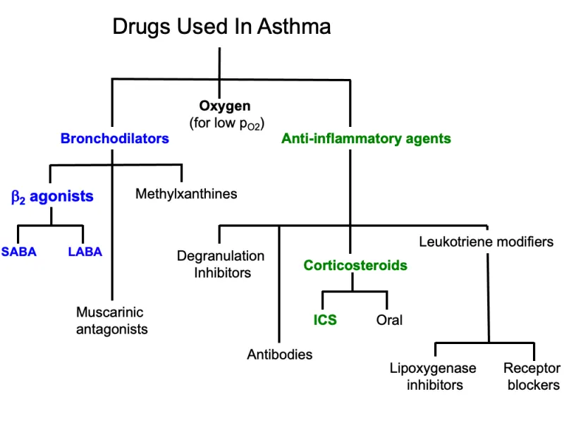
Medical management of asthma
https://www.aafp.org/pubs/afp/issues/1998/0701/p89.html
https://tmedweb.tulane.edu/pharmwiki/doku.php/treatment_of_asthma
Surgical procedure
Bronchial thermoplasty is a treatment option for people with severe asthma they can’t control with other therapies. The procedure uses heat to shrink the smooth muscles in the lungs so they can’t tighten and cause asthma symptoms.
Surgical management of asthma
The bronchi, bronchioles, and respiratory bronchioles are tremendously more susceptible to the effects of asthma. During an asthma attack the diameters of the bronchi, bronchioles, and respiratory bronchioles get smaller, reflecting roughly the same percentage of reduction noted in flow rate testing.
An attack occurs when the muscles surrounding the bronchial tubes — the airways that usher air into and out of our lungs — begin to tighten as they become inflamed and swollen. Inside the bronchial tubes, those inflamed membranes secrete thick mucus
https://www.anthem.com/blog/your-health-care/the-anatomy-of-an-asthma-attack/
Bronchial thermoplasty is a novel bronchoscopic treatment for patients with severe asthma refractory to maximal medical therapy.
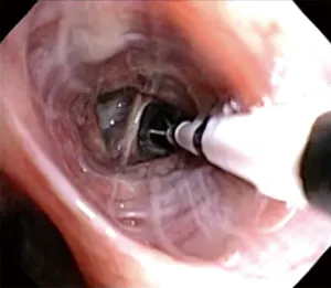
It is an FDA-approved technology that has demonstrated safety and efficacy in reducing the frequency of asthma exacerbations and improving quality of life.
It involves several bronchoscopic treatment of airways using heat therapy, which ablates airway smooth muscle, resulting in less bronchoconstriction.
In most cases, bronchial thermoplasty is an outpatient procedure. Unless you have severe side effects after the treatment, you will not have to spend the night in the hospital.
Patient will be under anesthesia during bronchial thermoplasty.
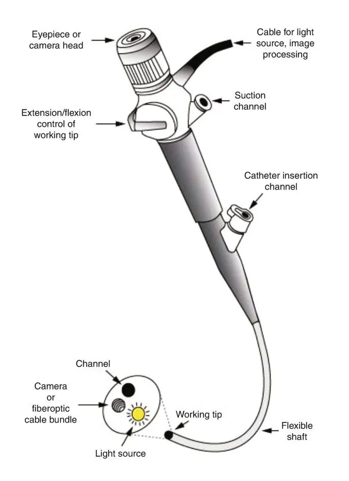
https://link.springer.com/chapter/10.1007/978-3-030-64661-5_9
Doctor inserts a thin, flexible tube called a bronchoscope through your nose or mouth and down into your lungs. The doctor then inserts a catheter (thin tube) through the bronchoscope and into your lungs. The catheter delivers the thermal heat to the smooth muscle in your lungs.
Bronchial thermoplasty doesn’t cure asthma. After the procedure, you will still need to take asthma medication to help control symptoms.
Short-term side effects can be severe. They include: Coughing up blood. Respiratory infections. Worsening asthma symptoms.
https://my.clevelandclinic.org/health/treatments/16811-bronchial-thermoplasty
Bronchial thermoplasty is a treatment option for people with severe asthma they can’t control with other therapies.
The procedure uses heat to shrink the smooth muscles in the lungs so they can’t tighten and cause asthma symptoms. Most experts agree the slight benefits of bronchial thermoplasty do not outweigh the risks for most people.
https://amj.amegroups.com/article/view/4573/html
Immediate post-procedure care is depended on individual institution guidelines. However, it is prudent to monitor patients carefully and discharged only when stable vital signs with adequate lung function (via spirometry), mental status, and able to take liquids.
Post-procedure assessment should include evaluation of gag reflex, vital signs, breath sounds, and spirometry to assess FEV1.
Typically, a 2 to 4 hours recovery or monitoring period following each procedure is needed.
An objective measure to use for discharge after BT is when post-bronchodilator FEV1 is within 80% of the pre-procedure value and patient is feeling well clinically.
Finally, it is important to stress the use of prednisone after the procedure and the adherence to prior asthma regimen.
Recent advances in severe asthma
https://www.webmd.com/asthma/newer-asthma-medications-treatments
Biologic therapy i.e., A type of treatment that uses substances made from living organisms to treat disease.
- Benralizumab (Fasenra)
- Dupilumab (Dupixent)
- Mepolizumab (Nucala)
- Omalizumab (Xolair)
- Reslizumab (Cinqair)
Eosinophils as biomarkers: Sputum eosinophilia is the most useful biomarker in asthma
https://onlinelibrary.wiley.com/doi/full/10.1111/all.14607
Seed storage proteins (Ara h 1, 2, 3, 6), uteroglobin from cat (Fel d 1), and lipocalin from dog (Can f 1) demonstrated the strongest linkage to clinical markers of asthma severity. These results suggest their potential contribution as biomarkers in preschool asthma.
Exhaled Biomarkers: Measurement of biomarkers in exhaled breath (ie, FeNO) and exhaled breath condensate (ie, volatile organic compounds) are noninvasive and safe.
FeNO is a widely accepted biomarker for type 2-driven airway inflammation.
PART B
Case Study
A 25 year old biker indulged in illegal racing met with a road traffic accident and severed head injury and skull fracture. After resuscitation and operation done for the hematoma it was resorted to do skull implant for his fracture by the orthopaedician
Type of bone the skull belongs to
- Type of bone the skull belongs to is Flat bones e., Flat bones are made of a layer of spongy bone between two thin layers of compact bone.
Microscopic anatomy of bone
- Osteon (Harversian system)
Unit of bone
- Central (Harversian) canal
Opening in the centre of an osteoma that carries blood vessels and nerves.
- Perforating (Voekmann) canal
Perforating canal is perpendicular to the central canal that also carries blood vessels and nerves.
- Lacunae
Lacunae are cavities containing bone cells i.e., Osteocytes which are arranged in concentric rings called lacunae. These are the rings around the central canal.
- Canaliculi
Canaliculi are tiny canals radiate from the central canal to lacunae that forms a transport system.
Blood supply of bone
- Nutrient artery
- Metaphyseal artery
- Epiphyseal artery
- Periosteal artery
The blood supply to bone is delivered to the endosteal cavity by nutrient arteries, then flows through marrow sinusoids before exiting via numerous small vessels that ramify through the cortex.
Bone remodeling and new bone formation
https://www.tobig.eu/bone-101/bone-101/bone-formation-and-remodeling/
Bone formation takes place to support mechanical forces, growth and reparative process. Bone remodeling is a process of constantly resorbing bone by osteoclasts and replace it by osteoblast.
Bone remodeling consists of four phases i.e.,
- Activation phase
- Resorption phase
- Reversal phase
- Bone formation by osteoblast
Mechanical stimulation stimulates bone formation and inhibits bone resorption.
Resting conditions inhibit bone formation. The remodeling cycle concludes when an equal amount of resorbed bone has been replaced.
Remodeling are most of the time develop randomly but also occur in areas that need repair. According to the Wolff’s law, bone architecture is determined by the mechanical stress on the bone.
The process of constant bone resorption and bone formation is called bone remodeling
The osteoclastic resorption of dead bone from the allograft and its replacement by new living bone made by osteoblasts from the host.
Gradual penetration across a fracture site by osteogenic tissue followed by bone formation
Bone formation is of two processes i.e.,
- Endochondral bone formation
- Intramembranous bone formation
Endochondral bone formation having following steps :
- Formation of bone collar around hyaline cartilage
- Cavitation of the hyaline cartilage
- Invasion of internal cavities by the periosteal bud and spongy bone formation
- Formation of the modullary cavity as ossification continues
- Ossification of the epiphysis
Intramembranous bone formation involves mesenchymal progenitor cells differentiating directly into osteoblasts, followed by development of the flat bones of the skull, mandible, maxilla, and clavicles.
The steps in intramembranous ossification are as follows:
- Ossification center formation
- Calcification
- Trabeculae formation
- Periosteum development
Anatomical basis of bone implant
Bone’s intricate hierarchy
- Bone consists mostly of collagen protein molecules and the mineral hydroxyapatite (HA).
- The collagen molecules intertwine to form strands of tropocollagen, which in turn align to form overlapping regions periodically separated by small gaps.
- Nanocrystals of HA populate the gaps with their axes aligned along the length of the tropocollagen strands. Many mineralized strands combine to make a fibril, the building block for higher-level architectures such as the fibers and cylindrical lamellae that form osteon structures in compact bone.
- Along the center of each osteon runs a Haversian canal, responsible for supplying the tissue with blood.
- At macroscopic length scales, two types of bone tissue are evident: compact bone, which forms an outer shell, and trabecular or spongy bone, found in the interior.
- That hierarchical organization makes bone simultaneously strong and lightweight.
(Adapted from ref. 2 and G. J. Tortora, Principles of Human Anatomy, 9th ed., Wiley, New York, 2002.)
https://physicstoday.scitation.org/doi/10.1063/PT.3.2748
Bone bonding at an implant surface
New bone tissue grows along an implant when undifferentiated cells are recruited to the implant’s surface, roughened at nanometer length scales to improve bone–implant bonding.
There, the cells become bone-producing osteogenic cells, which sequentially secrete a mineralized matrix that forms the so-called cement line and a bone precursor substance known as osteoid (red). Ensuing mineralization of the osteoid produces woven bone.
Indications and contraindications for the bone implant
Bone is a dynamic tissue, undergoing constant remodeling, and in the case of an injury, bone has the potential to regenerate with restorations of its biological and mechanical properties prior to damage.
The implants used for internal fixation are made from stainless steel and titanium, which are durable and strong. If a joint is to be replaced, rather than fixed, these implants can also be made of cobalt and chrome. Implants are compatible with the body and rarely cause an allergic reaction.
https://www.sciencedirect.com/science/article/pii/S2666138120300037
https://www.guidedessoins.com/en/contraindications-to-implantology/
Absolute contraindications
Relative contraindications
Local contraindications
Absolute contraindication means that event or substance could cause a life-threatening situation. A procedure or medicine that falls under this category must be avoided.
Absolute contraindications to implant rehabilitation include recent myocardial infarction and cerebrovascular accident, valvular prosthesis surgery, immunosuppression, bleeding issues, active treatment ofmalignancy, drug abuse, psychiatric illness, as well as intravenous bisphosphonate use.