Project - 2
Introduction: The case for additive manufacturing in orthopedics Within the healthcare and medical sector, it is the orthopedic sector that continues to adopt additive manufacturing, or 3D printing, at an astonishing rate. Industry analyst SmarTech estimates that the orthopedic segment of the additive manufacturing (AM) market generated almost $200 million in 2016 and it is forecast to grow by around 30 percent CAGR until at least the middle of this decade. Additive technology is expected to play a growing role in the ongoing evolution of more personalized medical treatment. Today, it is used for a wide range of orthopedic implants, helping designers and specialists to reimagine medical instruments and increasingly being incorporated into other types of medical equipment. Using additive technology, an implant is built layer-by-layer using a high-powered laser or electron beam to selectively melt titanium alloy powder according to computer aided design data. Complex geometries, such as porous structures, or windows can be produced in one manufacturing step. This offers the opportunity to increase the value of implants without adding cost or manufacturing time, due to the ability to design, and manufacture, unique and smart geometries with multiple design features.
Literature survey :
Mok et al., 2016 in their article “From the printer: Potential of 3D printing for orthopedic applications” has reviewed the way that 3D printing can be used for patients and the way it can be used to print PSI. The authors have also discussed the techniques and the technologies used, the materials needed for an implant based on their cell-material interactions, processing methods, mechanical properties, and chemical properties. Titanium (Ti) has excellent mechanical properties and is considered biocompatible for implantation. They have also discussed other materials that can also be used in 3D printing and bioprinting, such as natural polymers, bioceramics, synthetic, and metals. Materials for 3D printing are needed to be carefully chosen based on the proposed application of the construct. The article also discussed the various dimensions of application of 3D printing in orthopedics, including implants and prostheses for support, designing and printing customized equipment for surgical planning, and regenerating musculoskeletal tissues, including bone, cartilage, and soft tissues such as tendon, ligament, and muscles.
Wong and Scheinemann, 2018[17] reviewed the metallic biomedical materials used for fabricating orthopedic implants such as Ti-6Al-V4 (Ti-6 aluminium-4Vanadium) and CoCrMo (Cobalt-Chromium-Molybdenum) alloys in their paper “additive manufactured metallic implants for orthopedic applications.” That paper also described the processing chain in metallic implants and the AM technology based on metallic implants. Metallic implants are PSI which can be used in various orthopedic surgeries, such as spinal instrumentation, bone tumor surgery, joint replacement, and fracture fixation.
Tai et al., 2018[18] in their paper “3D printed composite for simulating thermal and mechanical responses of the cortical bone in orthopedic surgery” introduce about namely 3D polymer-infiltrated composite (3DPIC), which is a newly discovered bone-mimicking material fabricated by the binder-jetting technology and epoxy is used for the post-strengthening process. Different experiments were carried out to test 3DPIC, including the four-point bending, which is used for measuring the mechanical properties and the measurement of thermal properties. For the comparison of 3DPIC to cortical bone, thermal responses and drilling haptic of it are being tested.
Applications of Additive Manufacturing in Orthopaedics.
In orthopaedic the patient-specific analysis is essential to obtain an accurate medical imaging data of the individual patient. Magnetic resonance imaging (MRI), modern multi-row detector Computer Tomography (MDCT), Computer tomography (CT) scan, X-rays and 3D scanners provide an accurate, fast, high-resolution data and easily prepare a 3D view of patient's anatomy. AM convert the digital medical image into a 3D physical model.
| S No | Applications | Description | References |
|---|---|---|---|
| 1 | Anatomic models for the planning of surgery |
|
Pacione et al16; Vaish and Vaish17; Pietrabissa et al18; Souzaki et al8 |
| 2 | Cost-effectiveness for orthopaedics |
|
Trace et al12; Salmi et al19; Javaid et al20; Kumar et al4 |
| 3 | Surgical guides |
|
Yap et al21; Wong et al22; Javaid and Haleem1 |
| 4 | Patient-specific instruments |
|
Trace et al12; Negi et al2; Han et al23 |
| 5 | Bone tissue engineering |
|
Zein et al24; Peltola et al25 |
| 6 | 3D-printed custom implants |
|
Jardini et al11; Wong et al7 |
| 7 | Bone Defect |
|
Han et al23; Pietrabissa et al18 |
| 8 | Improved patient care |
|
Salmi et al19; Boonen et al26 |
| 9 | Precision for surgeons |
|
Yap et al21; Choi and Kim3 |
| 10 | Weight reduction of implants |
|
Wang et al27; Peltola et al25 |
| 11 | Osteochondral and Chondral defect |
|
Boonen et al26; Pacione et al16 |
The applications of additive manufacturing in orthopaedic surgery provide reproducible, safe and reliable models that improve patient outcomes and reduced operating time as compared to traditional surgical techniques. This technology can work successfully for preoperative planning, education and custom manufacturing. For Custom manufacturing applications it is used for prosthetics, surgical guides and implants. 3D printed models of anatomy have assisted in the education of students, trainees, patients and surgeons. Surgeon and doctors can use 3D printed bone model for practising of surgery and explain the patient or students about the surgery. It also has potential to increase the number of tools for the surgeon and easily undertake the redesigning. It becomes a valuable tool that has an impact on every area of medicine. Surgeons can now develop new tools and techniques for surgical procedures.
scope :
steps in AM :
- Design 3D Model Using Computer Aided Design (CAD)
- STL File Creation and File Manipulation
- Machine Setup
- Building Layer by Layer
- Part Removal
- Post-Processing
types of orthopedic implants :
- screws,
- plates,
- prostheses
Important:Contour the plate with a slight pre-bend; that is, there should be a small gap between the central part of the plate and the bone. Apply the plate and press it firmly against the bone by inserting a standard bicortical screw in the hole closest to the fracture in buttress mode.
Bone plates are the most common internal fixation implants used for fixating fractures [28]. They are attached to bone fragments with screws and function to reduce the fracture and prevent any movement, while also shielding the fracture site from stress to allow healing.
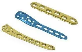
Materials :Materials involved in bone-plate design. The biocompatible materials used for bone plates are: stainless steel (SS), cobalt base alloys, bioceramics, titanium alloys, pure titanium, composite materials, and polymers (non-resorbable and bioresorbable).
Testing : Mechanical testing of these orthopedic implants can involve analysis of the implant rigidity, testing how many cycles it takes until it breaks, how the implant influences the rest of the body around it, or a multitude of other needs.
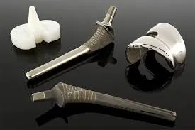
Materials used in Orthopedic implants Manufacturing
Implant Material
Metals
- Titanium
- Titanium Alloy
- Stainless steel
Orthopedic implants making process/machinery
CNC/VMC machines:
CNC machining is the manufacturing procedure where pre-modified programming computers software dictates the movements of factory tools and machinery. The procedure can be utilized to control the scope of complex machineries, from processors and machines to plants and switches. With CNC machining, the three-dimensional cutting undertaking can be cultivated in a solitary arrangement of prompts.
VMC the Vertical Machine is preferred where three-axis works are done on a single face as in molding and die work. Vertical machining occurs on a vertical machining center, (VMC), which employs a spindle with a vertical orientation. With a vertically oriented spindle, tools stick straight down from the tool holder and often cut across the top of a workpiece.
Buffing Process:
Buffing process is used to shine the metal, or any composites by the use of a cloth wheel soaked with cutting compounds or rouges. The buffing cloth holds the compound, by the time the compound does the cutting. The industry names this as the “polishing” process. Buffing normally requires two things, a cutting buff and a finishing buff. The cutting buff, which is the roughest buffing operation is good for removing pits, polishing lines or scratches.
Electropolishing process:
Electropolishing process also called as the electrochemical polishing, is a process that removes material from a metallic workpiece it reduces the roughness from the surface by levelling up the micro-peaks and the hollowness improvising the surface finish. It is used for polishing the metal parts and is also stated as the electroplating.
Laser marking process:
The laser marking process is used for marking the materials using a spot diameter laser beam and is generally used for adding marks and or brand names. A laser beam is used on a material to change and colour it.
Ultrasonic cleaning:
Ultrasonic cleaners are improving productivity in modern purifying for about 70 years; it\’s simply developed progressively compelling and solid over the long haul. More organizations and government offices than any other time in recent memory are exploiting the inventive and quick developing cleaning advancements, and they\’re receiving the rewards.
conclusion : This chapter provided a review of AM technologies, materials, design processes and survey of reported clinical applications of AM in orthopedics. Despite recent advancements in AM of orthopedic implants, there are several challenges that need to be addressed for its widespread adoption in orthopedics. In the realm of patient-specifc implants, these challenges include a need to semi-automate the data collec-tion and processing of patient’s data (DICOM data post processing, anatomic reconstruction and rendering), optimize custom design and AM process planning, and concurrent planning of design-AM-implantation due to surgical and clinical implications.
Improvements :
Immobilization of anti-bacterials for long-term release :
Due to the relatively short releasing profile and potential development of pathogen resistance of controlled antibiotics/bioactive molecules releasing coatings, there is increasing interest in immobilization of antibiotics/antimicrobial peptides to the implant surface by permanent covalent tethering for long-term prevention of implant-associated infections . Unlike other non-covalent elution coating systems, covalently immobilized antibiotics such as vanomycin can be permanently tethered to the implant surface and therefore could remain functional over the lifetime of the coating. This may be beneficial by decreasing local and systemic toxicity, as well as antibiotic resistance. Despite the advantages for non-eluting systems, this concept is limited by the fragility of the coatings and killing activity potential of bacteria which might not be directly adjacent to the implant. The best use of such systems might be in combination treatments that include systemic or locally delivered antibiotics.
Multifunctional coatings :
Osseointegration is very important to the success of orthopaedic devices implanted within bone. However, biomaterial surfaces that facilitate host cell adhesion, spreading, and growth also favor similar processes by bacteria. Infecting microorganisms share many of the same adhesive mechanisms as host tissue cells, such as extracellular matrix protein fibronectin (Fn) . This molecule is frequently used to coat implants to improve the immobilization rate of antibiotics/antimicrobial peptides, but can also be recognized by Staphylococci by its Fn-binding proteins . On the other hand, surfaces and coatings designed to inhibit bacterial colonization frequently do not effectively integrate with host tissues. Recently, the concept of multiple functionalities for surface coating of implants has been explored . However, this approach is still in the initial stage of development. These multifunctional coatings should be easily applied, efficacious, have optimal temporal and dosing release profiles, demonstrate no local and systemic toxicity, not interfere with (or possibly even facilitate) adjacent tissue integration and be cost-effective. While no one strategy has dominated the marketplace, active ongoing research will undoubtedly produce a coating technology that will mitigate the occurrence of commonly found implant infections.
reference : https://www.researchgate.net/publication/327073227_Additive_Manufacturing_of_Orthopedic_Implants_Progress_in_Biology_Manufacturing_and_Industry_Perspectives