The most common cause is infection by the bacteriumHelicobacter pylori, which accounts for more than 60% of cases. Certain types of H. pylori have greater risks than others. Smoking, dietary factors such as pickled vegetables and obesity are other risk factors. About 10% of cases run in families, and between 1% and 3% of cases are due to genetic syndromesinherited from a person's parents such as hereditary diffuse gastric cancer. Most of the time, stomach cancer develops in stages over years. Diagnosis is usually by biopsy done during endoscopy. This is followed by medical imaging to determine if the disease has spread to other parts of the body.
Signs and symptoms
Endoscopic image of linitis plastica, a type of stomach cancer where the entire stomach is invaded, leading to a leather bottle-like appearance with blood coming out of it
Endoscopic images of the stomach cancer in early stage. Its histology was poorly differentiated adenocarcinoma with signet ring cells. Left above: normal, right above: FICE, left low: acetate stained, right low: AIM stained
Stomach cancer is often either asymptomatic (producing no noticeable symptoms) or it may cause only nonspecific symptoms (which may also be present in other related or unrelated disorders) in its early stages. By the time symptoms are recognized, the cancer has often reached an advanced stage (see below) and may have metastasized (spread to other, perhaps distant, parts of the body), which is one of the main reasons for its relatively poor prognosis. Stomach cancer can cause the following signs and symptoms: Unexplained nausea, vomiting, diarrhoea and constipation. Patients also can experience unexplained weight loss.
Early cancers may be associated with indigestion or a burning sensation (heartburn). However, fewer than one in every 50 people referred for endoscopy due to indigestion has cancer. Abdominal discomfort and loss of appetite, especially for meat, can occur.
Gastric cancer can occur as a result of many factors. It occurs twice as commonly in males as females. Estrogen may protect women against the development of this form of cancer.
Infections
Helicobacter pylori infection is an essential risk factor in 65–80% of gastric cancers, but only 2% of people with H. pylori infections develop stomach cancer. The mechanism by which H. pylori induces stomach cancer potentially involves chronic inflammation, or the action of H. pylori virulence factors such as CagA. It was estimated that Epstein–Barr virus is responsible for 84,000 cases per year. AIDS is also associated with elevated risk.
Smoking
Smoking increases the risk of developing gastric cancer significantly, from 40% increased risk for current smokers to 82% increase for heavy smokers. Gastric cancers due to smoking mostly occur in the upper part of the stomach near the esophagus.
Diet
Sequence of 123-iodine human scintiscans after an intravenous injection: (from left) after 30 minutes, 20 hours, and 48 hours - a high and rapid concentration of radio-iodine is evident in gastric mucosa of the stomach, in salivary glands, oral mucosa, and the periencephalic and cerebrospinal fluid (left). In the thyroid gland, I-concentration is more progressive, also in the reservoir (from 1% after 30 minutes to 5.8% after 48 hours, of the total injected dose).
Dietary factors are not proven causes, and the association between stomach cancer and various foods and beverages is weak. Some foods including smoked foods, salt and salt-rich foods, red meat, processed meat, pickled vegetables, and brackens are associated with a higher risk of stomach cancer. Nitrates and nitrites in cured meats can be converted by certain bacteria, including H. pylori, into compounds that have been found to cause stomach cancer in animals.[citation needed]
Fresh fruit and vegetable intake, citrus fruit intake, and antioxidant intake are associated with a lower risk of stomach cancer. A Mediterranean diet is associated with lower rates of stomach cancer, as is regular aspirin use.
Obesity is a physical risk factor that has been found to increase the risk of gastric adenocarcinoma by contributing to the development of gastroesophageal reflux disease (GERD). The exact mechanism by which obesity causes GERD is not completely known. Studies hypothesize that increased dietary fat leading to increased pressure on the stomach and the lower esophageal sphincter, due to excess adipose tissue, could play a role, yet no statistically significant data have been collected. However, the risk of gastric cardia adenocarcinoma, with GERD present, has been found to increase more than 2 times for an obese person. There is a correlation between iodine deficiency and gastric cancer.
A genetic risk factor for gastric cancer is a genetic defect of the CDH1 gene known as hereditary diffuse gastric cancer (HDGC). The CDH1 gene, which codes for E-cadherin, lies on the 16th chromosome. When the gene experiences a particular mutation, gastric cancer develops through a mechanism that is not fully understood.This mutation is considered autosomal dominant, meaning that half of a carrier's children will likely experience the same mutation. Diagnosis of hereditary diffuse gastric cancer usually takes place when at least two cases involving a family member, such as a parent or grandparent, are diagnosed, with at least one diagnosed before the age of 50. The diagnosis can also be made if at least three cases occur in the family, in which case age is not considered.
The International Cancer Genome Consortium is leading efforts to identify genomic changes involved in stomach cancer.] A very small percentage of diffuse-type gastric cancers (see Histopathology below) arise from an inherited abnormal CDH1gene. Genetic testing and treatment options are available for families at risk.[49]
Diagnosis
To find the cause of symptoms, the doctor asks about the patient's medical history, does a physical examination, and may order laboratory studies. The patient may also have one or all of these exams:
Gastroscopic exam is the diagnostic method of choice. This involves insertion of a fibre optic camera into the stomach to visualise it.
Computed tomography or CT scanning of the abdomen may reveal gastric cancer. It is more useful to determine invasion into adjacent tissues or the presence of spread to local lymph nodes. Wall thickening of more than 1 cm that is focal, eccentric, and enhancing favours malignancy.
In 2013, Chinese and Israeli scientists reported a successful pilot study of a breathalyzer-style breath test intended to diagnose stomach cancer by analyzing exhaled chemicals without the need for an intrusive endoscopy. A larger-scale clinical trial of this technology was completed in 2014.
Abnormal tissue seen in a gastroscope examination is biopsied by the surgeon or gastroenterologist. This tissue is then sent to a pathologist for histological examination under a microscope to check for the presence of cancerous cells. A biopsy, with subsequent histological analysis, is the only sure way to confirm the presence of cancer cells.
Various gastroscopic modalities have been developed to increase yield of detected mucosa with a dye that accentuates the cell structure and can identify areas of dysplasia. Endocytoscopy involves ultra-high magnification to visualise cellular structure to better determine areas of dysplasia. Other gastroscopic modalities such as optical coherence tomography are being tested investigationally for similar applications.
A number of cutaneous conditions are associated with gastric cancer. A condition of darkened hyperplasia of the skin, frequently of the axilla and groin, known as acanthosis nigricans, is associated with intra-abdominal cancers such as gastric cancer. Other cutaneous manifestations of gastric cancer include "tripe palms" (a similar darkening hyperplasia of the skin of the palms) and the Leser-Trelat sign, which is the rapid development of skin lesions known as seborrheic keratoses.
Gastric adenocarcinoma is a malignant epithelial tumour, originating from glandular epithelium of the gastric mucosa. Stomach cancers are about 90% adenocarcinomas. Histologically, there are two major types of gastric adenocarcinoma (Lauren classification): intestinal type or diffuse type. Adenocarcinomas tend to aggressively invade the gastric wall, infiltrating the muscularis mucosae, the submucosa and then the muscularis propria. Intestinal type adenocarcinoma tumour cells describe irregular tubular structures, harbouring pluristratification, multiple lumens, reduced stroma ("back to back" aspect). Often, it associates intestinal metaplasia in neighbouring mucosa. Depending on glandular architecture, cellular pleomorphism and mucosecretion, adenocarcinoma may present 3 degrees of differentiation: well, moderate and poorly differentiated. Diffuse type adenocarcinoma (mucinous, colloid, linitis plastica or leather-bottle stomach) tumour cells are discohesive and secrete mucus, which is delivered in the interstitium, producing large pools of mucus/colloid (optically "empty" spaces). It is poorly differentiated. In signet-ring cell carcinomas, the mucus remains inside the tumour cell and pushes the nucleus to the periphery, giving rise to signet-ring cells.[citation needed]
If cancer cells are found in the tissue sample, the next step is to stage, or find out the extent of the disease. Various tests determine whether the cancer has spread, and if so, what parts of the body are affected. Because stomach cancer can spread to the liver, pancreas, and other organs near the stomach, as well as to the lungs, the doctor may order a CT scan, a PET scan, an endoscopic ultrasound exam, or other tests to check these areas. Blood tests for tumor markers, such as carcinoembryonic antigen and carbohydrate antigen may be ordered, as their levels correlate to extent of metastasis, especially to the liver, and the cure rate.
Staging may not be complete until after surgery. The surgeon removes nearby lymph nodes and possibly samples of tissue from other areas in the abdomen for examination by a pathologist.
The clinical stages of stomach cancer are:
Stage 0 – Limited to the inner lining of the stomach, it is treatable by endoscopic mucosal resection when found very early (in routine screenings), or otherwise by gastrectomy and lymphadenectomy without need for chemotherapy or radiation.
Stage I – Penetration to the second or third layers of the stomach (stage 1A) or to the second layer and nearby lymph nodes (stage 1B): Stage 1A is treated by surgery, including removal of the omentum. Stage 1B may be treated with chemotherapy (5-fluorouracil) and radiation therapy.
Stage II – Penetration to the second layer and more distant lymph nodes, or the third layer and only nearby lymph nodes, or all four layers but not the lymph nodes, it is treated as for stage I, sometimes with additional neoadjuvant chemotherapy.
Stage III – Penetration to the third layer and more distant lymph nodes, or penetration to the fourth layer and either nearby tissues or nearby or more distant lymph nodes, it is treated as for stage II; a cure is still possible in some cases.
Stage IV – Cancer has spread to nearby tissues and more distant lymph nodes, or has metastasized to other organs. A cure is very rarely possible at this stage. Some other techniques to prolong life or improve symptoms are used, including laser treatment, surgery, and/or stents to keep the digestive tract open, and chemotherapy by drugs such as 5-fluorouracil, cisplatin, epirubicin, etoposide, docetaxel, oxaliplatin, capecitabine, or irinotecan.
In a study of open-access endoscopy in Scotland, patients were diagnosed 7% in stage I, 17% in stage II, and 28% in stage III. A Minnesota population was diagnosed 10% in stage I, 13% in stage II, and 18% in stage III.However, in a high-risk population in the Valdivia Province of southern Chile, only 5% of patients were diagnosed in the first two stages and 10% in stage III.
Getting rid of H. pylori in those who are infected decreases the risk of stomach cancer, at least in those who are Asian A 2014 meta-analysis of observational studies found that a diet high in fruits, mushrooms, garlic, soybeans, and green onions was associated with a lower risk of stomach cancer in the Korean population. Low doses of vitamins, especially from a healthy diet, decrease the risk of stomach cancer.A previous review of antioxidant supplementation did not find supporting evidence and possibly worse outcomes.
Management
Treatment for stomach cancer may include surgery, chemotherapy, or radiation therapy. New treatment approaches such as immunotherapy or gene therapy and improved ways of using current methods are being studied in clinical trials.
Surgery
Anatomy before Roux-en-y surgery to resect stomach cancer
Surgery remains the only curative therapy for stomach cancer. The different surgical techniques, endoscopic mucosal resection (EMR) is a treatment for early gastric cancer (tumor only involves the mucosa) that was pioneered in Japan and is available in the United States at some centers. In EMR, the tumor, together with the inner lining of stomach (mucosa), is removed from the wall of the stomach using an electrical wire loop through the endoscope. The advantage is that it is a much smaller operation than removing the stomach. Endoscopic submucosal dissection is a similar technique pioneered in Japan, used to resect a large area of mucosa in one piece. If the pathologic examination of the resected specimen shows incomplete resection or deep invasion by tumor, the patient would need a formal stomach resection. A 2016 Cochrane review found low-quality evidence of no difference in short-term mortality between laparoscopic and open gastrectomy (removal of stomach), and that benefits or harms of laparoscopic gastrectomy cannot be ruled out. Post-operatively, up to 70% of people undergoing total gastrectomy develop complications such as dumping syndrome and reflux esophagitis. Construction of a "pouch", which serves as a "stomach substitute", reduced the incidence of dumping syndrome and reflux esophagitis by 73% and 63% respectively, and led to improvements in quality-of-life, nutritional outcomes, and body mass index.
Those with metastatic disease at the time of presentation may receive palliative surgery, and while it remains controversial, due to the possibility of complications from the surgery itself and because it may delay chemotherapy, the data so far are mostly positive, with improved survival rates being seen in those treated with this approach.Chemotherapy
The use of chemotherapy to treat stomach cancer has no firmly established standard of care Unfortunately, stomach cancer has not been particularly sensitive to these drugs, and chemotherapy, if used, has usually served to palliatively reduce the size of the tumor, relieve symptoms of the disease, and increase survival time. Some drugs used in stomach cancer treatment have included: fluorouracil or its analog capecitabine, BCNU (carmustine), methyl-CCNU (semustine) and doxorubicin (Adriamycin), as well as mitomycin C, and more recently cisplatin and taxotere, often using drugs in various combinations. The relative benefits of these different drugs, alone and in combination, are unclear. Clinical researchers are exploring the benefits of giving chemotherapy before surgery to shrink the tumor, or as adjuvant therapy after surgery to destroy remaining cancer cells.
Targeted therapy
treatment with human epidermal growth factor receptor 2 (HER2) inhibitor, trastuzumab, has been demonstrated to increase overall survival in inoperable locally advanced or metastatic gastric carcinoma over-expressing the HER2/neu gene. In particular, HER2 is overexpressed in 13–22% of patients with gastric cancer.Of note, HER2 overexpression in gastric neoplasia is heterogeneous and comprises a minority of tumor cells (less than 10% of gastric cancers overexpress HER2 in more than 5% of tumor cells). Hence, this heterogeneous expression should be taken into account for HER2 testing, particularly in small samples such as biopsies, requiring the evaluation of more than one bioptic sample.
2. Urinary system :
Cystitis usually occurs when bacteria that typically live harmlessly on the skin or in the bowel enter the urethra and bladder. The bacteria then fastens to the lining of the bladder and causes the area to become irritated and inflamed. This is known as a bladder infection.
Cystitis can affect anyone, regardless of sex or age. But it is more commonTrusted Source among females. One reason for this is that females have a shorter urethra that is closer to the anus. This means that bacteria can enter the bladder more easily.
Cystitis may be either acute or interstitial. Acute cystitis is cystitis that happens suddenly. Interstitial cystitis is longer term, and is also known as chronic cystitis. The exact cause of interstitial cystitis is yet unknown, but researchers are working to understand it.
There are several types of cystitis. The different types are determined based on their causes, and include:
Bacterial cystitis: This occurs when bacteria enter the bladder through the urethra and irritate the bladder walls.
Drug-induced cystitis: Some medications can irritate the bladder as they exit the body. Certain chemotherapy drugs can causeTrusted Source a person to develop cystitis.
Radiation cystitis: Radiation therapy can shrink tumors and kill cancer cells, but it can also damage healthy cells. Radiation cystitis typicallyTrusted Source occurs following radiation therapy that affects the pelvic region.
Foreign body cystitis: This can occur if a person receives treatment using a catheter, stent, or other endoscopic instruments. Bacteria may enter the urethra through the insertion or removal of the catheter or stent, or damage from the insertion or removal can cause inflammation.
Chemical cystitis: This is the inflammation of the bladder that results from some forms of treatment, such as chemotherapeutic agents. It can also developTrusted Source as a result of soaps, spermicide, gels, and dyes.
A person can also develop asymptomatic bacteriuria. This is when there is the presence of bacteria in the urine, but a person does not present with any symptoms of a UTI. MostTrusted Source people will not require treatment, but those who are pregnant may benefit from treatment.
Causes
Acute cystitis typicallyTrusted Source occurs as a result of bacteria called Escherichia coli. Other bacterial causes include:
Proteus mirabilis
Klebsiella pneumoniae
Staphylococcus saprophyticus
Group B streptococci
Lactobacillus
S. saprophyticus
enterococci
In males, cystitis can also occur due to problems that restrict typical urine flow.
An example of this is an enlarged prostate, which could prevent someone from being able to urinate normally.
inserting, changing, or prolonged use of a urinary catheter
using a diaphragm for birth control
difficulty fully emptying the bladder
sexual activity and friction from sexual activity
radiotherapy
diabetes, due to the high sugar levels in the urine providing a good environment for the bacteria to grow
Males who have unprotected anal sex have a slightly higher chance of cystitis.
People going through menopause also have a higher chance of developing cystitis. During menopause, estrogen levels drop and the lining of the urethra gets thinner. The thinner the lining becomes, the higher the chances are of infection and damage.
Symptoms
The symptoms of cystitis may vary depending on what type of cystitis a person has.
Acute cystitis
For cases of acute cystitis, the symptoms may include:
This is where a person develops cystitis with the presenceTrusted Source of blood in the urine.
A person will experience the same symptoms of acute cystitis, and the blood may or may not be visible in the urine. In some cases, the blood is only visible under a microscope.
Hemorrhagic cystitis
This is where the blood vessels in the lining of the bladder are also damaged.
Symptoms will include blood in the urine, along with:
pain when urinating
burning or stinging when passing urine
urinating more often
feeling a strong sense of urgency to urinate
loss of bladder control
Hemorrhagic cystitis can occur as a complication of radiation therapy. Without treatment, hemorrhagic cystitis will progress through stagesTrusted Source:
Grade I: This involves microscopic bleeding.
Grade II: The bleeding is visible.
Grade III: The bleeding contains small clots.
Grade IV: There is bleeding with large clots that may obstruct the flow of urine.
Interstitial cystitis
Interstitial cystitis may cause:
pain in the lower abdominal, urethral, or vaginal area
frequent need to urinate
urgent need to urinate
According to the Urology Care Foundation, males who have interstitial cystitis may also experience pain in their testicles, scrotum, and perineum, as well as pain when they ejaculate.
People with interstitial cystitis may experience acute pain or chronic pain. Acute pain is sudden pain for a short period of time, whereas chronic pain is pain that may recur frequently, last longer, or both.
Vs. UTI
Cystitis is a type of UTI. UTIs are infections of the urinary tract, meaning that they can include anything from the urethra to the kidneys, as well as the bladder. An infection in the bladder is cystitis. Bacterial cystitis is a type of UTI, but other types of cystitis may not be.
Lower tract UTIs affect the urethra and bladder, whereas upper tract UTIs affect the kidneys. An upper tract UTI can be life threatening if the bacteria move from the infected kidney to the bloodstream.
Symptoms of a lower tract UTI are the symptoms of bacterial cystitis. Symptoms of an upper tract UTI may also include:
pain in the upper back
fever
nausea
vomiting
chills
Treatment
Most cases of mild cystitis will resolve themselves within a few days. If cystitis lasts more than 3 days, a person should discuss this with a doctor.
A doctor may prescribe antibiotics. The type and duration of antibiotic treatment will depend on the individual. For example, a doctor may prescribe:
Nitrofurantoin: A person may take 100 milligrams (mg) twice a day for 5–7 days.
Sulfamethoxazole-trimethoprim: A person may take this twice a day for 3 days.
Fosfomycin: This entails a single dose of 4 mg.
If symptoms do not ease after taking the antibiotics, a person should return to the doctor.
Home remedies
To help ease symptoms at home, a person can take pain relievers such as acetaminophen or ibuprofen. They should also drink plenty of water.
Cranberries contain an active ingredientTrusted Source that helps prevent bacteria from sticking to the bladder wall. But cranberry juice or capsules may not contain enough of the active ingredient to prevent symptoms.
A person should also avoid alcohol.
Diagnosis
A doctor will ask the person some questions, carry out an examination, and do a urine test. The urine test will either be sent to a laboratory, or the doctor may use a dipstick. Urine dipstick results come back quickly while the person is still in the office.
A urine culture or catheterized urine specimen may be needed to determine the type of bacteria in the urine. After finding out which specific bacterium is causing the infection, the doctor will prescribe an oral antibiotic.
People who regularly develop cystitis may need further tests. These could include an ultrasound scan, X-ray, or cystoscopy of the bladder using a fiber-optic camera.
CT scans can be performed on every region of the body for a variety of reasons (e.g., diagnostic, treatment planning, interventional, or screening). Most CT scans are performed as outpatient procedures. Computed tomography was originally known as the "EMI scan" as it was developed at a research branch of EMI, a company best known today for its music and recording business. It was later known as computed axial tomography (CAT or CT scan) and body section röntgenography.
The cross-sectional images generated during a CT scan can be reformatted in multiple planes, and can even generate three-dimensional images which can be viewed on a computer monitor, printed on film or transferred to electronic media.
Prevention
Cystitis is often not preventable, but the following measures may help:
practicing good hygiene after sex
using neutral, unperfumed soaps around the genitals
emptying the bladder completely when urinating
not postponing urination
avoiding tight underwear and tight pants
wearing cotton underwear
wiping from front to back
using a lubricant during sex
Those who use catheters should ask a doctor or nurse how to avoid damage when changing the catheter.
3. repiratory system :
Lung cancer
Non-small cell lung cancer is a disease in which malignant (cancer) cells form in the tissues of the lung.
There are several types of non-small cell lung cancer.
Smoking is the major risk factor for non-small cell lung cancer.
Signs of non-small cell lung cancer include a cough that doesn't go away and shortness of breath.
Tests that examine the lungs are used to diagnose and stage non-small cell lung cancer.
If lung cancer is suspected, a biopsy is done.
Certain factors affect prognosis (chance of recovery) and treatment options.
For most patients with non-small cell lung cancer, current treatments do not cure the cancer.
Non-small cell lung cancer is a disease in which malignant (cancer) cells form in the tissues of the lung.
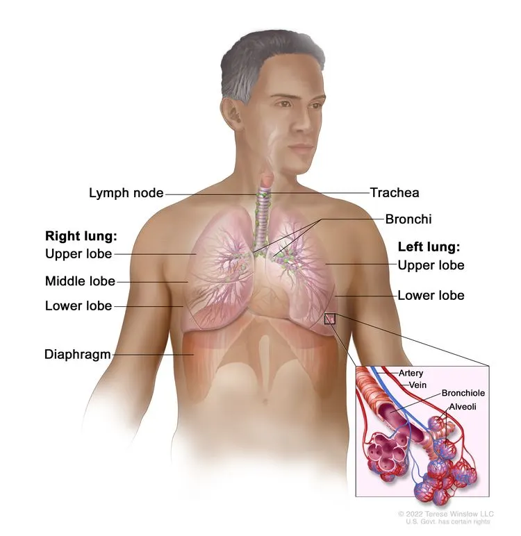
The lungs are a pair of cone-shaped breathing organs in the chest. The lungs bring oxygen into the body as you breathe in. They release carbon dioxide, a waste product of the body’s cells, as you breathe out. Each lung has sections called lobes. The left lung has two lobes. The right lung is slightly larger and has three lobes. Two tubes called bronchi lead from the trachea (windpipe) to the right and left lungs. The bronchi are sometimes also involved in lung cancer. Tiny air sacs called alveoli and small tubes called bronchioles make up the inside of the lungs.
ENLARGEAnatomy of the respiratory system showing the trachea, the right and left lungs and their lobes, and the bronchi. The lymph nodes and the diaphragm are also shown. Oxygen is inhaled into the lungs and passes through the alveoli (the tiny air sacs at the end of the bronchioles) and into the bloodstream (see inset), where it travels to the tissues throughout the body.
A thin membrane called the pleura covers the outside of each lung and lines the inside wall of the chest cavity. This creates a sac called the pleural cavity. The pleural cavity normally contains a small amount of fluid that helps the lungs move smoothly in the chest when you breathe.
There are several types of non-small cell lung cancer.
Each type of non-small cell lung cancer has different kinds of cancer cells. The cancer cells of each type grow and spread in different ways. The types of non-small cell lung cancer are named for the kinds of cells found in the cancer and how the cells look under a microscope:
Squamous cell carcinoma: Cancer that forms in the thin, flat cells lining the inside of the lungs. This is also called epidermoid carcinoma.
Smoking is the major risk factor for non-small cell lung cancer.
Anything that increases your chance of getting a disease is called a risk factor. Having a risk factor does not mean that you will get cancer; not having risk factors doesn't mean that you will not get cancer. Talk to your doctor if you think you may be at risk for lung cancer.
Risk factors for lung cancer include the following:
Smoking cigarettes, pipes, or cigars, now or in the past. This is the most important risk factor for lung cancer. The earlier in life a person starts smoking, the more often a person smokes, and the more years a person smokes, the greater the risk of lung cancer.
Older age is the main risk factor for most cancers. The chance of getting cancer increases as you get older.
When smoking is combined with other risk factors, the risk of lung cancer is increased.
Signs of non-small cell lung cancer include a cough that doesn't go away and shortness of breath.
Sometimes lung cancer does not cause any signs or symptoms. It may be found during a chest x-ray done for another condition. Signs and symptoms may be caused by lung cancer or by other conditions. Check with your doctor if you have any of the following:
Chest discomfort or pain.
A cough that doesn’t go away or gets worse over time.
Tests that examine the lungs are used to diagnose and stage non-small cell lung cancer.
Tests and procedures to diagnose and stage non-small cell lung cancer are often done at the same time. Some of the following tests and procedures may be used:
Physical exam and health history: An exam of the body to check general signs of health, including checking for signs of disease, such as lumps or anything else that seems unusual. A history of the patient’s health habits, including smoking, and past jobs, illnesses, and treatments will also be taken.
Laboratory tests: Medical procedures that test samples of tissue, blood, urine, or other substances in the body. These tests help to diagnose disease, plan and check treatment, or monitor the disease over time.
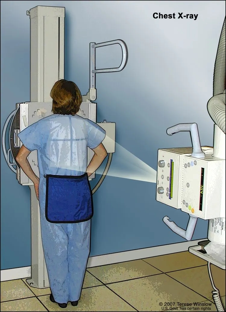
Chest x-ray: An x-ray of the organs and bones inside the chest. An x-ray is a type of energy beam that can go through the body and onto film, making a picture of areas inside the body.
ENLARGEX-ray of the chest. X-rays are used to take pictures of organs and bones of the chest. X-rays pass through the patient onto film.
CT scan (CAT scan): A procedure that makes a series of detailed pictures of areas inside the body, such as the chest, taken from different angles. The pictures are made by a computer linked to an x-ray machine. A dye may be injected into a vein or swallowed to help the organs or tissues show up more clearly. This procedure is also called computed tomography, computerized tomography, or computerized axial tomography.
Sputum cytology: A procedure in which a pathologist views a sample of sputum (mucus coughed up from the lungs) under a microscope, to check for cancer cells.
Thoracentesis: The removal of fluid from the space between the lining of the chest and the lung, using a needle. A pathologist views the fluid under a microscope to look for cancer cells.
If lung cancer is suspected, a biopsy is done.
One of the following types of biopsies is usually used:
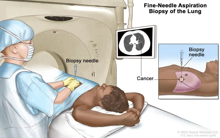
Fine-needle aspiration (FNA) biopsy of the lung: The removal of tissue or fluid from the lung using a thin needle. A CT scan, ultrasound, or other imaging procedure is used to locate the abnormal tissue or fluid in the lung. A small incision may be made in the skin where the biopsy needle is inserted into the abnormal tissue or fluid. A sample is removed with the needle and sent to the laboratory. A pathologist then views the sample under a microscope to look for cancer cells. A chest x-ray is done after the procedure to make sure no air is leaking from the lung into the chest.
ENLARGEFine-needle aspiration biopsy of the lung. The patient lies on a table that slides through the computed tomography (CT) machine, which takes x-ray pictures of the inside of the body. The x-ray pictures help the doctor see where the abnormal tissue is in the lung. A biopsy needle is inserted through the chest wall and into the area of abnormal lung tissue. A small piece of tissue is removed through the needle and checked under the microscope for signs of cancer.
An endoscopic ultrasound (EUS) is a type of ultrasound that may be used to guide an FNA biopsy of the lung, lymph nodes, or other areas. EUS is a procedure in which an endoscope is inserted into the body. An endoscope is a thin, tube-like instrument with a light and a lens for viewing. A probe at the end of the endoscope is used to bounce high-energy sound waves (ultrasound) off internal tissues or organs and make echoes. The echoes form a picture of body tissues called a sonogram.
4. cardiovascular disorder
A stroke is a serious life-threatening medical condition that happens when the blood supply to part of the brain is cut off.
Strokes are a medical emergency and urgent treatment is essential.
The sooner a person receives treatment for a stroke, the less damage is likely to happen.
If you suspect that you or someone else is having a stroke, phone 999 immediately and ask for an ambulance.
Symptoms of a stroke
The main symptoms of stroke can be remembered with the word FAST:
Face – the face may have dropped on 1 side, the person may not be able to smile, or their mouth or eye may have dropped.
Arms – the person with suspected stroke may not be able to lift both arms and keep them there because of weakness or numbness in 1 arm.
Speech – their speech may be slurred or garbled, or the person may not be able to talk at all despite appearing to be awake; they may also have problems understanding what you're saying to them.
Time – it's time to dial 999 immediately if you see any of these signs or symptoms.
Sort by:
Popular
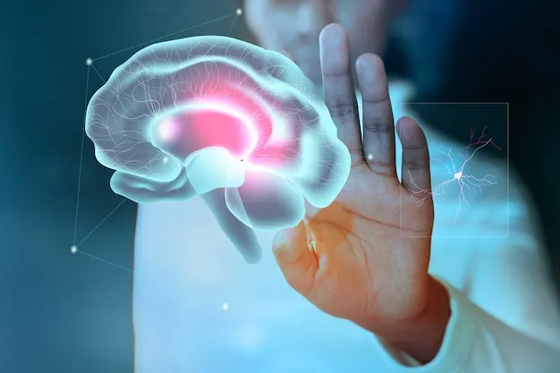
Brain study background for mental health care medical technology
The first step in assessing a stroke patient is to determine whether the patient is experiencing an ischemic or hemorrhagic stroke so that the correct treatment can begin. A CT scan or MRI of the head is typically the first test performed.
Computed tomography (CT) of the head: CT scanning combines special x-ray equipment with sophisticated computers to produce multiple images or pictures of the inside of the body. Physicians use CT of the head to detect a stroke from a blood clot or bleeding within the brain. To improve the detection and characterization of stroke, CT angiography (CTA) may be performed. In CTA, a contrast material may be injected intravenously and images are obtained of the cerebral blood vessels. Images that detect blood flow, called CT perfusion (CTP), may be obtained at the same time. The combination of CT, CTA and CTP can help physicians decide on the best therapy for a patient experiencing a stroke.
MRI of the head: MRI uses a powerful magnetic field, radio frequency pulses and a computer to produce detailed pictures of organs, soft tissues, bone and virtually all other internal body structures. MR is also used to image the cerebral vessels, a procedure called MR angiography (MRA). Images of blood flow are produced with a procedure called MR perfusion (MRP). Physicians use MRI of the head to assess brain damage from a stroke.
To help determine the type, location, and cause of a stroke and to rule out other disorders, physicians may use:
Blood tests.
Electrocardiogram (ECG, EKG): An electrocardiogram, which checks the hearts' electrical activity, can help determine whether heart problems caused the stroke.
Carotid ultrasound/Doppler ultrasound: Ultrasound imaging involves exposing part of the body to high-frequency sound waves to produce pictures of the inside of the body. Physicians use a special ultrasound technique called Doppler ultrasound to check for narrowing and blockages in the body's two carotid arteries, which are located on each side of the neck and carry blood from the heart to the brain. Doppler ultrasound produces detailed pictures of these blood vessels and information on blood flow.
Cerebral angiography. Angiography is a medical test that is performed with one of three imaging technologies—x-rays, CT or MRI, and in some cases a contrast material, to produce pictures of major blood vessels in the brain. Cerebral angiography helps physicians detect or confirm abnormalities such as a blood clot or narrowing of the arteries.
Doctor tracking patient evolution during neurology headset test. neorological center
DCStudio
Patient woman with scanning device on her head and doctor scheduling it with tablet in hand. neurological clinic.
Treating a stroke
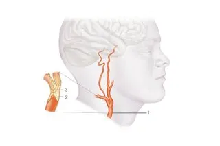
Carotid endarterectomy is a surgical procedure to remove a build-up of fatty deposits (plaque), which cause narrowing of a carotid artery.
The carotid arteries are the main blood vessels that supply blood to the neck, face and brain.
Carotid endarterectomies are carried out when 1 or both carotid arteries become narrowed because of a build-up of fatty deposits (plaque).
This is known as carotid artery disease or carotid artery stenosis.
There is a carotid artery on either side of the neck.
Carotid artery
Area of plaque blocking the artery
Area of blood clot in the artery
If a narrowed carotid artery is left untreated, blood flow to the brain may be affected.
This is usually because the carotid artery becomes blocked or a blood clot forms and a piece breaks off and goes to the brain.
This can result in either:
a stroke – a serious medical condition that can cause brain damage or death
a transient ischaemic attack (TIA) – sometimes known as a "mini-stroke", a TIA is similar to a stroke but the signs and symptoms are temporary and usually disappear within 24 hours
About a quarter of strokes in the UK are caused by a narrowing of the carotid arteries.
A carotid endarterectomy can significantly reduce the risk of a stroke in people with severely narrowed carotid arteries.
In people who have previously had a stroke or a TIA, surgery can significantly reduce their risk of having another stroke or TIA.
It's now thought the operation should be carried out as soon as possible after symptoms appear.
It's therefore important to get immediate medical advice if you experience symptoms such as:
The advantage of local anaesthetic is it allows the surgeon to monitor brain function while you're awake. But there's no evidence that either is safer or better.
During the procedure, a 7 to 10cm cut is made on the side of the neck and then a small cut is made along the narrowed section of artery, so the fatty deposits can be removed.
The artery is closed with stitches or a patch and your skin is also closed with stitches.
What happens after the procedure
You'll usually be moved to the recovery area of the operating theatre for monitoring for about 3 hours, before returning to the vascular ward.
Most people are well enough to go home within about 48 hours of the procedure.
In most cases, the only problems experienced after the operation are temporary numbness or discomfort in the neck.
But there's a small risk of more serious complications, which can include stroke or death in 2 to 3% of cases.
Nevertheless, this risk is much lower than in people with carotid artery disease who haven't chosen to have the operation.
Are there any alternatives?
Carotid endarterectomy is the main treatment for narrowing of the carotid arteries, but sometimes an alternative procedure called carotid artery stent placement may be available.
It's a less invasive procedure than a carotid endarterectomy because there's no need to make a cut in the neck.
Instead, a thin flexible tube is guided to the carotid artery through a small cut in the groin.
A mesh cylinder (stent) is then placed into the narrowed section of artery to widen it and allow blood to flow through it more easily.
Current guidelines recommend that a carotid endarterectomy should be the first line of treatment for most people.
This is because carotid stenting is associated with a higher risk of stroke during the procedure, particularly if it's carried out in the first few days after symptoms appear.
But it's an important alternative for some people who may otherwise be considered to be high risk because of other medical problems.
Treatment depends on the type of stroke you have, including which part of the brain was affected and what caused it.
Strokes are usually treated with medicine. This includes medicines to prevent and dissolve blood clots, reduce blood pressure and reduce cholesterol levels.
In some cases, procedures may be required to remove blood clots. Surgery may also be needed to treat brain swelling and reduce the risk of further bleeding if this was the cause of your stroke.
Recovering from a stroke
People who survive a stroke are often left with long-term problems caused by injury to their brain.
Some people need a long period of rehabilitation before they can recover their former independence, while many never fully recover and need ongoing support after their stroke.
Local authorities should provide free reablement services for anyone assessed as needing them. These services help the person recovering from a stroke learn or relearn the skills they need to live at home independently.
Some people will continue to need some form of care or help with their daily activities.
For example, a care worker may come to the person's home to help with washing and dressing, or to provide companionship.
If you have a condition that increases your risk of a stroke, it's important to manage it effectively. For example, taking medicine you've been prescribed to lower high blood pressure or cholesterol levels.
If you've had a stroke or TIA in the past, these measures are particularly important because your risk of having another stroke is greatly increased.
Stroke in children
Stroke does not just affect adults. Every year around 400 children in the UK have a stroke, according to the Stroke Association.

_H%26E_magn_400x.jpg)
.jpg)

 rawpixel.com
 kjpargeter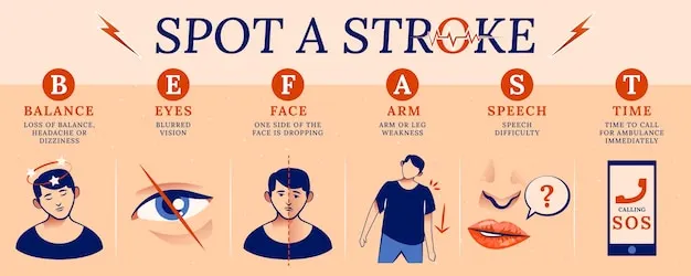
 freepik
 DCStudio