Project 2
1. A kidney transplant is a surgery to place a healthy kidney from a living or deceased donor into a person whose kidneys no longer function properly. When kidneys lose this filtering ability, harmful levels of fluid and waste accumulate in the body, which can raise blood pressure and result in kidney failure (end-stage renal disease). End-stage renal disease occurs when the kidneys have lost about 90% of their ability to function normally. End-stage renal disease occurs when the kidneys have lost about 90% of their ability to function normally.
Common causes of end-stage kidney disease include:
- Diabetes
- Chronic, uncontrolled high blood pressure
- Chronic glomerulonephritis — an inflammation and eventual scarring of the tiny filters within the kidneys
- Polycystic kidney disease
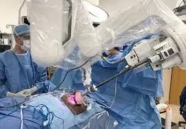
Kidney transplantation can treat advanced kidney disease and kidney failure, but the surgery isn't a cure. Some forms of kidney disease may return after a transplant.
The health risks associated with a kidney transplant include those associated directly with the surgery and rejection of the donor organ. Risks also include the side effects of taking anti-rejection medications (immunosuppressants) needed to prevent the body from rejecting the donated kidney.
Deciding whether a kidney transplant is right for you is a personal decision that deserves careful thought and consideration of the serious risks and benefits. Talk through your decision with your family, friends and other trusted advisers.
Kidney transplant surgery carries a risk of significant complications, including:
- Blood clots and bleeding
- Leaking from or blockage of the tube that links the kidney to the bladder (ureter)
- Infection
- Failure or rejection of the donated kidney
- An infection or cancer that can be passed on from the donated kidney
- Death, heart attack and stroke
2. https://www.ncbi.nlm.nih.gov/ Pranjal Modi, Bipinchandra Pal, Jayesh Modi, Suresh Kumar, Akshay Sood,1 and Mani Menon1
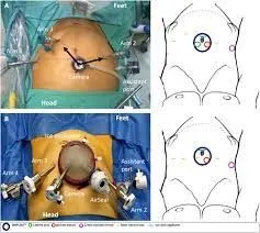
The kidney was placed in the retroperitoneum through an open incision (7.7 ± 1.04 cm) in the iliac fossa. Dissection was carried out till the exposure of the iliac vessels from the umbilical level to the urinary bladder. The robot was docked from behind the patient's back and the assistant surgeon stood between the two legs of the patient. The table was tilted 15° to the left side for right iliac fossa transplantation. Two working ports were placed at the umbilicus and in the anterior axillary line. The anterior abdominal wall was lifted up 3 cm by dVSS robotic arm ports. The kidney was placed in the wound after controlling the iliac vessels and the wound was left open. Vascular anastomosis was carried out by the robot and the robot was undocked. The console surgeon scrubbed to perform ureteric reimplantation by the open method. This method is similar to one described in year 2002.
https://journals.lww.com/ Case Report
A 63-year-old man with end-stage renal disease managed by hemodialysis for 4 years and arterial hypertension and diabetes mellitus under pharmacological control underwent robotic-assisted dual KT. He provided his written informed consent to the procedure. The patient had not undergone prior surgical treatment. The kidneys came from a marginal donor, a 70-year-old man who had died of cerebral hemorrhage. Pretransplant donor biopsy demonstrated a bilateral Karpinski13 score greater than 5. The organs did not exhibit malformations and had an artery and a vein each. Once removed, they were perfused with Viaspan (University of Wisconsin Solution; Bristol-Myers Squibb, Chester, UK) and stored in ice.
Living donor kidney transplantation [https://www.intechopen.com/]
RAKT has recently been introduced to reduce the morbidity of open KT. Since the first RAKT in 2010 by Giulianotti et al. in the USA, this technique has been standardized and evaluated in highly experienced robot and KT centers around the world. In 2014 Menon et al. standardized the technique with the transperitoneal approach and regional hypothermia known as the Vattikuti-Medanta technique. The authors highlighted that RAKT is a safe technique with possible advantages such as low intra- and postoperative complications, better cosmetic results, and superlative vision that could result in better quality of the vascular and ureteral anastomoses. The first two European pure RAKTs were performed in July 2015 by Breda et al. and Doumerc et al.. In 2016, the European Association of Urology (EAU) formed the EAU Robotic Urology Section (ERUS) RAKT working group in order to prospectively follow the outcomes of RAKT. Breda et al. published the largest multicenter series of RAKT to date (120 patients). Angelo Territo et al. addressed the functional results of RAKT from living donors at 1-year follow-up, and Vignolini et al. developed a RAKT program with grafts from deceased donors. Siena et al. described the technique for RAKT in grafts with multiple vessels. The feasibility of RAKT in children was described by the Ghent University group by Spinoit et al..
3.