Project 2
Project(a)
1. Pathological Condition 1- Gallstone in the Digestive system
The normal function of the gallbladder is to store bile. The bile helps the digestive system to breakdown fats and is a mixture of mainly bilirubin, cholesterol and bile salts
Pathology stage- The gallstones block the tubes known as ducts through which the bikes flow from the gallbladder or liver to the small intestine
Reasons-
- Too much cholesterol in the bile
- If the bile contains too much bilirubin or not enough bile salts
- The gallbladder doesn't empty correctly
Homeostasis change
- Severe pain, bile duct infection and jaundice might occur due to the blockage of the pancreatic duct caused by the gallstones
- Bile acid homeostasis changes as the biliary secretion of cholesterol in increased while the bile acid synthesis and the pool size are decreased
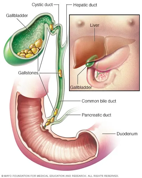
Fig1: Gallstones
2. Diagnostic methods
- Abdominal ultrasound
- Endoscopic ultrasound
- Imaging tests -
- ERCP (Endoscopic Retrograde Cholangiopancreatography)
- Magnetic resonance Cholangiopancreatography (MRCP)
- CT scan
- Blood tests
I have chosen two methods from the above to discuss briefly for assessing the pathological conditions
Endoscopic ultrasound
- Data- The data is collected by the doctor during the Endoscopic ultrasonography procedure as we can see from the above referring image
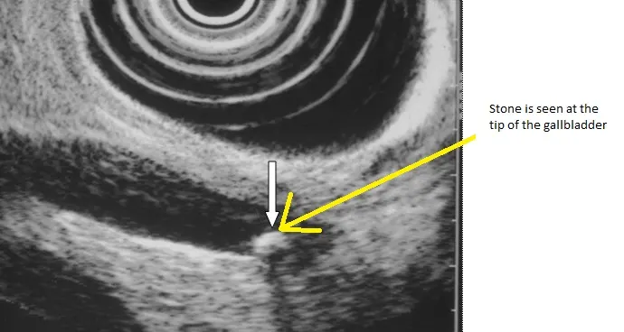
Fig2: Endoscopic ultrasound image shows stones in the gallbladder
- The doctor identifies the Gallstone from imaging of the gallbladder lesions. This method provides high-resolution images of small lesions with high ultrasound frequencies
ERCP
- Data- Endoscope and x-rays are used to inject the dyes to view the images of the pancreatic and bile ducts
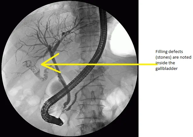
Fig3: ERCP image shows gall stones inside the gallbladder
- Doctors check for signs of blockage, gallstones or any other medical problems such as inflamed bladders, pancreatic cancer etc.
Result to the patient -
- The results will be given to the patient within 1 week where the findings of the Gallstone is written
3. Medications-
- Doctors provide medication for gallstones lesser than 1.5 cm which isn't causing any symptoms
- Anti-inflammatory non-steroidal tablets or drugs are given to dissolve the gallstones
Surgery-
- For gallstones, more than 1.5 cm in size surgery is needed.
- Laparoscopic Cholecystectomy surgery is performed
4. Surgical solution-
- Laparoscopic Cholecystectomy
Fig4: Laparoscopic Cholecystectomy
- Surgical position-Supine position
- Procedure
- A tiny video camera is inserted
- A small 4 incision is done with the surgical tool
- After that, the inside of the abdomen can be seen and the Gallstone is removed
- A larger incision is used to remove the gallbladder
- Medical instruments used in laparoscopic cholecystectomy are Graspers, Dissectors, Clipping, Kelly camps, Kocher forceps, retractors, needle holders, scissors, and suctions.
- Video link for laparoscopic cholecystectomy-https://jomi.com/article/251/Laparoscopic-Cholecystectomy
5. Post-surgical care-
- The doctor might suggest not to do strenuous activities
- A high fibre diet is suggested by the doctor to normalise the bowel movements
6. Medical devices used in the gallstone removal
- Laparoscope
- To access the inside of the abdomen and pelvis
Fig5: Flexible video laparoscope
Fig6: Rigid laparoscopepe
- Cholangiography equipment
- To find the size of gallbladder cancer which has spread or not
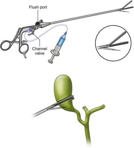
Fig7: Cholangiography kit
- X-ray
- Image acquisition
2. Pathological condition 2- Urinary tract obstruction in the urinary system
The normal function of the urinary tract is to filter the blood and create urine as a waste by-product
Pathology stage-
- Blockage in one or both the tubes of the ureters which carry urine from the kidney to the bladder
Reasons-
- Pregnancy
- An enlarged prostate also called Benign prostatic hyperplasia
- Tumours, cysts or scar tissue in the abnormal area
- Uterine prolapse or endometriosis
Homeostasis change
- The acid-base homeostasis and blood pH are regulated in the renal system of the urinary tract and the blood pressure homeostasis is too regulated in the urinary system which keeps the blood pressure normal
- If the condition is not treated pain, fever infection might occur
- The blockage or obstruction near the urinary tract can cause loss of kidney function, sepsis and even death
Fig1: Urinary tract obstruction
2. Diagnostic method
- Ultrasound, CT scan, MRI
- Blood and urine tests
- Bladder catheterization
- Renal nuclear scan
- Cystoscopy
From the above, I have chosen 2 diagnostic methods to discuss briefly-
Blood and urine tests
- Data acquisition - The blood and urine are collected from the patient
- The doctor identifies the problem by checking the signs of infection in the urine and the presence of creatinine. This indicates that the kidneys of the patient are not working properly
Bladder catheterization
- Data acquisition - A dye is injected into the bladder and an x-ray is used to take an image of the kidneys, ureters, bladder and urethra before and during the urination of the patient
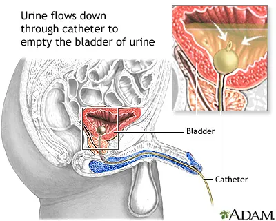
Fig2: Bladder catheterization
- The doctor inserts a small tube called a catheter through the urethra to test for the blocked or incomplete urine flow
Result to the patient
- The result of these diagnostic procedures is given to the patient within 1 week to let them know whether the clinical condition is severe or not.
3. Medication
- Anti-inflammatory non-steroidal drugs or tablets are given for normal pain
Surgery
For severe pain immediate surgical procedure is obtained to remove the urine from the body such as-
- Endoscopic surgery
- Open surgery
- Laparoscopic surgery
- Robot-assisted laparoscopic surgery
4. Surgical solution
- Endoscopic surgery
- Surgical position- supine, left lateral, and prone positions
- Procedure -
- A lighted scope is passed through the urethra into the bladder and other parts of the urinary tract
- A cut is made by the surgeon into the damaged or blocked part of the ureter to basically widen the area
- A hollow tube stent is then placed in the ureter to keep it open
- Medical instruments used in the endoscopic surgery are-Grasping and biopsy forceps, hook electrode, the knife of the triangular tip, Scissors, Suction instruments, Endobag, endo loop,endoclip, suture material
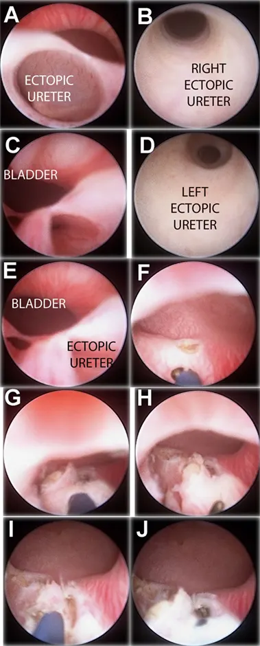
Fig3: Endoscopic urinary diversion is done through the endoscopic surgery
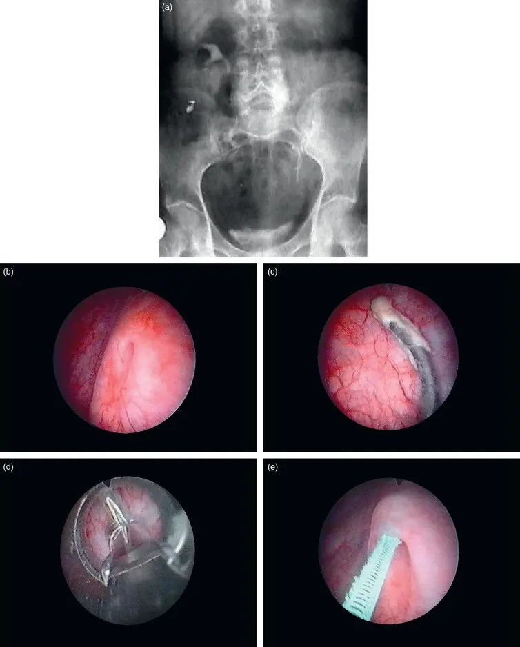
Fig4: Stent placement during the endoscopic surgery
5. Post-surgical care-
- Ureteral stent-
- It is used to help the patient to have an easy flow of the urine
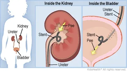
Fig5: Ureteral stent placement in the bladder
- Nephrostomy tube-
- It is placed through the back directly into the renal pelvis to drain urine directly from the kidney
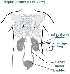
Fig6: Nephrostomy tube
6. Medical devices used for endoscopic surgery-
- Endoscope- It gives a proper view of the urethra into the bladder and other parts so that the person can operate on the damaged or blocked part of the ureter
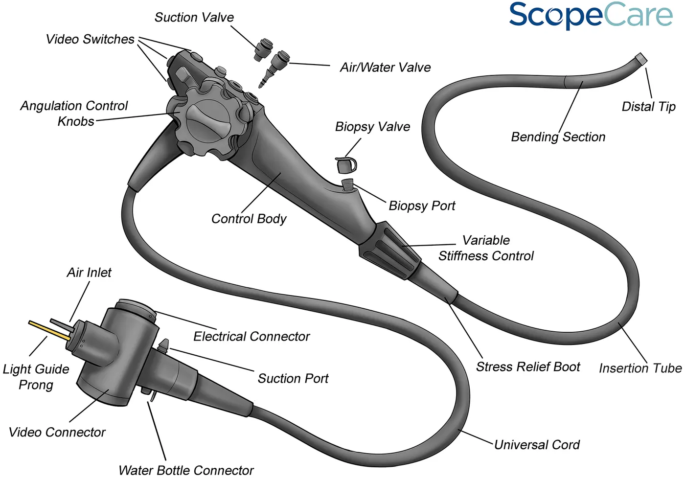
Fig7: Endoscope
- Ureteroscope-To access the bladder site
Fig8: Ureteroscope
- X-ray, CT scan, Ultrasound
- Image acquisition
- Video monitor
- It displays the ongoing surgical procedure
- Laser system
- Energy source
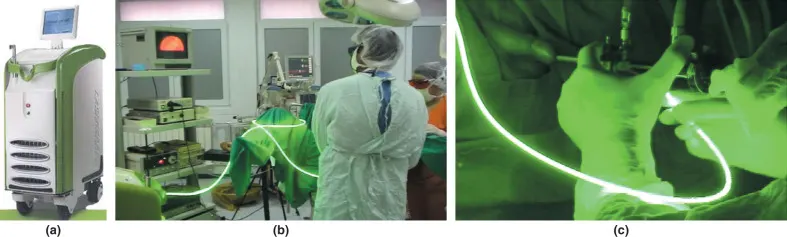
Fig9: Surgical laser system
- Laparoscope
- To access the site
Fig10: Laparoscope
Project (B)
1. Pathological Condition1-Lung cancer in the respiratory system
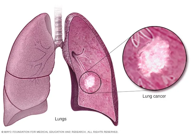
Fig1: Lung cancer
The normal function of the lungs
- Make oxygen available to the body and remove waste products like carbon dioxide from the body
Pathology stage-
- The lung cancer grows and blocks the major arteries
- It also causes fluid to accumulate around the lungs and makes it harder for the lungs to expand fully while inhaling.
Reasons
- The second smoke exposure
- If a person got previous radiation therapy
- Radon gas exposure
- Asbestos exposure and other carcinogens
- A person having a family history of lung cancer
Homeostasis change
- The cancer cells maintain protein homeostasis which affects the tumour evolution
- The continuous exposure to protein helps the cancer cells to expand more.
- Lung cancer basically spreads or metastasizes to the lymph nodes and other parts of the body which can affect any part of the respiratory system
2. Diagnostic methods
- Chest X-ray
- CT scan
- PET CT scan
- MRI
- Other types of biopsy
- Bronchoscopy and biopsy
- Sputum cytology
- Thoracentesis
From the above, I have chosen 2 diagnostic methods of lung cancer to discuss briefly
CT scan
- Data acquisition- The image is acquired by using the CT scan which creates detailed cross-sectional images of the body
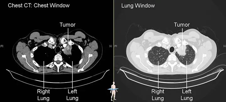
Fig2: CT scan image shows tumors in the right and left lung
Fig3: Stage 4 lung cancer CT image
- Identification of the lung cancer
- The doctor uses the image of the CT scan to identify long tumours and helps in finding the enlarged lymph nodes which might contain information regarding cancer that has spread
- CT scan also shows adrenal glands, liver, brain and other organs where the lung cancer has spread
Result to the patient
- The quick imaging of the CT scan test shows layers of the body structures in detail
Sputum cytology
Data acquisition-
- A sputum sample is collected from the patient and sent to the lab to see the cancer cells
Identification of the lung cancer
- This test help find the cancers that start in the squamous cell of the lungs or the major airways of the lung
Fig4: The arrow in the images showing the presence of the squamous carcinoma cells in the sputum cytology
Result to the patient
- The result of the presence of lung cancer can be found using this sputum cytology test
3. Medications
Anti-angiogenesis therapy
- Anti-angiogenic drugs are given for lung cancer
- Chemotherapy and immunotherapy drugs are given to shrink the cancer cells before the surgery.
Surgery
For the severe spread of lung cancer surgery is obtained, such as-
- Pneumonectomy
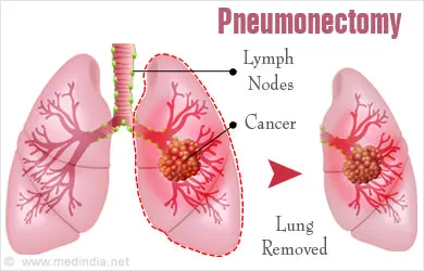
Fig: Pneumenoctomy
- Lobectomy
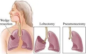
Fig5: Image showing the two different procedures-Lobectomy and pneumonectomy
4. Surgical solution
- Lobectomy
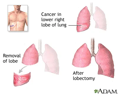
Fig6: Lobectomy surgery
- Surgical solution-Lateral decubitus position
- Procedure -
- This procedure is done when the problem is found in just one part of the lung
- The remaining healthy lung tissues can work normally and the affected lobe is removed using lobectomy surgery
- Instruments used in lobectomy are-
- Electric hook
- Vascular clamp
- Oval forceps
- Separating pliers
- Articulated stapler
- Non-invasive grasping forceps
- Ultrasound scalpal
- Plastic Endobag
- Utility port for wound protector
- Video link for lobectomy-https://youtu.be/4IMenyA74ds
5. Post-surgical care
- Coughing methods and deep breathing exercises are recommended by the doctor to help expand the lungs to re-expand after the surgery
- This helps in preventing pneumonia and breathing
6. Medical devices used in the lobectomy
- Video system
- To monitor the surgical procedure
Fig7: Surgical video system
- Video thoracoscope-
- To assist in monitoring the inside of the lungs
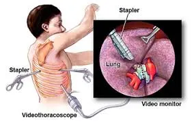
Fig8: Videothoracoscope
- Light source power
- Ultrasonic dissector-coagulator or bipolar electrocautery device
- Energy dissection device
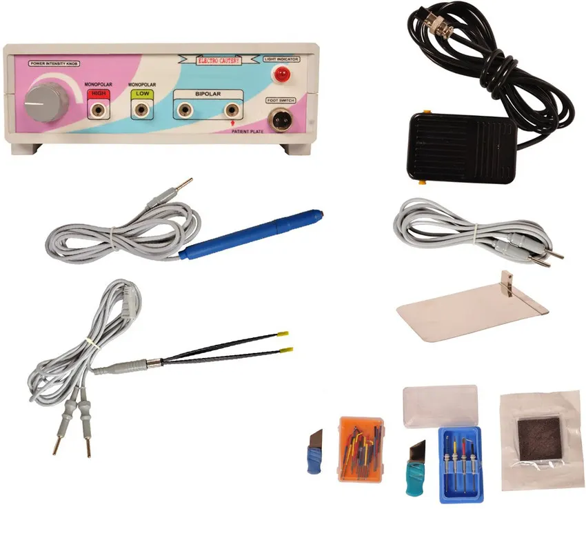
Fig9: Ultrasonic dissector-coagulator or bipolar electrocautery device
2. Pathological condition2-Myocardial infarction or heart attack in the cardiovascular system
The normal function of the cardiovascular system is to give oxygen, nutrients and other essentials through the blood and also remove unwanted waste
Pathological stage-
- When a part of the heart muscle doesn't get enough blood due to a blood clot that occurs to block the blood flow to the heart
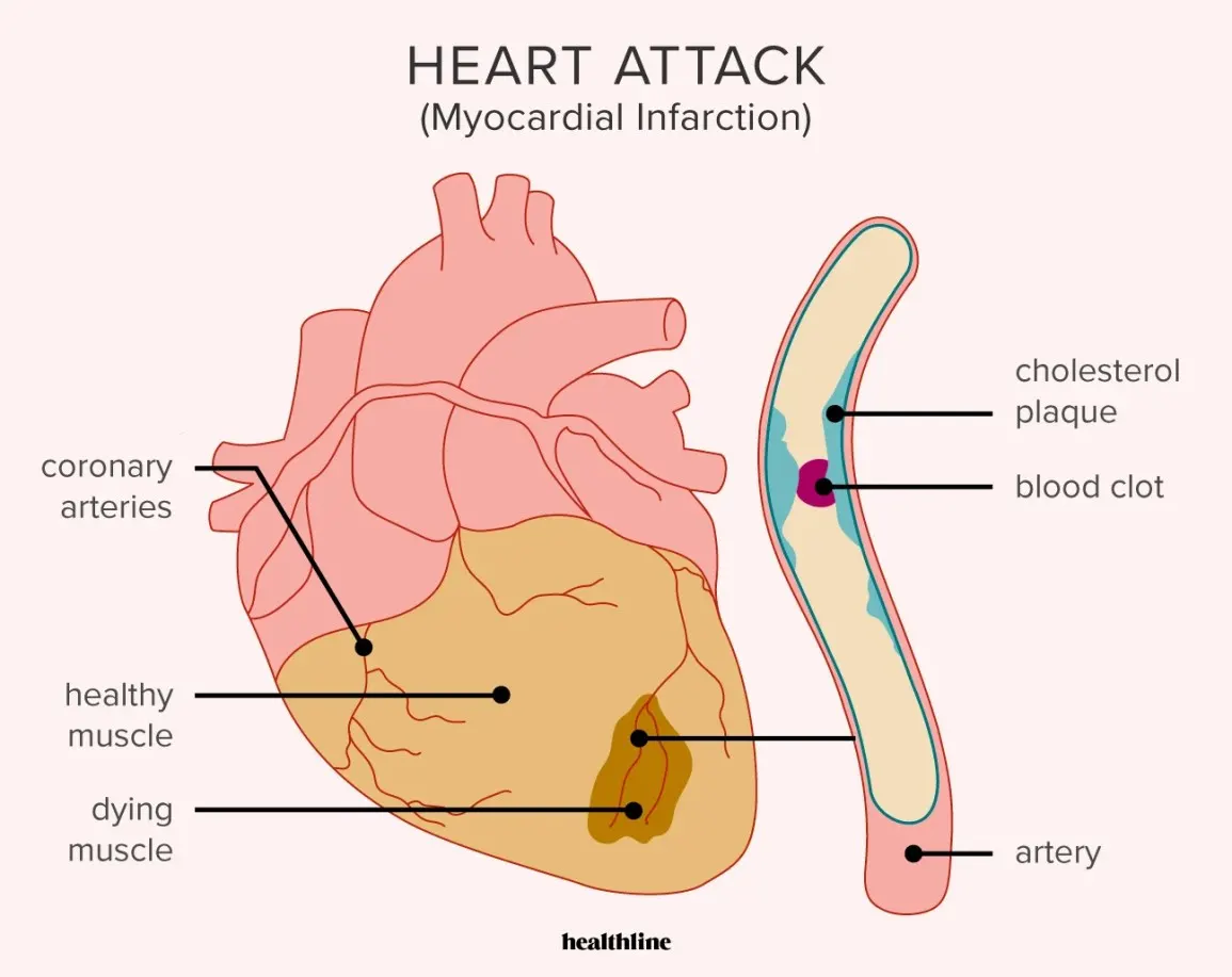
Fig1: Diagram showing blood clot blocking the blood flow to cause the heart attack
Reasons for heart attack
- High-fat diet
- Diabetes and
- High blood pressure
- Smoking
- High cholesterol
- Overweight or obese
Homeostasis change
- Reduction in cardiac output and renal blood flow
- The heart fails and damage caused to the systolic or diastolic function
- This whole process causes kidneys to initiate a homeostatic hormonal response which can be compared to the contracted volume of intravascular muscle due to salt and water deprivation or food loss.
2. Diagnostic method
- ECG or EKG
- Blood tests
- Echocardiogram
- Chest x-ray
- Coronary catheterization
- Cardiac CT or MRI
From the above, I have chosen 2 diagnostic methods to discuss briefly for diagnosing a heart attack-
ECG or EKG
- Data acquisition method
- The data is acquired by signals received from the electrode attached to the chest
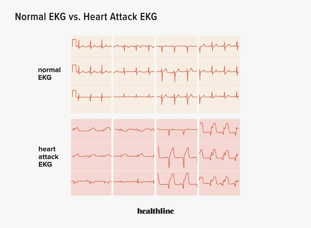
Fig2: Normal ECG and heart attack ECG
- The doctor studies the patterns of the signals and analyses whether the patient is having or has had a heart attack previously
Result to the patient
- The result is given in a printed paper where the recorded waves printed on it show if the person has the upcoming presence of a heart attack or not.
Cardiac CT or MRI
- Data acquisition method
- The data is acquired by using radio waves to create images of the heart in a CT scan
- While in the MRI magnetic field and radio waves are used to create the image of the heart
Fig3: MRI after a heart attack
- The doctor identifies the severity of the heart damage by looking at the anatomy and function of the heart chambers, size of heart valves, blood flow through major vessels and the structure of the pericardium on the cardiac CT scan or MRI
Results to the patient
- The obtained result is used to diagnose the heart problems of the patients because it gives an inner view of the heart which helps the doctor to give proper medical care to the patient
3. Medications
- To break up the blood clot formation clot blusters drugs are given
- Beta-blocker
- Anticoagulant and antianginal
- Narcotic
Surgery
When the patient's condition becomes severe like shortness of breath or inability to breathe, surgical options have opted as the last option. Such as-
- Coronary angioplasty and stenting
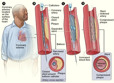
Fig4: Coronary angioplasty and stenting
- Coronary artery bypass graft surgery
4. Surgical solution
- Coronary artery bypass graft surgery
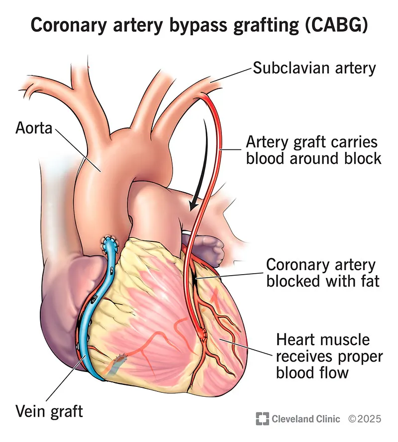
Fig5: Coronary artery bypass graft surgery
- Surgical position-supine position
- Procedure
- A blood vessel is taken from another part of the body such as the chest, leg or arm and it is attached to the coronary artery below and above the blockage or narrowed area
- The new blood vessel is called a graft and that's why this procedure is also known as coronary artery bypass graft surgery.
- Instruments used in CABG are-
- Coronary scissors
- Dissecting scissors
- Needle holders
- Forceps
- Clamps - partial occlusion
- Chest tube passer
- Vessel dilators and probes
- Occlusion clips
- Sterilisation trays
- Vessel loops
- Vein holder
- Aorta/vein punches
- Video link for coronary artery bypass graft surgery-https://youtu.be/PO6ioZXVBvs
5. Post-surgical care for the CABG
- Airway patency is maintained
- Monitoring continuously along the bedside
- Provide relief to the pain levels by assessing daily
- Surgical incisions assessment for infection
- Monitor the complications
- Monitor the drainage and avoid any kinks in tubing - chest tube management
- Edema and impaired circulation signs should be monitored
6. Medical devices used in the coronary artery bypass graft surgery
- Heart-lung bypass machine
- Provides blood and oxygen to the body when the heart is stopped during the surgical procedure
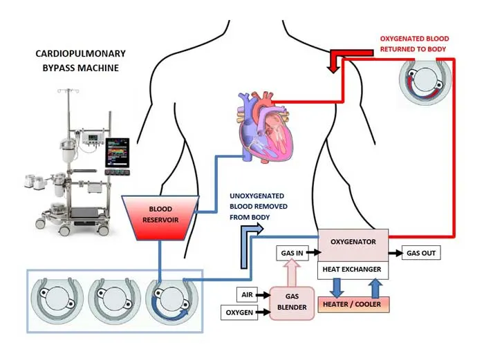
Fig6: Heart-lung bypass machine
- Heater cooler
- To regulate the blood during the surgery
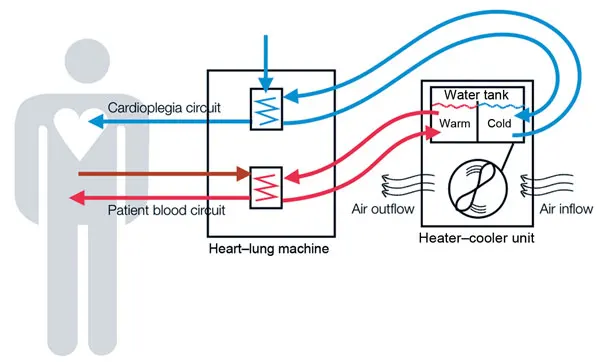
Fig7: Heater cooler unit and heart-lung machine helping in the cardiac surgery
Fig8: Heater cooler unit
- Defibrillators with internal paddles
- It restores the normal heartbeat if the heartbeat of a patient stops
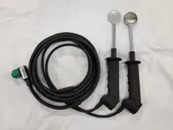
Fig9: Defibrillators with internal paddles
- Sternum saw
- A vital machine through which the sternum is precisely cut.
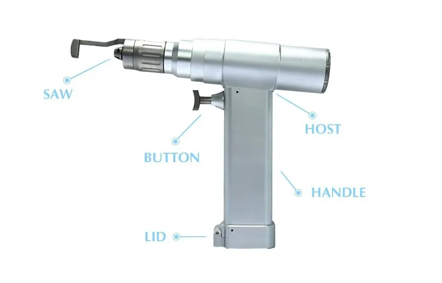
Fig10: Sternum saw
Reference
Image credits
- https://images.app.goo.gl/th5LSfaP4wpG5PBp7
- https://images.app.goo.gl/jyBXZoEf8CbDm8u59
- https://images.app.goo.gl/CC54DqXNzkArAP4X6
- https://images.app.goo.gl/grsiagZ7MYhutm5H7
- https://images.app.goo.gl/gfTi5MTzfpmNEHDq7
- https://images.app.goo.gl/gfTi5MTzfpmNEHDq
- https://images.app.goo.gl/MBFccXkxsFBLudks6
- https://images.app.goo.gl/zcpPc9sHzTDaXqB99
- https://images.app.goo.gl/H1KS2V345kxfaee18
- https://images.app.goo.gl/tVynMYDZaynRzKi67
- https://images.app.goo.gl/WkNft8cHZya9HLgr9
- https://images.app.goo.gl/K7uAFQ1XKo23oRNVA
- https://images.app.goo.gl/gMJCeNWYoUEZJs1M6
- https://images.app.goo.gl/n7NyyYNq37ji1vxz7
- https://images.app.goo.gl/PSq8NQ5urGuEaNGX7
- https://images.app.goo.gl/7wBZugaeuGYB85mi7
- https://images.app.goo.gl/V5QsN92Zi7DXaanF9
- https://images.app.goo.gl/4xgYqkwM7Syyprt49
- https://images.app.goo.gl/cmYhQYUnGTDmBpUdA
- https://images.app.goo.gl/itKsz7xwcWbBMKYT9
- https://images.app.goo.gl/pN5vZQKFrP8Fi48P7
- https://images.app.goo.gl/aMNV5GvsJVybb8kNA
- https://images.app.goo.gl/jEG6TzCjsutRGnYg6
- https://images.app.goo.gl/oExq2ZiFVV196LYVA
- https://images.app.goo.gl/kN4mbbySsH4k8uyJ6
- https://images.app.goo.gl/pBekvW7zbaGt5czw9
- https://images.app.goo.gl/vvpBzKXtdfp9JehZ8
- https://images.app.goo.gl/UK4cftc41HeBeNmFA
- https://images.app.goo.gl/gP4KHzzzy9DHG1Pr9
- https://images.app.goo.gl/2nmbXFPMDQ1sVWG76
- https://images.app.goo.gl/SJ7Mih2QexPREh796
- https://images.app.goo.gl/iHBJwGBtiWeST1x59