Project 2
Design a robotic surgery procedure to remove a healthy kidney from the donor using an AI guided robot.
The idea of robotic kidney transplantation surgery is that a healthy kidney is removed from a donor using a surgical robot.
Title: Robotic Surgery Procedure for Donor Nephrectomy: An AI-Guided Approach
Introduction:
Kidney disease is a major public health problem in part because of its common etiology caused by diabetes, hypertension, obesity, and aging; the incidence of these conditions is increasing.
Robotic surgery has revolutionized many surgical procedures, including kidney transplantation. The use of AI-guided robots in the field of surgery offers enhanced precision, improved outcomes, and reduced invasiveness. Robotic-assisted donor nephrectomy, which involves removing a healthy kidney from the donor, has become a standard approach in kidney transplantation.
The da Vinci robot provides several advancements that aid in expediting the learning process for surgeons without laparoscopic experience, particularly through its three-dimensional (3-D) vision and standard surgical manipulation capabilities. The robot's six degrees of freedom can also reduce the learning curve for proficient open surgeons who lack laparoscopic expertise. On the other hand, the laparoscopic approach may present challenges in tumor dissection and intracorporal suturing, whereas the robotic approach offers benefits such as magnified stereoscopic vision and fully articulating instruments, potentially mitigating these technical difficulties. However, it is important to note that these procedures come with higher costs compared to alternative approaches.
The emphasis of robotic renal surgery lies more in technically demanding procedures rather than simple uncomplicated nephrectomies. The robotic technology is expected to bring advantages in the management of renal procedures, particularly in complex cases, making robot-assisted surgery increasingly relevant in the future.
History: The development of robotic surgery can be traced back to the late 20th century. In 1985, the PUMA 560 robotic system was used in a neurosurgical biopsy procedure. Since then, advancements in technology and surgical techniques have led to the introduction of robotic platforms specifically designed for various surgical procedures, including kidney transplantation.
Another goal of robotic surgery was the elimination of unwanted motion. The first surgical robot, PUMA 560, was used in 1985 in a stereotaxic operation, in which computed tomography was used to guide the robot as it inserted a needle into the brain for biopsy, a procedure previously subject to error from hand tremors during needle placement. In 1988 PROBOT, developed at Imperial College London, was used to perform transurethral prostate surgery, a procedure that required numerous repetitive cutting motions. Also in 1992 ROBODOC, developed by the American companies Integrated Surgical Systems, Inc. (ISS), and IBM, was used to successfully prepare a cavity in the femur for hip replacement in human patients. It carried out the task more precisely and more quickly than human surgeons.
By the late 1990s three systems designed for minimally invasive surgery had been tested: the da Vinci Surgical System, developed by California-based Intuitive Surgical, Inc., and the AESOP and Zeus Robotic Surgical systems, both developed by Computer Motion, Inc., another California company.As a result, the da Vinci Surgical System became the most widely used robotic surgical system worldwide.
Improvements from PUMA 560 Robotic system to AI-guided robots:
fig: Robot Puma 560
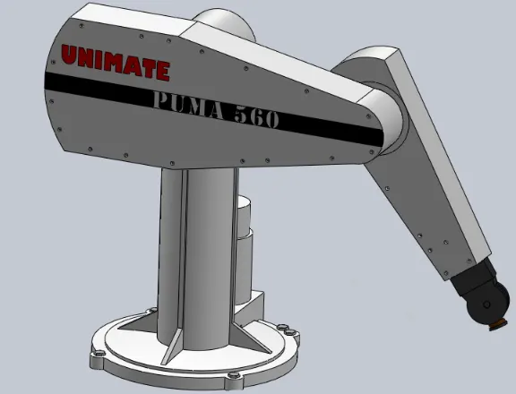
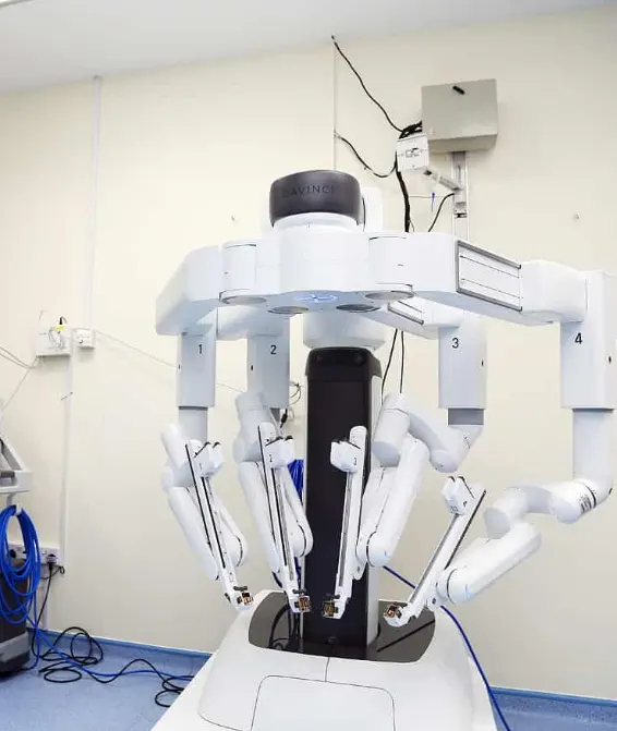
fig : Da Vinci Xi surgical robot
The advancements from the PUMA 560 robotic system to AI-guided robotic systems in kidney surgery have brought significant improvements in terms of precision, capabilities, and outcomes. Here are some key improvements:
1. Enhanced Precision: AI-guided robotic systems offer enhanced precision and accuracy compared to the earlier PUMA 560 system. The integration of artificial intelligence algorithms allows for more precise movements and control of robotic instruments, reducing the risk of human error during the surgery.
2. Improved Visualization: AI-guided robotic systems provide improved visualization through advanced imaging technologies. High-definition cameras and 3D imaging capabilities offer surgeons a detailed and magnified view of the surgical site, enabling better identification of anatomical structures and precise surgical maneuvers.
3. Intelligent Surgical Planning: AI algorithms can analyze preoperative imaging data, such as CT scans or MRIs, to assist surgeons in surgical planning. This technology helps surgeons identify the optimal surgical approach, plan the trajectory of robotic instruments, and anticipate potential complications or challenges specific to the patient's anatomy.
4. Real-time Feedback: AI-guided robotic systems can provide real-time feedback to surgeons during the procedure. They can monitor physiological parameters, analyze surgical data, and offer insights to assist surgeons in making informed decisions during the surgery.
5. Adaptive and Learning Capabilities: AI-guided robotic systems have the ability to adapt and learn from previous surgical experiences. By analyzing large datasets of surgical procedures, these systems can continuously improve their performance and provide personalized assistance to surgeons based on their preferences and patient-specific factors.
6. Integration of Machine Learning and Computer Vision: AI-guided robotic systems often incorporate machine learning algorithms and computer vision techniques. These technologies enable the system to recognize and interpret visual information, track surgical instruments, and assist in complex tasks such as tissue identification, vessel segmentation, and precise suturing.
7. Telesurgery and Remote Assistance: AI-guided robotic systems have the potential for remote surgical collaboration and teleoperation. Surgeons can receive guidance and support from experts located remotely, allowing for knowledge transfer and access to specialized expertise, especially in underserved areas.
Overall, the transition from the PUMA 560 robotic system to AI-guided robotic systems in kidney surgery has brought substantial improvements in terms of precision, visualization, surgical planning, real-time feedback, adaptive capabilities, and the integration of advanced technologies. These advancements have contributed to improved surgical outcomes, reduced complications, and expanded the possibilities for complex kidney surgeries.
When kidney transplantation employs surgical robots, there are several specific requirements beyond ease of procedure and reduction in procedure time. These additional requirements include:
1. Precision and Accuracy: Surgical robots used in kidney transplantation must offer precise and accurate movements to ensure successful transplantation. The robot should enable surgeons to perform delicate maneuvers with high precision, minimizing the risk of damage to the kidney or surrounding tissues.
2. Minimally Invasive Approach: One of the key advantages of surgical robots is their ability to perform minimally invasive procedures. The robot should facilitate small incisions and provide dexterity to maneuver within the limited space of the surgical site. This leads to reduced trauma, less post-operative pain, shorter hospital stays, and faster recovery for the patient.
3. Enhanced Visualization: Clear and magnified visualization is essential during kidney transplantation. The surgical robot should provide high-definition imaging capabilities, including 3D visualization, to enable the surgeon to accurately identify anatomical structures and perform precise surgical maneuvers.
4. Ergonomics and Comfort for Surgeons: The robotic system should offer ergonomic features to minimize surgeon fatigue during long procedures. It should provide adjustable positioning, intuitive control interfaces, and comfortable hand controllers, allowing the surgeon to maintain dexterity and control throughout the surgery.
5. Integrated Instrumentation: The robotic system should have versatile instrument integration capabilities, allowing seamless integration of various surgical tools and devices required for kidney transplantation. This includes specialized instruments for tissue dissection, vessel suturing, and organ manipulation.
6. System Stability and Reliability: Given the complexity and critical nature of kidney transplantation, the robotic system must exhibit high stability and reliability. The system should have robust hardware, software, and communication interfaces to ensure uninterrupted and accurate performance throughout the procedure.
7. Compatibility with Imaging and Navigation Systems: Integration with preoperative imaging and intraoperative navigation systems is crucial for precise surgical planning and guidance. The robotic system should be compatible with advanced imaging modalities, such as CT scans or MRI, and support real-time navigation feedback to aid the surgeon in accurately locating and accessing the transplant site.
8. Support for Telesurgery and Collaborative Surgery: In certain cases, remote surgical assistance or collaboration may be required during kidney transplantation. The robotic system should have capabilities for telesurgery, enabling experienced surgeons to provide guidance and assistance from remote locations. It should also facilitate collaborative surgeries, allowing multiple surgeons to work together seamlessly.
Overall, the successful employment of surgical robots in kidney transplantation requires a comprehensive robotic system that combines precision, minimally invasive capabilities, enhanced visualization, ergonomic design, stability, compatibility, and support for advanced surgical techniques. By meeting these requirements, robotic-assisted kidney transplantation can offer improved outcomes, reduced complications, and enhanced patient care.
The da Vinci Surgical System is one of the most commonly used surgical robots in various minimally invasive procedures, including kidney transplantation.
The robotic platform, the classic model of the da Vinci Surgical System, was used by the team for two main reasons. Firstly, the precision of surgery could be enhanced through motion-scaling and increased dexterity of the robot. Secondly, the prevalence of blood-borne infections including HIV, hepatitis B and hepatitis C among hemodialysis patients can be significantly high. The risk of potential viral transmission could be reduced through dissociation of the surgeon from the operating field by the use of telepresence surgery. This may well be beneficial for the protection of surgeons from contracting blood-borne infections. For these reasons, the team aimed to demonstrate the feasibility of robotic-assisted kidney transplantation.
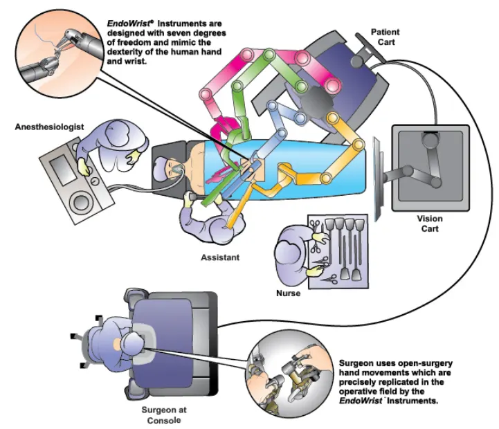
fig : Operating theatre set up Credit: ©2017 Intuitive Surgical, Inc.
A Da Vinci is a medical robot, and more precisely a machine run by a surgeon to perform operations, mainly at the level of the abdomen.
It is sold by the American company Intuitive Surgical. In April 2011, it reported that 1,750 copies were in service worldwide.
APPLICATIONS
Cardiac surgery
Gastro-intestinal surgery
Gynecology
Neurosurgery
Orthopedics
Pediatrics
Radio surgery
Urology
Components of the robot:
1.The Patient Cart:
It is the operative component of the da Vinci Si System and its primary function is to support the instrument arms and camera arm. It contains five main components:
the setup joints;
instrument arms;
camera arm;
EndoWrist instruments;
and an endoscope.
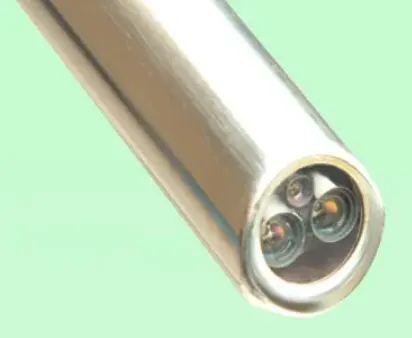
Da Vinci® stereo-endoscope.
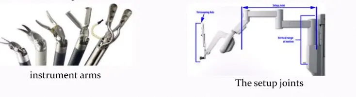
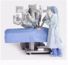
fig : The patient cart
2. The Surgical Console
The surgical console is the workplace of the robotic surgeon and contains the following core elements:
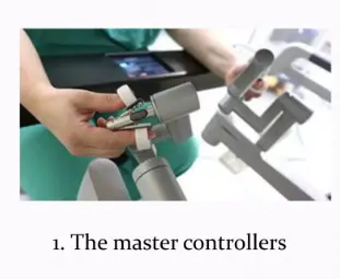
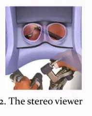

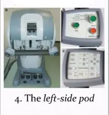
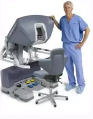
fig : The Surgical console
3. The Vision Cart
- It includes a 24" touchscreen monitor used to control system settings and view the surgical image.
- A camera control unit on the vision cart is connected to the camera and controls the acquisition and processing of the image from the camera.
- It also provides adjustable shelves for optional ancillary surgical equipment such as insufflators and electrosurgical generators.

fig: The vision cart
key components and features:
-
Robotic Arms: The da Vinci Surgical System consists of multiple robotic arms that mimic the movements of the surgeon's hands. Typically, there are four robotic arms, three of which hold and manipulate surgical instruments, while the fourth arm holds a high-definition 3D camera for visualization.
-
Surgical Instruments: The robotic arms of the da Vinci system hold a variety of specialized surgical instruments. These instruments include graspers, scissors, forceps, needle drivers, and others, which are used for tasks like tissue dissection, suturing, and organ manipulation during kidney transplantation.
-
EndoWrist Technology: The instruments used in the da Vinci system are equipped with EndoWrist technology, enabling them to mimic the movements of the surgeon's wrists with enhanced dexterity and precision. This allows for intricate movements and delicate tissue handling.
-
Teleoperation: The da Vinci Surgical System allows for teleoperation, which means that the surgeon can control the robotic arms remotely from a console. The surgeon sits at the console, which provides a magnified 3D view of the surgical site, and manipulates the instruments using hand controllers and foot pedals.
-
Enhanced Visualization: The system includes a high-definition 3D camera that provides a magnified and detailed view of the surgical site. This allows the surgeon to have a clear and precise visualization of the anatomy during the kidney transplantation procedure.
-
Computerized Control System: The robotic system is controlled by a computerized control system, which translates the surgeon's hand movements at the console into precise movements of the robotic arms. The control system ensures accurate and synchronized movements of the instruments.
-
Safety Features: The da Vinci Surgical System incorporates several safety features to prevent accidents or unintended movements. These include collision detection and automatic retraction of the instruments to avoid collisions with sensitive structures
- The surgeon's console allows the surgeon to control the robotic arms and provides a magnified 3D view of the surgical site. The robotic arms hold and manipulate the surgical instruments, while the 3D camera provides enhanced visualization of the procedure.
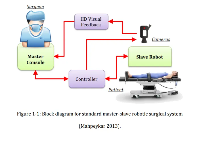
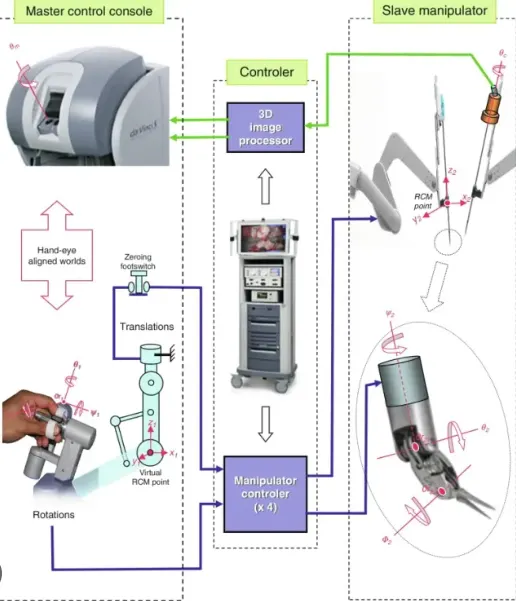
FLOWCHART: Representation of the whole kidney transplantation process
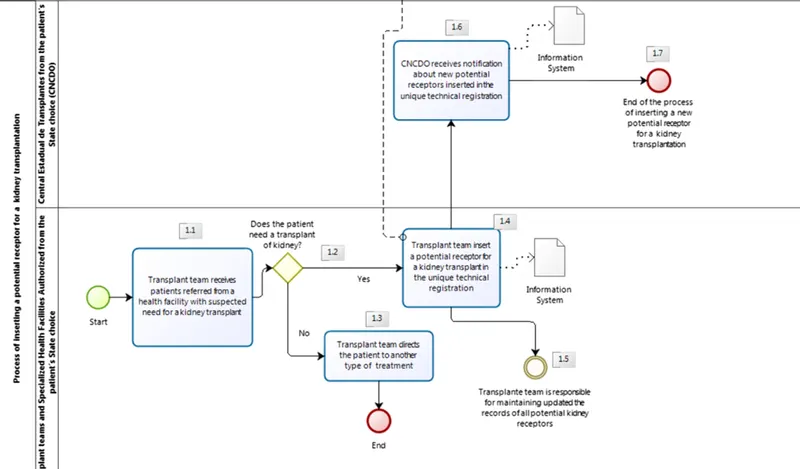
Live kidney donors are healthy individuals who intentionally undergo major surgery to improve the well-being of another individual; as such, maximizing the donor safety during this procedure is of paramount importance.
Several surgical techniques have been described for living donor nephrectomy (LDN), including open, pure or hand-assisted laparoscopic, natural orifice transluminal endoscopic surgical (NOTES), and robotic approaches.
In this view, minimally invasive techniques are increasingly being performed worldwide with the aim to further limit the morbidity of surgery for the donors while ensuring optimal grafts for kidney transplantation . While laparoscopic living donor nephrectomy (LDN) has become a common procedure in most Transplant Centers and has been shown to be associated with shorter hospital stay, less pain, and faster recovery as compared to open surgery, the use of robotic surgery in this setting might further improve its perioperative outcomes providing distinct benefits for both donors and surgeons . This is mainly due to the advantages of the robotic platform as compared to standard laparoscopy (improved ergonomics, Endowrist technology, magnification and 3D vision). Consequently, robotic LDN has been implemented and is being increasingly performed at referral Centers with expertise in both living donor kidney transplantation and robotic surgery, with excellent outcomes, comparable to those of pure laparoscopic LDN.
Robotic Living Donor Nephrectomy:
Robotic LDN was performed with a transperitoneal access using the da Vinci Si robotic platform (Intuitive Surgical Inc)
Step 1: Patient Positioning and Port Placement
After general anesthesia, the patient was placed in the right 70° lateral decubitus, for left nephrectomy, or in left 70° lateral decubitus for the right kidney.
Port placement was performed according to the principles employed for radical nephrectomy for oncological reasons. In case of right LDN, one additional trocar (5 mm) was placed for liver retraction.
Our technique involves the use of two robotic arms, a 12 mm trocar for the camera and two assistant ports (5 and 12 mm, respectively).
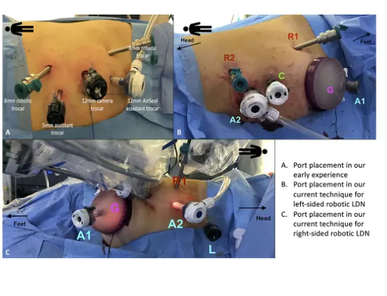
figure :Port placement for robotic living donor nephrectomy (LDN). (A) Overview of the port placement for robotic LDN in the early phase of our experience. This included one 12 mm trocar for the camera, two 8 mm ports for the robotic arms and two assistant ports (one 12 mm and one 5 mm). The site for the Pfannenstiel incision was marked at the beginning of surgery but the incision (and the subsequent placement of the GelPOINT device) was made only after the kidney was entirely mobilized, just before controlling the renal vessels. (B,C) Overview of the current port placement for robotic left-sided (B) and right-sided (C) LDN (from 2017). The Pfannenstiel incision and placement of the GelPOINT device is performed at the beginning of surgery to save time and facilitate the kidney extraction. In this way, there is no need to re-dock the robot during the procedure. The AirSeal system is used routinely in all cases to maintain a constant pneumoperitoneum pressure of 10–12 mmHg.
Step 2: Identification of the Retroperitoneal Anatomic Landmarks of Dissection
The procedure began with the medialization of the colon along the line of Toldt. Then, the classical retroperitoneal landmarks were identified (psoas muscle, ureter, and gonadal vein).
On both sides, after the medialization of the gonadal vein, the ureter was lateralized and freed from all surrounding tissues. Following the gonadal vein, the renal hilum was identified.
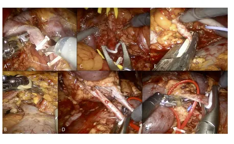
figure : Overview of selected steps of robotic living donor nephrectomy (A) Dissection and control of the left gonadal vein. (B) Isolation and dissection of the left adrenal vein. (C) Control of the left renal artery using Hem-o-Lok clips (at the beginning of our experience). (D) Control of the right renal artery using Hem-o-Lok clips (at the beginning of our experience). (E) Control of the right renal artery using a EndoGIA vascular stapler (current technique). (F) Control of the right renal vein using Hem-o-Lok clips (for the right renal vein).
Step 3: Isolation and Dissection of the Renal Hilum and the Kidney
Once the renal hilum was identified, the renal vein and artery (and all potential accessory vessels) were carefully isolated. The ascending lumbar vein was routinely identified on the left side, isolated and transected.
On the left side, the adrenal vein was always carefully dissected and ligated with two Hem-o-Lok clips. Any additional gonadal or lumbar vessels were also controlled in order to free the entire length of the renal vein.
On the right side, the renal vein is shorter but there is usually no need to control the adrenal, gonadal, and lumbar veins as they drain directly into the inferior cava vein.
After hilar dissection, the kidney was entirely mobilized and isolated from the perirenal fat tissue.
The use of artificial intelligence (AI) in robotic kidney transplantation can assist in various tasks throughout the procedure. Here are some of the tasks performed by AI :
1. Surgical Planning: AI algorithms can analyze preoperative imaging data, such as CT scans or MRI, to assist in surgical planning. By segmenting and analyzing the anatomical structures, AI can help identify the optimal locations for incisions, determine the best trajectory for vessel anastomosis, and plan the placement of the transplanted kidney.
2. Image Analysis and Recognition: AI can analyze intraoperative images, such as real-time ultrasound or fluoroscopy, to provide assistance in identifying key anatomical landmarks and structures during the surgery. It can help surgeons locate blood vessels, differentiate tissues, and navigate the surgical site with increased accuracy.
3. Surgical Guidance and Navigation: AI algorithms can provide real-time guidance and navigation assistance during the procedure. By integrating with the surgical robot's system, AI can help the surgeon in maintaining the planned trajectory, ensuring precise incisions, and avoiding critical structures.
4. Instrument Tracking and Control: AI algorithms can track and analyze the movements of surgical instruments used during the kidney transplantation procedure. By monitoring instrument positions, AI can provide feedback to the surgeon, alerting them to potential collisions or deviations from the planned path.
5. Decision Support: AI can analyze patient-specific data, such as medical history, laboratory results, and physiological parameters, to provide decision support to the surgical team. AI algorithms can help in evaluating the compatibility of the donor kidney, assessing the risk factors, and suggesting the most appropriate surgical approach based on the patient's condition.
6. Surgical Performance Evaluation: AI can analyze surgical data, including instrument movements, time taken for specific tasks, and surgical outcomes, to evaluate the performance of the surgical team. This feedback can be used to identify areas for improvement, optimize surgical techniques, and enhance overall surgical outcomes.
7. Postoperative Monitoring and Analysis: AI can assist in postoperative monitoring of the patient's recovery progress. By analyzing postoperative data, such as vital signs, laboratory values, and imaging results, AI algorithms can detect early signs of complications, provide alerts to healthcare providers, and facilitate timely intervention.
It's important to note that while AI can provide valuable assistance in robotic kidney transplantation, it does not replace the expertise and decision-making of the surgical team. AI is designed to augment the surgeon's skills and provide valuable insights and support throughout the procedure, ultimately leading to improved surgical outcomes and patient care.
Perioperative care refers to the comprehensive care provided to a patient throughout the entire surgical process, including preoperative, intraoperative, and postoperative stages. Here are the key steps taken during the perioperative period to ensure a successful surgery:
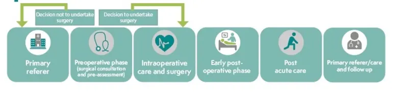
Preoperative Phase:
1. Patient Preparation: The patient is prepared for the surgery by ensuring proper fasting, administering preoperative medications (if needed), and confirming the patient's identity, surgical site, and consent.
2. Anesthesia Administration: The anesthesiologist administers the chosen anesthesia technique, such as general anesthesia, regional anesthesia, or local anesthesia, based on the patient's condition and the nature of the surgery.
3. Patient Monitoring: Throughout the surgery, the patient's vital signs (blood pressure, heart rate, oxygen saturation, etc.) are closely monitored to ensure their stability and response to anesthesia. Monitoring devices such as electrocardiogram (ECG), pulse oximeter, and blood pressure monitor are used.
Intraoperative Phase:
1. Sterile Environment: The operating room is maintained in a sterile condition to minimize the risk of infection. Surgical team members wear sterile gowns, gloves, masks, and caps, and surgical drapes are used to cover the patient and create a sterile field.
2. Surgical Procedure: The surgical team performs the planned surgical procedure while adhering to aseptic techniques and following established surgical protocols. Surgeons use specialized instruments and techniques appropriate for the specific surgery, ensuring precision and safety.
3. Anesthesia Management: The anesthesiologist closely monitors the patient's vital signs, administers additional medications or adjustments as necessary, and ensures the patient's comfort and stability throughout the procedure.
4. Team Communication: Effective communication and teamwork among the surgical team members are crucial during the intraoperative phase. Clear and concise communication ensures smooth coordination and efficient execution of the surgical plan.
Postoperative Phase:
1. Recovery and Stabilization: After the surgery, the patient is transferred to the recovery room or post-anesthesia care unit (PACU). Here, they are closely monitored until they recover from the effects of anesthesia and their vital signs stabilize.
2. Pain Management: Adequate pain management strategies, such as administering pain medications, regional anesthesia techniques, or non-pharmacological approaches, are implemented to ensure the patient's comfort and promote early mobilization.
3. Wound Care and Infection Prevention: Proper wound care techniques are employed to minimize the risk of infection. Sterile dressings, antibiotics (if required), and regular wound assessments are performed to promote healing and prevent complications.
4. Patient Education and Discharge Planning: Patients and their caregivers are provided with postoperative instructions, including medication schedules, wound care guidelines, and signs of potential complications. Discharge planning is initiated, considering the patient's recovery progress and their ability to continue care at home or in a rehabilitation facility.
Equipment-related steps:
1. Equipment Sterilization and Maintenance: All surgical instruments, equipment, and devices used during the procedure are sterilized according to established protocols. Regular maintenance and calibration of equipment ensure their proper functioning and reliability.
2. Equipment Availability and Readiness: The surgical team ensures that all necessary equipment and supplies are readily available in the operating room. This includes surgical instruments, anesthesia equipment, monitoring devices, surgical drapes, and disposables.
By following these perioperative steps and providing comprehensive care to the patient, healthcare providers aim to ensure a successful surgery with optimal patient outcomes, minimized complications, and a smooth recovery process.
Ethical Concerns and Regulations in Kidney Transplantation Procedure:
1. Organ Donation and Allocation: The ethical concern revolves around ensuring fair and equitable organ allocation, addressing the shortage of available organs, and preventing exploitation or coercion in the organ donation process. Ethical guidelines and regulations are in place to establish transparent and ethical practices for organ procurement and allocation.
2. Informed Consent: Obtaining informed consent from the donor and recipient is essential. It involves providing detailed information about the risks, benefits, alternatives, and expected outcomes of the procedure. Ethical guidelines emphasize the importance of patient autonomy, voluntary decision-making, and ensuring that consent is free from coercion or undue influence.
3. Organ Trafficking and Commercialization: There is a need to combat illegal organ trafficking and prevent the commercialization of organs. Ethical principles prioritize the prohibition of organ sales and the protection of vulnerable populations from exploitation.
4. Medical and Surgical Ethics: Ensuring patient safety, minimizing risks, and adhering to professional standards are fundamental ethical considerations. Surgeons and healthcare professionals must maintain high standards of surgical competence, follow established protocols, and prioritize patient welfare throughout the transplantation process.
5. Equity and Access: Ethical concerns arise regarding equitable access to transplantation for all eligible patients, irrespective of socioeconomic status, race, ethnicity, or other factors. Efforts are made to address disparities in access to transplantation and ensure fairness in patient selection and evaluation.
Regulations and Standards:
1. Machine Classifications: Medical devices and equipment used in kidney transplantation are categorized into different classes based on their risk level. The classification systems vary across different regions, but they generally include classes such as Class I (low risk), Class II (moderate risk), and Class III (high risk). The classification determines the level of regulatory scrutiny and conformity assessment required for the equipment.
The da Vinci Surgical System is classified as a Class II medical device by the U.S. Food and Drug Administration (FDA). Specifically, it is classified as a robotic surgical system under the product code "NAY" (Surgical robot system). This classification is based on the intended use, design features, and level of risk associated with the device. Class II devices are considered to have a moderate level of risk and require general controls, as well as special controls to ensure their safety and effectiveness.
2. IEC 60601 Standards: The IEC 60601 series of standards provides specific requirements for the safety and effectiveness of medical electrical equipment, including devices used in kidney transplantation. Some relevant standards within the series include:
- IEC 60601-1: General requirements for basic safety and essential performance of medical electrical equipment.
- IEC 60601-1-2: Electromagnetic compatibility requirements for medical electrical equipment.
- IEC 60601-2-xx: Specific standards for particular types of medical electrical equipment. For example, IEC 60601-2-77 covers safety requirements for kidney dialysis equipment.
These standards ensure that medical devices used in kidney transplantation meet essential safety and performance criteria. Compliance with these standards helps in mitigating risks, ensuring patient safety, and facilitating regulatory approvals.
It's important to note that specific regulations and standards may vary across countries or regions. Regulatory bodies, such as the U.S. Food and Drug Administration (FDA) or the European Medicines Agency (EMA), provide guidelines and oversight to ensure compliance with ethical and safety considerations in kidney transplantation procedures. Healthcare professionals and organizations involved in kidney transplantation adhere to these regulations and standards to ensure the highest standards of patient care and safety.
Here is a detailed flowchart of the robotic kidney transplantation surgery, outlining the steps involved in the procedure:
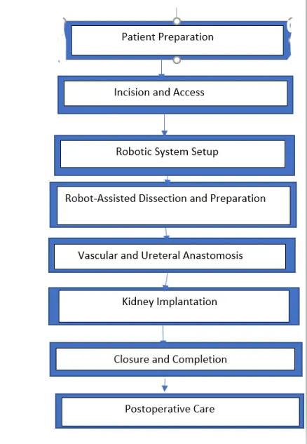
Patient Preparation:
- Preoperative assessment and evaluation
- Administer anesthesia
Incision and Access:
- Make small incisions in the abdomen
- Insert trocars for robotic instruments and camera
Robotic System Setup:
- Position and dock robotic arms and camera system
- Establish connection to surgeon's console
Robot-Assisted Dissection and Preparation:
- Use robotic instruments to dissect and prepare the surgical area
Vascular and Ureteral Anastomosis:
- Identify and prepare recipient's blood vessels and bladder
- Connect donor kidney's blood vessels and ureter to recipient's blood vessels and bladder
Kidney Implantation:
- Place donor kidney in recipient's pelvic area
- Connect renal artery and vein of donor kidney to recipient's blood vessels
- Connect donor kidney's ureter to recipient's bladder
Closure and Completion:
- Ensure hemostasis at surgical site
- Close incisions with sutures or staples
- Apply sterile dressings
Postoperative Care:
- Transfer patient to recovery area
- Monitor vital signs and kidney function
- Manage pain and administer medications
Benefits of a Robotic Kidney Transplant:
Robotic transplant of the kidney has many advantages over the conventional surgical method.
• The robotic hands are able to make better and précised incisions compared to a human. This is especially useful when the operation site is narrow and requires a smooth dissection.
• For patients who have reached the final stage of renal disorders, traditional open surgery may not be very successful. In that case, RAKT is a better solution that increases the chances of survival.
• This technique is safer for obese patients for whom open surgery is not applicable.
• In the robotic transplant method, there is minimal blood loss and scarring.
• The recovery period is short.
• Possibilities of postoperative complications are reduced.
Risks associated with RAKT:
Robot-assisted kidney transplant, like any surgical procedure, carries certain risks. It's important to note that the risks can vary depending on factors such as the patient's overall health, the complexity of the surgery, and the specific robotic system being used. Here are some potential risks associated with robot-assisted kidney transplant:
1. Surgical Complications: There is a risk of surgical complications, such as bleeding, infection, blood vessel or ureter injury, organ damage, or hernia formation. While robotic systems aim to minimize these risks, they still exist.
2. Anesthesia Risks: Anesthesia carries its own set of risks, including allergic reactions, respiratory problems, or adverse reactions to medication. An anesthesiologist carefully monitors the patient during the procedure to minimize these risks.
3. Robotic System Malfunction: Although rare, there is a possibility of technical issues or malfunctions with the robotic system, such as mechanical failure or electrical errors. Adequate system maintenance and regular checks are performed to minimize these risks.
4. Longer Procedure Time: Robot-assisted surgeries can take longer than traditional open surgeries due to the setup and the need for precise robotic movements. Prolonged surgery may increase the risk of complications and potential fatigue for the surgical team.
5. Conversion to Open Surgery: In some cases, it may be necessary to convert a robot-assisted procedure to open surgery if complications arise or if the surgeon determines it is the safest course of action. This decision is made in the best interest of the patient's well-being.
6. Postoperative Complications: After the surgery, there is a risk of postoperative complications such as pain, infection, blood clots, delayed graft function, rejection of the transplanted kidney, or complications related to immunosuppressive medications.
It's important to note that robotic surgery is generally considered safe, and the benefits often outweigh the risks. Surgeons and medical teams undergo specialized training to minimize complications, and patients are thoroughly evaluated to ensure they are suitable candidates for the procedure. Prior to undergoing a robot-assisted kidney transplant, patients should have detailed discussions with their healthcare providers to understand the risks and benefits specific to their situation.
Future perspectives:
Most published cases of RKT have been in living donors. A further step in development will be in performing the procedure in deceased donors. The majority of the technique will be the same as for living donors but there will be some subtle differences such as the use of thicker sutures for suturing stiffer vessels. Logistical considerations will need to be made and significant investment in these programs to ensure round-the-clock availability of robotic theatres and appropriately trained staff.
The technology itself also has scope for improvement. There is currently no haptic feedback built into the da Vinci Surgical System. If haptic feedback was available, there would be potential for
shortening the time needed to become fluent in the procedure. Another limitation to robotic surgery is the inability to perform multi-quadrant abdominal surgery. This will require development in future robotic platforms. There is also room for improvement in the ergonomics of the robot with 56.1% of robotic surgeons still regularly experiencing physical symptoms or discomfort related to utilizing the robot.There have already been four generations of the da Vinci Surgical System and it is likely that as technology improves, the technique will evolve simultaneously.
Conclusions:
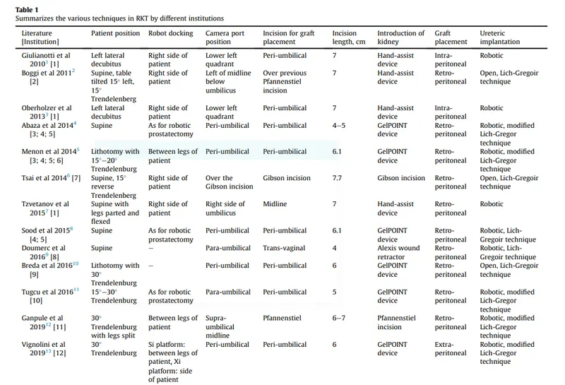
Robotic surgery has emerged as a transformative approach in donor nephrectomy for kidney transplantation. The integration of AI-guided robotic systems offers surgeons enhanced precision, improved visualization, and increased surgical capabilities.The evolution of techniques in RKT has been encouraging and the establishment of RKT as a safe and effective alternative to OKT appears likely. A variety of techniques have already been published in the literature with variations based on differing patient and graft characteristics. As techniques continue to be innovated in the future, the words of John Wickham, founding father of robotic urology, should always be borne in mind "at all time patients are more important than techniques".
REFERENCES:
1. Hiess M, Seitz C. Robot-assisted renal surgery: current status and future directions. Robot Surg. 2016 May 24;3:1-12. doi: 10.2147/RSRR.S71328. PMID: 30697551; PMCID: PMC6193442.
2. A. Peres Penteado, F. Molina Cohrs, A. Diniz Hummel, J. Erbs, R.F. Maciel, C.L. Feijó Ortolani, B. de Aguiar Roza, I. Torres Pisa,
Kidney Transplantation Process in Brazil Represented in Business Process Modeling Notation, Transplantation Proceedings,Volume 47, Issue 4, 2015, Pages 963-966, ISSN 0041-1345, https://doi.org/10.1016/j.transproceed.2015.03.044.
3. Serni S, Pecoraro A, Sessa F, Gemma L, Greco I, Barzaghi P, Grosso AA, Corti F, Mormile N, Spatafora P, Caroassai S, Berni A, Gacci M, Giancane S, Tuccio A, Sebastianelli A, Li Marzi V, Vignolini G, Campi R. Robot-Assisted Laparoscopic Living Donor Nephrectomy: The University of Florence Technique. Front Surg. 2021 Jan 15;7:588215. doi: 10.3389/fsurg.2020.588215. PMID: 33521044; PMCID: PMC7844329.
4. Rosales A, Vidal-Mora I, Plata M, et al. Robotic-assisted surgery in living-donor nephrectomy: a systematic review and meta-analysis. Transpl Int. 2018;31(12):1318-1331.
5.Ghani KR, Sukumar S, Sammon JD, et al. Practice patterns and outcomes of open and minimally invasive partial nephrectomy since the introduction of robotic partial nephrectomy: results from the nationwide inpatient sample. J Urol. 2014;191(4):907-912.
6. Thomas Fonseka, Evolution of techniques in robotic kidney transplantation with perspectives on future developments. 2021 March.Volume 4, Issue 1: 3-8.
7. https://www.lybrate.com/topic/robotic-kidney-transplant-all-about-it/9c56f7854c899fbc6f0a8b95345718dc
Image sources:
1. https://www.siasat.com/da-vinci-xi-surgical-robot-perform-kidney-surgery-in-dubai-2330469/
2. https://blog.sciencemuseum.org.uk/robot-surgery-the-da-vinci-robot/
3. https://entokey.com/the-da-vinci-system-technology-and-surgical-analysis/
4. https://www.cpoc.org.uk/about-cpoc/what-perioperative-care