Project 2
- Pathological Condition: COVID-19 in respiratory system
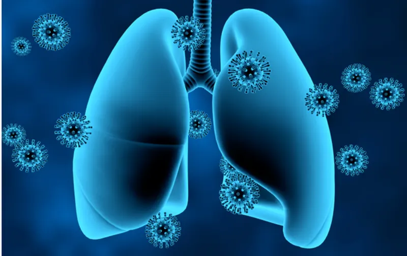
https://www.cedars-sinai.org/newsroom/improving-lung-capacity-pre--and-post-covid-19/
- (a) Normal function of the lungs is to bring in air from the atmosphere and pass oxygen into the bloodstream. Also, it circulates to the rest of the body and sends carbon dioxide out. Removes waste gases, including nitrogen and other volatile organic compounds (VOCs). These VOC compounds consist of methanol, ethanol, isoprene, acetone, and other alcohol.
respiratory zone is found deep inside the lungs and is made up of the respiratory bronchioles, alveolar ducts, and alveoli.
The conducting zone consists of all of the structures that provide passageways for air to travel into and out of the lungs: the nasal cavity, pharynx, trachea, bronchi, and most bronchioles
(b) Pathology stage: coronavirus can infect the upper or lower part of the respiratory tract. coronavirus travels down the airways. The lining can become irritated and inflamed by the virus. In some cases, the infection can reach all the way down into the alveoli. The lungs and airways swell and become inflamed. This can start in one part of the lung and spread. In the most critical cases, your lungs need a ventilator for oxygen supply.
(c) Reasons: lower immunity and poor sanitation and hygiene, not wearing masks.
(d) Homeostasis change: SARS-CoV-2 can affect the expression of blood pressure regulators(ACE2). ACE2 is a regulator of blood pressure homeostasis and is present on the surface of different cell types. ACE2 catalyzes the proteolysis of Angiotensin II (Ang II) into Angiotensin-(1-7).In critical cases, it will result in an oxygen imbalance and you may need a ventilator to breathe. Change in ph happens when too much carbon dioxide can cause the body to become acidic.
2.Description of the diagnostic methods selected by the surgeon for assessing the condition. Points to be considered are
(a) Diagnostic method
There are two main types of viral tests: nucleic acid amplification tests (NAATs) and antigen tests - laboratory test is taken based on the sample collected from the patients
- NAATs: This is a PCR-based tests, mostly performed in a laboratory. They are the most reliable tests for people with or without covid symptoms. These tests detect viral genetic material, which may stay in your body for up to 90 days after you have tested positive.
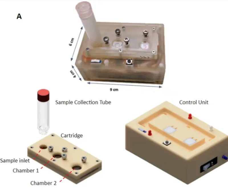
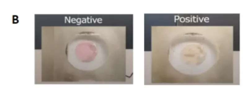
- SOURCE-https://www.nature.com/articles/s41598-021-94652-0/figures/1
- (A) A photograph of the actual device and a rendered image of its various components which include a collection tube, a disposable cartridge and a reusable control unit. (B) The readout of the result in Chamber 2. Pink indicates negative and yellow indicates positive result.
- Antigen test: It is a rapid test that produces results in 15-30 minutes. They are less reliable than NAATs, especially for people who do not have symptoms. A single, negative antigen test result does not rule out infection. For the detection of the virus in your body, a negative antigen test should be repeated at least 48 hours apart (known as serial testing). Sometimes a follow-up NAAT may be recommended to confirm an antigen test result.
The most widely used method to examine patients with COVID-19 are chest X-ray(CXR) and CT scan. These scan can only be taken after the your doctor consultation. These techniques help clinicians identify the effects of COVID-19 on different organs at various stages of the disease. They are used on the chest and lungs because respiratory symptoms are known to be among the first signs of COVID-19.
Lung ultrasound is also a diagnostic tool for covid-19.
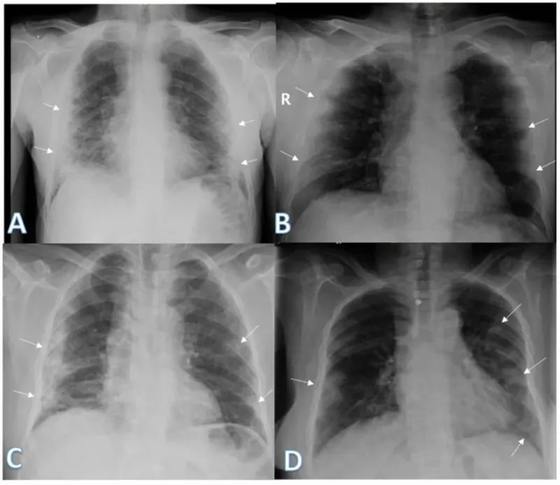
SOURCE: (https://ejrnm.springeropen.com/articles/10.1186/s43055-020-00296-x)
(b)Data acquisition method:
Fast and accurate diagnosis of COVID-19 is possible with chest X-ray (CXR) and computed tomography (CT) scan images.
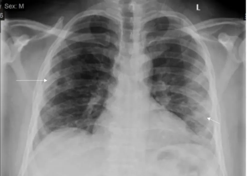
source: (https://ejrnm.springeropen.com/articles/10.1186/s43055-020-00296-x)
Above is the image of a 54-year-old male patient who was found positive for COVID-19. He was having a cough and fever. Follow-up chest X-ray showed a right mid-zonal lesion with central lucency giving an atoll sign (long arrow) of organizing pneumonia. Also, left lower zonal small air space consolidation opacities with reticular thickening was seen (short arrow). The severity score was 1 for each lung, so the total severity score was 2.
(c) Doctors considered the patients who were admitted with fever, cough, shortness of breath, and other symptoms. Patients were scanned using computed tomography (CT), which revealed varied opacities (denser, more profuse, and confluent) in comparison to images of healthy lungs. This finding led to the initial diagnosis of pneumonia. Additional nucleic acid analysis using multiplex real-time polymerase chain reaction (PCR) was performed.
The most common hallmark features of COVID-19 identified by doctors include bilateral and peripheral ground-glass opacities (areas of hazy opacity) and consolidations of the lungs (fluid or solid material in compressible lung tissue) by using a CT SCAN.
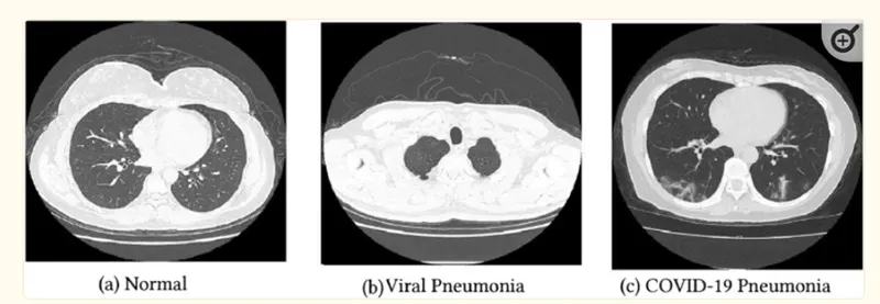
source - (https://www.ncbi.nlm.nih.gov/pmc/articles/PMC7992299/)
(d) If the patient is found positive for covid-19 may have a dry cough, fever, or sore throat. Some people have pneumonia, a lung infection in which the alveoli are inflamed.
Doctors can see signs of respiratory inflammation on a chest X-ray or CT scan.
The current target oxygen saturation range for patients with COVID-19 recommended by the National Institutes of Health is 92–96%. Below this threshold, the patient should be hospitalized and need a ventilator for oxygen supply.
3.
- Below is the list of the drugs approved for the treatment of patients who are suffering from COVID-19.
(a) Nirmatrelvir with Ritonavir (Paxlovid)is a antiviral drug given for Adults; children ages 12 years and older.
(b) Molnupiravir (Lagevrio) is a Antiviral drug, it should be given to Adults only.
The infected patients can take these two drugs orally as soon as possible; must begin within 5 days of symptoms.
(c) Veklury (Remdesivir) - adults and pediatric patients (28 days of age and older and weighing at least 3 kilograms) with positive results of direct SARS-CoV-2 viral testing, who are: hospitalized, or not hospitalized and have mild-to-moderate and are at high risk for progression to severe condition, including hospitalization or death.
If you are hospitalized, your healthcare provider might use other types of treatments, depending on how sick you are.
The current COVID-19 vaccines provide strong protection against serious illness.
- Initially, surgery is not needed for patients who were infected with covid-19. In most critical cases, patients may need a ventilator. A ventilator pumps air—usually with extra oxygen—into patients' airways when they are unable to breathe adequately on their own. Even though COVID-19 is a disease that predominantly affects the lungs, it can also harm other body organs like the heart, kidneys, skin, and brain, including damage to the heart muscle, scar tissue in the lungs, strokes, and seizures. The liver usually suffers the biggest injuries from COVID-19. Acute liver injury and liver failure are life-threatening complications. So, if it affects the other organs then surgery is needed for that particular organ. Furthermore, recent studies have reported that over a third of infected patients develop a broad spectrum of neurological symptoms affecting the central nervous system, peripheral nervous system and skeletal muscles, including anosmia and ageusia. In few cases, lung transplant surgery may be done for the survival of patients.
4.
As mentioned above, initially surgery is not required for treating covid-19 in patients. Prescription of medications to patients who are having mild to moderate symptoms. If your oxygen level is below 92-95%, your lungs need help from a machine called a ventilator to do their job. Some people had a cough even after they recovered from COVID-19. Others had scarring in their lungs. Doctors are still studying whether these effects are permanent or might heal over time. A few people have even needed lung transplants because of severe tissue damage from COVID-19. Lung transplant is a complex, but often life-saving, surgery for people with lung failure. The procedure involves removing one or both diseased lungs and replacing them with new healthy ones.
- Surgical position: The patient is placed in the supine position.
- Surgical procedure in detail:
All patients receive general anesthesia through a double-lumen endotracheal tube.
During a double lung transplant, you may connect to a heart-lung machine.
This machine supports your heart and lungs during surgery. It oxygenates your blood outside your body and pumps it back into your body.
It is important to insert the venous neckline on the left side as post-procedure extracorporeal membrane oxygenation (ECMO) may be needed.
A Swan Ganz catheter is also placed to monitor PA pressures.
During lung transplantation (LTx), the close interrelation between heart and lung function makes TEE (trans-esophageal echocardiography) an invaluable tool for instantly monitoring the physiopathological situation in the subsequent steps of the intervention.
For a single lung transplant, anterolateral thoracotomy is used. Bilateral transplants are traditionally done using a clamshell incision with sternal splitting. Make an incision in your chest.
Cut off the airways and blood vessels to your diseased lung. Remove the diseased lung, beginning with the division of the inferior pulmonary ligament.
The pulmonary artery and pulmonary veins (PV) are subsequently divided using an endo stapler taking care to leave behind an adequate stump for anastomosis. Heparin is given systemically to keep the vessels patent.
The hilum is prepared by the circumferential opening of the pericardium. The bronchus is prepared centrally. Replace it with a healthy donor lung beginning with bronchial anastomosis.
Reattach the airways and blood vessels. Close up the incision. The surgical duration usually takes 6 to 12 hours for a double lung transplant.
- Implants or instruments required during the surgery with relevant images
heart-lung-machine
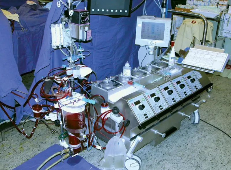
(https://www.britannica.com/science/heart-lung-machine)
Swan Ganz catheter
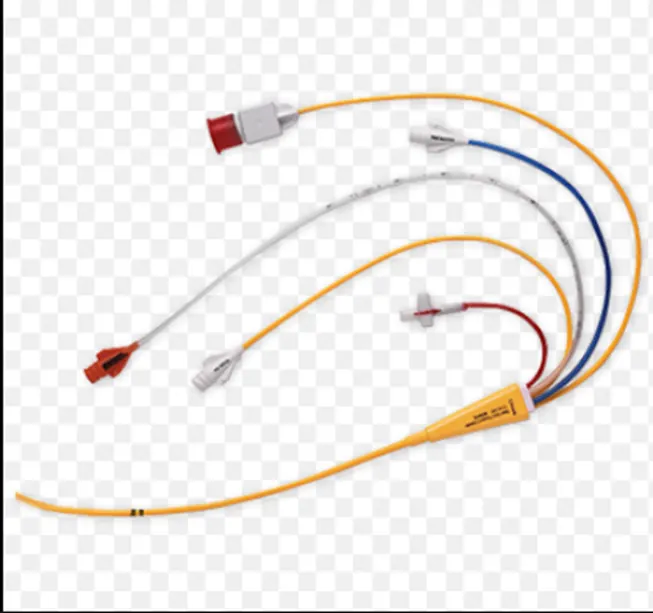
(https://www.edwards.com/gb/devices/Hemodynamic-Monitoring/swan-ganz-catheters)
endo stapler
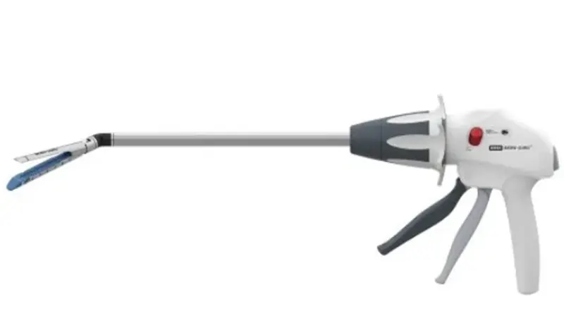
(https://www.indiamart.com/proddetail/endo-gia-stapler-25672604830.html)
Ventilator
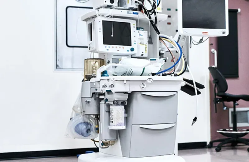
(https://www.roechling.com/industrial/healthcare/materials-for-ventilators)
- Post-operatively, patients are transferred to the critical care unit. Here they remain on a ventilator until they regain consciousness and lung function is appropriate to start weaning. Adequate pain control is provided and anti-rejection drugs right away. It is critical to managing fluid intake, hemodynamics, and optimizing the outcome of end-organ perfusion in the intensive care unit. The majority of recipients receive immunosuppression consisting of steroids, a cell cycle inhibitor, and a calcineurin inhibitor. All patients undergo routine postoperative surveillance bronchoscopy and maintenance immunosuppression. Once stable, they are shifted to the cardiothoracic nursing unit, based on the recovery. From here begins the post-transplant rehabilitation, which includes physical therapy and breathing and speech exercises. The expected length of stay is one to three weeks for an uncomplicated lung transplant.
- bronchoscopy - A bronchoscope is used to view the airways and check for any abnormalities.
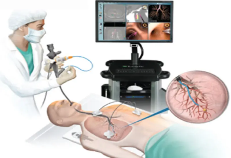
(https://www.yashodahealthcare.com/blogs/what-is-a-bronchoscopy-and-when-do-doctors-recommend-it/)
Chest X-ray machine is used to diagnose internal injuries and disorders. The patient stands in front of the apparatus at centre left, which contains the film. The box at right emits X-rays towards the patient's chest. The X-rays pass through the patient and cast an image onto the film. X-rays are electromagnetic rays that can penetrate soft tissues, but are absorbed by bones or opaque contrast media.
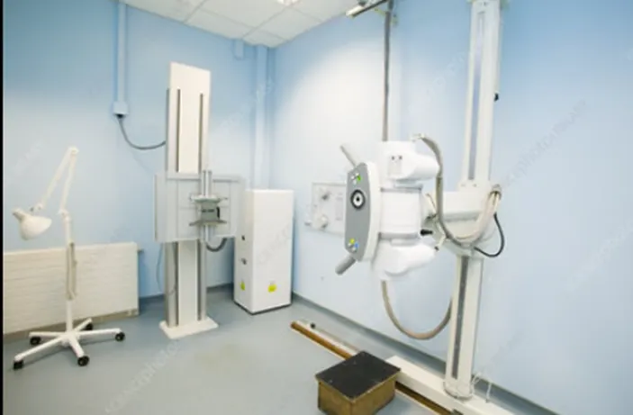
https://www.sciencephoto.com/media/271956/view/chest-x-ray-machine
A CT scan is a diagnostic imaging technique which combination of X-rays and computer technology to produce images of the inside of the body. It shows detailed images of any part of the body, including the muscles, bones, and blood vessels.
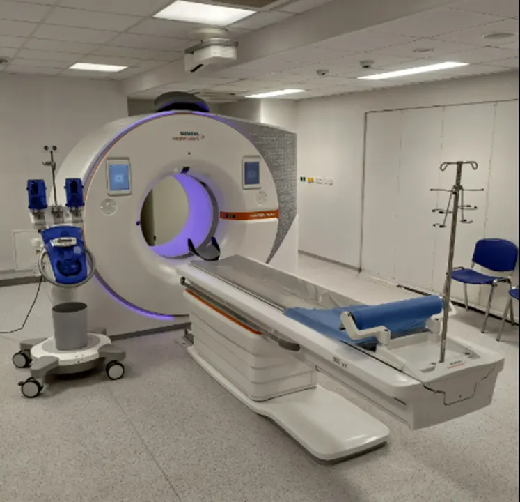
https://en.wikipedia.org/wiki/CT_scan
pulse oximeter
It is a device used to measure the oxygen level (oxygen saturation) of the blood.
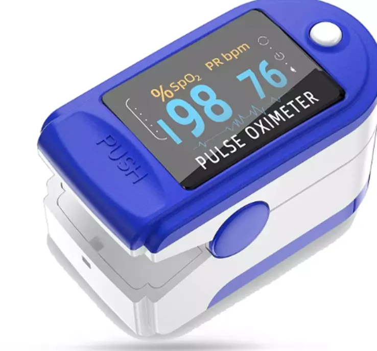
https://pharmeasy.in/health-care/products/jziki-jzk-301-pulse-oximeter-3129260
Oxygen Concentrator
The oxygen concentrator provides the higher amounts of oxygen needed for oxygen therapy. Concentrators may be large and stationary or small and portable.
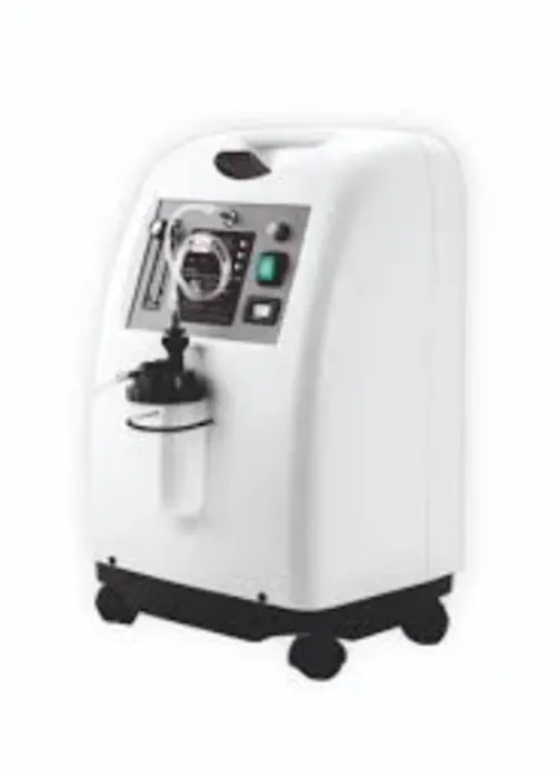
http://www.biopharmgroup.co.in/product/oxygen-concentrator-5-ltr/
real-time-fluorescent RT-PCR KIT- diagnostic kit for covid-19
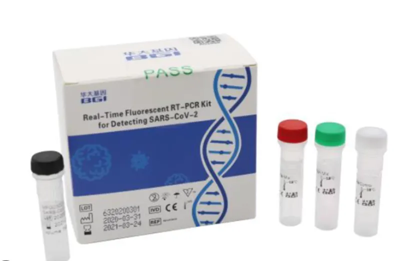
https://www.bgi.com/us/sars-cov-2-real-time-fluorescent-rt-pcr-kit-ivd/
REFERENCES:
1.https://www.ncbi.nlm.nih.gov/pmc/articles/PMC7383117/
- https://www.fda.gov/drugs/emergency-preparedness-drugs/coronavirus-covid-19-drugs
- https://www.ncbi.nlm.nih.gov/pmc/articles/PMC7144809/
- https://www.cdc.gov/coronavirus/2019-ncov/symptoms-testing/testing.html
- https://www.news-medical.net/news/20210622/Does-SARS-CoV-2-alter-the-expression-of-molecules-involved-in-the-homeostasis-of-blood-pressure.aspx