Project 1
Part A - Detailed study of the anatomical basis of surgical procedures or Clinical management in association with any of the organ system
Description:
1. Describe one common clinical condition or disorder associated with the system selected.
answer: Alzheimer's disease is a progressive neurologic disorder that causes the brain to shrink (atrophy) and brain cells to die. (system involved nervous system and organ is brain)
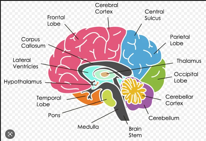
Brain is the central organ of the nervous system, and with the spinal cord makes up the central nervous system, which controls most of the activities of the body, processing, integrating, and coordinating the information it receives from the sense organs and determining the signals or instructions sent back to the rest of the body.
Factors to be remembered
- Describe the gross anatomy of the organ involved
Organ involved – brain
The brain consists of three main structural divisions
- The cerebrum (outer layer is the cerebral cortex)
Cerebrum
The cerebrum (front of brain) comprises gray matter (the cerebral cortex) and white matter at its center. The largest part of the brain, the cerebrum initiates and coordinates movement and regulates temperature. Other areas of the cerebrum enable speech, judgment, thinking and reasoning, problem-solving, emotions and learning. Other functions relate to vision, hearing, touch and other senses.The cerebral cortex is divided into two halves, or hemispheres. It is covered with ridges (gyri) and folds (sulci).
- Frontal Lobe
- Parietal lobe.
- Occipital lobe.
- Temporal lobe.
2.The cerebellum,
The cerebellum (“little brain”) is a fist-sized portion of the brain located at the back of the head, below the temporal and occipital lobes and above the brainstem.
3.The brain stem at the base of the brain (which extends from the upper cervical spinal cord to the diencephalon of the cerebrum
The brainstem (middle of brain) connects the cerebrum with the spinal cord. The brainstem includes the midbrain, the pons and the medulla.
4.Three layers of protective covering called meninges surround the brain and the spinal cord.
- The outermost layer, the dura mater, is thick and tough.
- The arachnoid mater is a thin, weblike layer of connective tissue.
- The pia mater is a thin membrane that hugs the surface of the brain and follows its contours.
5.The cerebrum consists of two cerebral hemispheres, the right hemisphere and the left hemisphere, connected by the corpus callosum which facilitates communication between both sides of the brain,
- Describe the microscopic and macroscopic structure of the organ involved with relevant labelled diagram
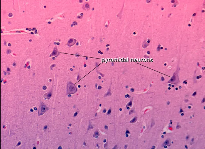
This is stained with Hematoxylin and Eosin (H&E), a routine, general purpose histological stain used for basic examination of tissue
macroscopic structure of the brain:
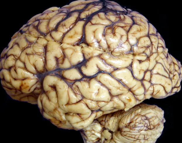
Here in this picture you can see the cerebral cortex is divided into two halves, or hemispheres. It is covered with ridges (gyri) and folds (sulci).
above two images are taken from the given website
https://renaissance.stonybrookmedicine.edu/pathology/neuropathology/chapter1)
- Describe the blood supply and nerve supply of the organ involved with suitable diagram
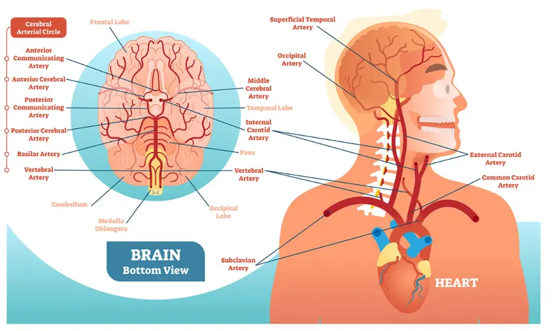
There are two sets of blood vessels supply blood and oxygen to the brain: the vertebral arteries and the carotid arteries.
The external carotid arteries extend up the sides of your neck, and are where you can feel your pulse when you touch the area with your fingertips. The internal carotid arteries branch into the skull and circulate blood to the front part of the brain.
The vertebral arteries follow the spinal column into the skull, where they join together at the brainstem and form the basilar artery, which supplies blood to the rear portions of the brain.
The circle of Willis, a loop of blood vessels near the bottom of the brain that connects major arteries, circulates blood from the front of the brain to the back and helps the arterial systems communicate with one another.
below you can see the image with various arteries
2. Describe the common disorders in association with the organ
Factors to be remembered
- Describe in detail about clinical presentation of the common disorder associated with the organ
- Alzheimer's Disease.
- Dementias.
- Brain Cancer.
- Epilepsy and Other Seizure Disorders.
- Mental Disorders.
- Parkinson's and Other Movement Disorders.
- Stroke and Transient Ischemic Attack
Here, I'm going to discuss about
- Alzheimer's Disease.
- Patients with mild Alzheimer disease usually have somewhat less obvious executive, language, and/or visuospatial dysfunction.
- In atypical presentations, dysfunction in cognitive domains other than memory may be most apparent.
- Patients with Alzheimer disease (AD) most commonly present with insidiously progressive memory loss, to which other spheres of cognition are impaired over several years.
- This loss may be associated with slowly progressive behavioral changes. After memory loss occurs, patients may also experience language disorders (eg, anomic aphasia or anomia) and impairment in their visuospatial skills and executive functions.
- Confusion with time or place.
Enumerate the various diagnostic techniques and investigations used in diagnosis of the disorder :
investigations used in diagnosis of the Alzheimer's Disease.
- Conduct tests of memory, problem solving, attention, counting, and language.
- Order blood, urine, and other standard medical tests that can help identify other possible causes of the problem.
- Administer a psychiatric evaluation to determine if depression or another mental health condition is causing or contributing to a person's symptoms.
- Collect cerebrospinal fluid (CSF) via a spinal tap and measure the levels of proteins associated with Alzheimer’s and related dementias.
various diagnostic techniques are:
- brain scans, such as computed tomography (CT), magnetic resonance imaging (MRI), or positron emission tomography (PET), to support an Alzheimer’s diagnosis or rule out other possible causes for symptoms.
- Describe one diagnostic technique in detail
Magnetic Resonance Imaging (MRI)
A non-invasive imaging technology used to investigate anatomy and function of the body in both health and disease without the use of damaging ionizing radiation. It is often used for disease detection, diagnosis, and treatment monitoring. It is based on sophisticated technology that excites and detects changes in protons found in the water that makes up living tissues.
MRIs employ powerful magnets which produce a strong magnetic field that forces protons in the body to align with that field. When a radiofrequency current is then pulsed through the patient, the protons are stimulated, and spin out of equilibrium, straining against the pull of the magnetic field. When the radiofrequency field is turned off, the MRI sensors are able to detect the energy released as the protons realign with the magnetic field. The time it takes for the protons to realign with the magnetic field, as well as the amount of energy released, changes depending on the environment and the chemical nature of the molecules. Physicians are able to tell the difference between various types of tissues based on these magnetic properties.
3. Describe the management and treatment plan utilized in the disorder
Factors to be remembered
- Medical management
Regular monitoring of patient's health and cognition, initiation of pharmacologic and nonpharmacologic treatments as appropriate, and evaluation of patient motivation to volunteer for a clinical trial.
There are several ways for treating Alzheimer’s disease.
Giving the patients proper medications:the Aducanumab is indicated for the treatment of Alzheimer’s disease.Aducanumab is the first therapy to demonstrate that removing amyloid, one of the hallmarks of Alzheimer’s disease, from the brain is reasonably likely to reduce cognitive and functional decline in people living with early Alzheimer’s.
The following medications are prescribed to treat symptoms related to memory and thinking.
The cholinesterase inhibitors most commonly prescribed are:
- Donepezil (Aricept®): approved to treat all stages of Alzheimer’s disease.
- Rivastigmine (Exelon®): approved for mild-to-moderate Alzheimer’s as well as mild-to-moderate dementia associated with Parkinson’s disease.
- Galantamine (Razadyne®): approved for mild-to-moderate stages of Alzheimer’s disease.
- Surgical procedure applicable for the disorder :
The following 5 categories of neurosurgical treatment were identified: cerebrospinal fluid shunting, intraventricular infusions, tissue grafting, gene therapy, and electrical neural stimulation and Omental transposition for Alzheimers disease
Cerebrospinal fluid (CSF) tests
Research suggests that Alzheimer's disease in early stages may cause changes in CSF levels of multiple markers such as tau and beta-amyloid, two markers that form abnormal brain deposits strongly linked to Alzheimer's.
cerebrospinal fluid shunting - These shunt systems drain excess fluid from the brain to another part of the body where the fluid is absorbed as part of the circulatory process.
Deep brain stimulation (DBS) is a neurosurgical procedure where electrodes are implanted in certain parts of the brain. These electrodes send electrical impulses to specific parts of the brain to change brain activity.
-------------------------------------------------------------------------------------------------
4. Describe the surgical management of the disorder
Factors to be remembered
- Anatomical basis of the surgery
- Organ structure and anatomical relation of the organ to be noted while doing the procedure
- Detailed description of the instruments and equipments used for surgery
- Write a brief note on the post operative care
- Elaborate on the equipments/ medical devices used to monitor in post operative care
answer:
The following assessments will be performed at baseline, then at 1, 3, 6, 12, and 24 months following surgery:
- Montreal Cognitive Assessment (MoCA)
- Alzheimer's Disease Assessment Scale-Cognition (ADAS-Cog)
- General Practitioner Assessment of Cognition (GPCOG)
- Eight-item Informant Interview to Differentiate Aging and Dementia (AD8) Subjects who have early stage AD confirmed by a neuropsychological test (MoCA) and who are healthy enough to undergo surgery.
Omental Transposition Surgery in clinical trial:
during this surgery which a general surgeon and a neurosurgeon work as a team to transpose a pedicle omental graft to the surface of the brain. The general surgeon is responsible for performing a laparotomy/laparoscopy, elongating the omentum into a long pedicle and developing an extensive subcutaneous tunnel up the chest and neck to the head. The neurosurgeon is responsible for performing the craniotomy and securing the omentum on the brain.
Creating an intact omental pedicled flap requires the services of a general surgeon and neurosurgeon. The first step in the operation is to obtain access to the abdominal cavity laparoscopically using four 5cm trocars, or via laparotomy through an upper midline incision. The omentum is then removed from the transverse colon, after which it is separated from its proximal and central attachments to the stomach. The omentum detachments are made directly on the greater curvature of the stomach, leaving the gastroepiploic arteries and veins within the still-connected omentum apron.
Final separation of the omentum from the proximal portion of the stomach is done by dividing the left gastroepiploic vessels, which at the highest proximal level on the stomach become the short gastric vessels. The vascular connections to and from the omentum are now maintained solely from the right gastric and right gastroepiploic vessels. In order for the omental pedicle graft to reach the brain without tension, further surgical tailoring of the omentum is carried out, with care being taken to ensure the preservation of a major omental artery and vein.
After the omentum has been lengthened to an appropriate extent to reach the head, several small (3- to 4-inch) transverse incisions are made on the chest wall slightly lateral to the midline, with the side of the midline to make the transverse incisions depending on which cerebral hemisphere the omentum is to be placed. These transverse incisions are connected subcutaneously, thus creating a tunnel that begins at the upper pole of the midline abdominal incision and travels subcutaneously, up the chest wall and neck to behind the ear.
Although the subcutaneous tissue behind the ear is extremely dense, it is essential that the tunnel at this location be at least 2-3 finger lengths in width so that there is no constriction on the omentum in this particular area within the tunnel. As the omentum in the tunnel passes behind the ear, it goes beneath the base of the scalp flap that had been previously dissected in making the initial craniotomy incision.
The neurosurgery portion of the operation involves removal of a single piece of bone over the temporal-frontal area, opening the dura mater, followed by the removal of patches of arachnoid membrane, with care being taken to avoid blood vessels on the surface of the brain.
At this point in the operation, the omentum is laid directly upon the parietal-temporal-frontal area, after which the dura mater is sutured to the omentum. It is not necessary that the edges of the omentum be applied to the cut edges of the dura since the omentum can be tucked under the edges of the dura for greater coverage of the brain. The edges of the dura are then sewn to the top surface of the omentum using absorbable sutures and the craniotomy is closed.
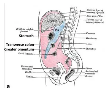
(a) anatomy of the greater omentum relative to the greater curve of the stomach and transverse colon
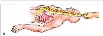
(b)Illustration of the omental delivery to the cranial compartment
post operative care:
Patient Surveillance
• First 24 hours after surgery:
∙ Patient is in the intensive care unit with regular neuro checks.
∙ Closely monitor hemodynamic status (fluid intake, output, electrolyte, and gastrointestinal function).
∙ Maintain thresholds for blood pressure (mean arterial pressure goals 90-110 to prevent hypoperfusion, TND, and hyperperfusion with potential postoperative hematoma).
Analgesia and antiemetic.
• Day 2: Mobilize, observe oral intake, and transfer to regular ward care.
• Discharge from hospital when deemed to be safe by the neurosurgical and general surgical team (4-day average hospital stay).
• Post discharge: Educate and inform about TND, wound care, and subsequent follow-up plans
--------------------------------------------------------------
During the stay, your healthcare providers will likely:
- elevate your head to manage swelling
- give you oxygen for several days
- teach you deep-breathing exercises to prevent pneumonia
- place compression devices on your legs to prevent blood clots
The urinary catheter will stay in your bladder for several days.
Your healthcare team will frequently do neurological checkups to ensure your brain and body are functioning properly.
Before you go home, your surgeon will provide home care instructions.
5. Self study on recent advances in the field
Factors to be remembered
- Describe the recent advances with respect to management of the clinical condition /disorder involved.
- Describe on the possible newer techniques (diagnostic or therapeutic) involved in managing the disorder
answers:
advances
Functional Neuromodulation – Deep brain stimulation
One of the more invasive medical devices recently designated a Breakthrough Device by the FDA is Functional Neuromodulation’s Vercise deep brain stimulation system, developed and manufactured by Boston Scientific Corporation to treat Alzheimer’s.
The technology requires a surgical procedure to imbed electrodes into certain areas of the brain – in this case the fornix – which are connected to an internal pulse generator installed under a layer of subcutaneous fat in the upper chest.
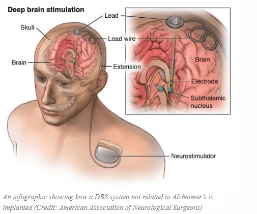
Small electric pulses are delivered into targeted areas of the brain to control abnormal brain activity, a process already shown to work in other neurological conditions, like advanced-stage Parkinson’s disease and essential tremor.
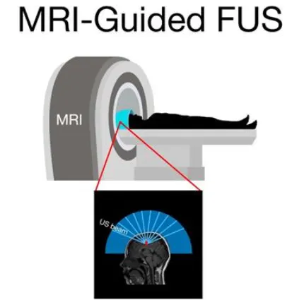
(b) magnetic resonance (MR)-guided low-intensity focused ultrasound (FUS)- The blood-brain barrier (BBB) presents a significant challenge for treating brain disorders. The hippocampus is a key target for novel therapeutics, playing an important role in Alzheimer's disease (AD), epilepsy, and depression. Preclinical studies have shown that magnetic resonance (MR)-guided low-intensity focused ultrasound (FUS) can reversibly open the BBB and facilitate delivery of targeted brain therapeutics.
--------------------------------------------------------------------------------------------------------------------------
New technologies based on structural and functional neuroimaging, and on the biochemical analysis of cerebrospinal fluid may depict correlates of intracerebral amyloidosis in individuals with mild, pre-dementia symptoms. These methods are commonly referred to as AD-related biomarkers, and the combination of clinical and biological information yields good diagnostic accuracy to identify individuals at high risk of AD. In other words, the characterization of pathogenic Aβ by means of biochemical analysis of biological fluids or by molecular neuroimaging are presented as diagnostic tools to help identify AD cases at the earliest stages of the disease process. The relevance of this early diagnosis of AD relies on the hypothesis that pharmacological interventions with disease-modifying compounds are more likely to produce clinically relevant benefits if started early enough in the continuum towards dementia. Therapies targeting the modification of amyloid-related cascades may be viewed as promising strategies to attenuate or even to prevent dementia. Therefore, the cumulative knowledge on the pathogenesis of AD derived from basic science models will hopefully be translated into clinical practice in the forthcoming years.
Part B – Case Study
A 25 year old biker indulged in illegal racing met with a road traffic accident and severed head injury and skull fracture. After resuscitation and operation done for the hematoma it was resorted to do skull implant for his fracture by the orthopaedician
Discuss the case under the following headings
- Describe the type of bone the skull belongs to
Flat bones are made up of a layer of spongy bone between two thin layers of compact bone. They have a flat shape, not rounded. Examples include the skull.The skull is composed of four types of bone i.e., cranial bones, facial bones, ear ossicles and hyoid bone. However two parts are more prominent: the cranium and the mandible. In humans, these two parts are the neurocranium and the viscerocranium (facial skeleton) that includes the mandible as its largest bone.
- Describe the microscopic anatomy of bone
Bones are composed of bone matrix, which has both organic and inorganic components. Bone matrix is laid down by osteoblasts as collagen, also known as osteoid. Osteoid is hardened with inorganic salts, such as calcium and phosphate, and by the chemicals released from the osteoblasts through a process known as mineralization.
The basic microscopic unit of bone is an osteon (or Haversian system). Osteons are roughly cylindrical structures that can measure several millimeters long and around 0.2 mm in diameter.
Each osteon consists of a lamellae of compact bone tissue that surround a central canal (Haversian canal). The Haversian canal contains the bone’s blood supplies. The boundary of an osteon is called the cement line. Osteons can be arranged into woven bone or lamellar bone.
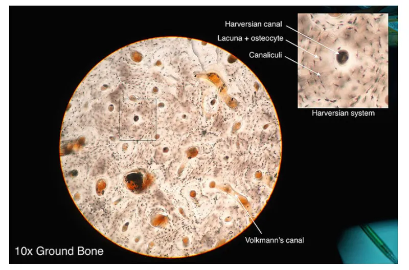
Osteon: A photo taken through a microscope that shows the anatomy of compact bone with a detailed view of an osteon
- Explain the blood supply of bone :
The blood supply to bone is delivered to the endosteal cavity by nutrient arteries, then flows through marrow sinusoids before exiting via numerous small vessels that ramify through the cortex.Healthy bone requires a substantial blood flow to supply the requisite oxygen and nutrients, and to eliminate carbon dioxide, acid and other metabolic waste products. Estimates of the proportion of the cardiac output received directly by the skeleton range from about 5.5% to 11% . The rich perfusion of bone reflects not only the requirements of the bone cells (osteoblasts, osteocytes and osteoclasts), but also of the marrow (hematopoietic lineage cells, stromal cells and adipocytes), as well as endothelial cells. The long bones form from embryonic cartilage rudiments that are invaded by blood vessels and bone cells. The vascular supply of bone enables rapid growth, remodelling (including mechanical responsiveness) that is not possible in cartilage, which is essentially avascular. For a detailed consideration of the functional anatomy of the blood supply to bone during growth, maturity and regeneration
- Elaborate on bone remodeling and new bone formation
Bone remodeling is a lifelong process where mature bone tissue is removed from the skeleton and new bone tissue is formed. These processes also control the reshaping or replacement of bone following injuries like fractures but also micro-damage, which occurs during normal activity.
There are five phases in the bone remodeling process: ACTIVATION, RESORPTION, REVERSAL, FORMATION, and QUIESCENCE. The total process takes about 4 to 8 months, and occurs continually throughout our lives.
Bone ossification, or osteogenesis, is the process of bone formation. This process begins between the sixth and seventh weeks of embryonic development and continues until about age twenty-five; although this varies slightly based on the individual
- Describe the anatomical basis of bone implant
several factors determine how well an implant will bond with native bone. They include physical and chemical factors, such as the chemical composition, porosity, and surface morphology of the implant, and patient-specific factors, such as age, bone quality, and overall health. The patient-specific factors often aren’t controllable. Still, a better understanding of how biomaterials integrate with bone can lead to improved device materials and fewer device failures.
At the simplest level, bone is approximately 35% collagen protein and 65% hydroxyapatite, a natural mineral rich in calcium and phosphate. Bone also contains water and noncollagenous proteins.
New bone tissue grows along an implant, when undifferentiated cells are recruited to the implant’s surface, roughened at nanometer length scales to improve bone–implant bonding. There, the cells become bone-producing osteogenic cells, which sequentially secrete a mineralized matrix that forms the so-called cement line (blue) and a bone precursor substance known as osteoid . Ensuing mineralization of the osteoid produces woven bone.
osseointegration
- Enumerate the indications and contraindications for the bone implant
indications:
biomaterials researchers have begun pursuing an alternative to replacement: endogenous regeneration, in which the implanted device is made of so-called resorbable materials that promote the natural healing and regrowth of the native tissue. Ideally, the implant is gradually replaced by the regenerating native tissue.
Resorbable materials include ceramics such as some calcium phosphates, polymers such as poly-caprolactone, and composites of the two. One well-known example is bioglass, a widely used, bioactive glass–ceramic composite developed in the 1960s. Like implant coatings, resorbable biomaterials can be augmented with growth factors, including bone morphogenetic protein, and with other biomolecules that modulate natural healing processes in the body.
permanent implant devices such as titanium and stainless steel still have a place in biomaterials, particularly in load-bearing applications.
Computer aided design (CAD) or computer aided manufacturing (CAM) systems allows for the design of implants that fit the unique anatomic features of a patient and the injury. Computed tomography or magnetic resonance imaging scans of the bone can be employed to create a model of the fracture or bone defect and used to create a suitable fracture fixation device or implant for bone defect repair via CAD or CAM. Early CAD/CAM systems used subtractive manufacturing, typically using computer numerical control (CNC) manufacturing, which cut away material from a block to get the desired shape. However, subtractive manufacturing has a limited resolution and limited geometries by the tool used to cut, and there is an associated waste of material.
contraindications for the bone implant :
- clinician contend with infection, implant failure, or even patient death. There are conditions that, if stabilized, do not seem to interfere perceptibly with repair; a subsequent paper concerns these relative contraindications.
- you may experience temporary side effects during the first few months, like headaches, nausea, breast tenderness and mood swings.
- Abdominal or back pain.
- An increased risk of noncancerous ovarian cysts.
- Changes in vaginal bleeding patterns, including absence of menstruation (amenorrhea)
- Decreased sex drive.
- Dizziness.
- Headaches.
- Mild insulin resistance.
- Mood swings and depression.