Project 2
project a:
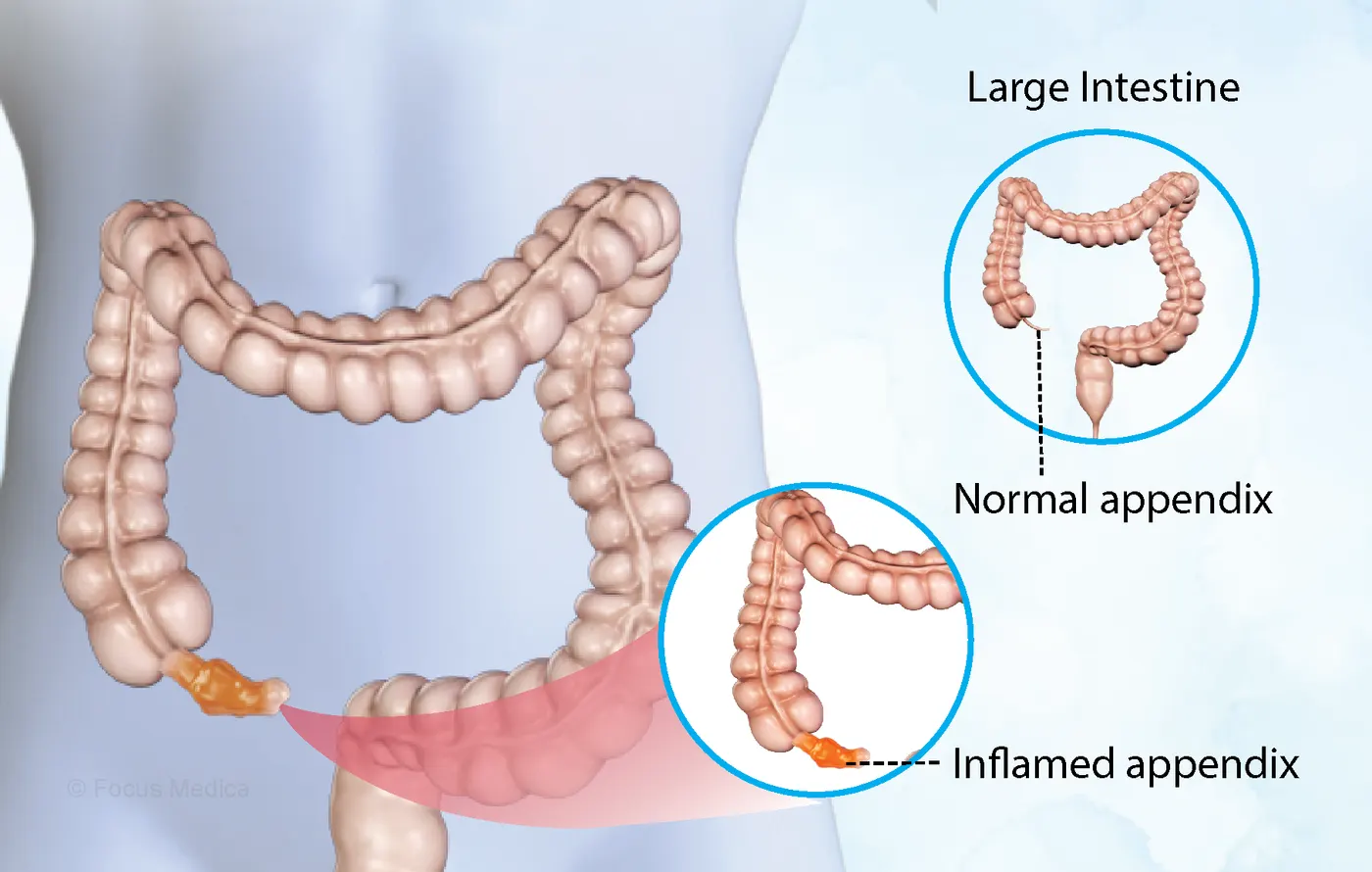
1) pathalogical condition:-appendictis of gastrological system
- Appendicitis is an inflammation of the appendix
- It is a finger-shaped pouch that projects from your colon on the lower right side of your abdomen
- Appendicitis causes pain in your lower right abdomen
- As inflammation worsens, appendicitis pain typically increases and eventually becomes severe.
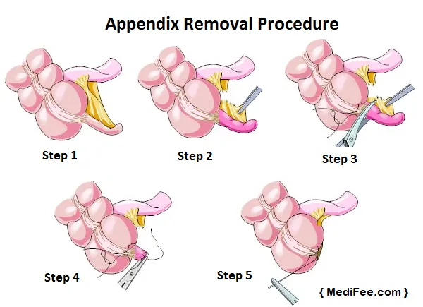
- Standard treatment is surgical removal of the appendix.
It is classified into 5 categories:
- Acute appendicitis: is a condition in which an individual experiences sudden inflammation of the appendix. This is usually caused by obstruction of the lumen resulting from invasion of the appendix wall by the gut flora.
-
Sub-acute appendicitis: mild inflammation is observed. In some cases, it subsides before reaching acute stage.
-
Chronic appendicitis: pathological examination of the appendix may reveal scarring and thickening suggesting an old, healed acute inflammation. This often leads to complaints of persistent right lower abdominal pain.
-
Recurrent appendicitis: repeated attacks of non-obstructive appendicitis without symptoms.
-
Acute non-obstructive appendicitis: this is less critical and occurs when there is an inflammation of the mucous membrane.
Signs and symptoms of appendicitis may include:
- Sudden pain that begins on the right side of the lower abdomen
- Sudden pain that begins around your navel and often shifts to your lower right abdomen
- Pain that worsens if you cough, walk or make other jarring movements
- Nausea and vomiting
- Loss of appetite
- Low-grade fever that may worsen as the illness progresses
- Constipation or diarrhea
- Abdominal bloating
Appendicitis can cause serious complications, such as:
- A ruptured appendix
- A rupture spreads infection throughout your abdomen (peritonitis). Possibly life-threatening, this condition requires immediate surgery to remove the appendix and clean your abdominal cavity.
- A pocket of pus that forms in the abdomen
- If your appendix bursts, you may develop a pocket of infection (abscess). In most cases, a surgeon drains the abscess by placing a tube through your abdominal wall into the abscess. The tube is left in place for two weeks, and you’re given antibiotics to clear the infection.
- Once the infection is clear, you’ll have surgery to remove the appendix. In some cases, the abscess is drained, and the appendix is removed immediately.
- Consult a doctor immediately if you have any of the above symptoms.
DAIGNOSIS:
To rule out any abdomen related problems, with similar symptoms, certain tests are recommended.
complete boold count(CBC): The WBC count may indicate if the body is fighting infection.
ultrasound:Done to rule out other conditions like kidney stones, pancreatitis, ovarian cysts, ectopic pregnancy and also to confirm appendicular mass or abscess.
CT scan:nCT scan of abdomen or pelvis helps in detecting the cause.
MRI:MRI scan of pelvis helps in diagnosis of appendicitis during pregnancy.
clinical diagnosis of appendictis:
- Appendicitis is peritonitis and shows the common signs which are abdominal tenderness and systemic inflammation. The disease history obtained from the patient provides only the time frame and progression of the episode. The surgeon must integrate the information from history, abdominal examination and laboratory tests of acute inflammation to reach the diagnosis (5, 6). The basic boundaries are disease progression over 8 to 48 hours, with distinct tenderness of the right lower quadrant associated with rebound tenderness, and increased temperature, leukocytosis with left shift and increased concentration of C-reactive protein. Outside such boundaries the chance that it is appendicitis is progressively reduced. For instance, it is uncommon that abdominal tenderness without systemic inflammation is appendicitis.
- Every surgeon will be able to relate appendicitis that had an atypical presentation where the traditional diagnostic signs were minimal or absent. It cannot be denied but it begs the question if patients with minimal responses need an operation.
ultrasonographic diagnosis of appendictisis:
- This diagnostic aid has been tried extensively with very differing results. In the hands of a dedicated radiologist US may have similar, or better, accuracy than clinical diagnosis alone .
- When used by surgeons as part of their clinical evaluation of the patient the results are disappointing . Apart from being dependent on the investigator there are other limitations of the technique.
- In particular it is difficult to exclude appendicitis as the normal appendix may be hard to visualize especially in obese patients. Unless substantial improvement of the technique becomes available US will remain a marginal investigation for use in patients under observation.
Computed tomography diagnosis of appendicitis
- The appendix is nearly always visible with a helical CT scan. It is claimed that an enlarged appendix with periappendiceal fat stranding is present in 93% of patients with appendicitis but there are additional signs of appendicitis .
- Both the sensitivity and the specificity can be higher than for the clinical diagnosis. In a recent study of 100 consecutive patients with suspected appendicitis reviewed in the emergency department before being admitted to hospital the diagnostic accuracy was 98% when checked against histology in the 59 operated patients and follow-up in those not operated.
- It was claimed that unnecessary operations could have been avoided, but also hospital admission for observation could be avoided based on the results of the CT scan .
- In addition a number of other diagnoses were established. In a following study some of these claims were supported in that the negative appendectomy rate was reduced from 20% to 7% and to only 3% in those who had had a CT before the operation .
- It seems quite clear that the CT scan can provide a diagnostic accuracy in excess of the best clinical diagnosis. The diagnosis can be established without observation. Its potential is therefore to change clinical management by reducing negative appendectomy to a minimum and to shorten or eliminate observation for possible appendicitis. Several independent studies that confirm the accuracy will be needed.
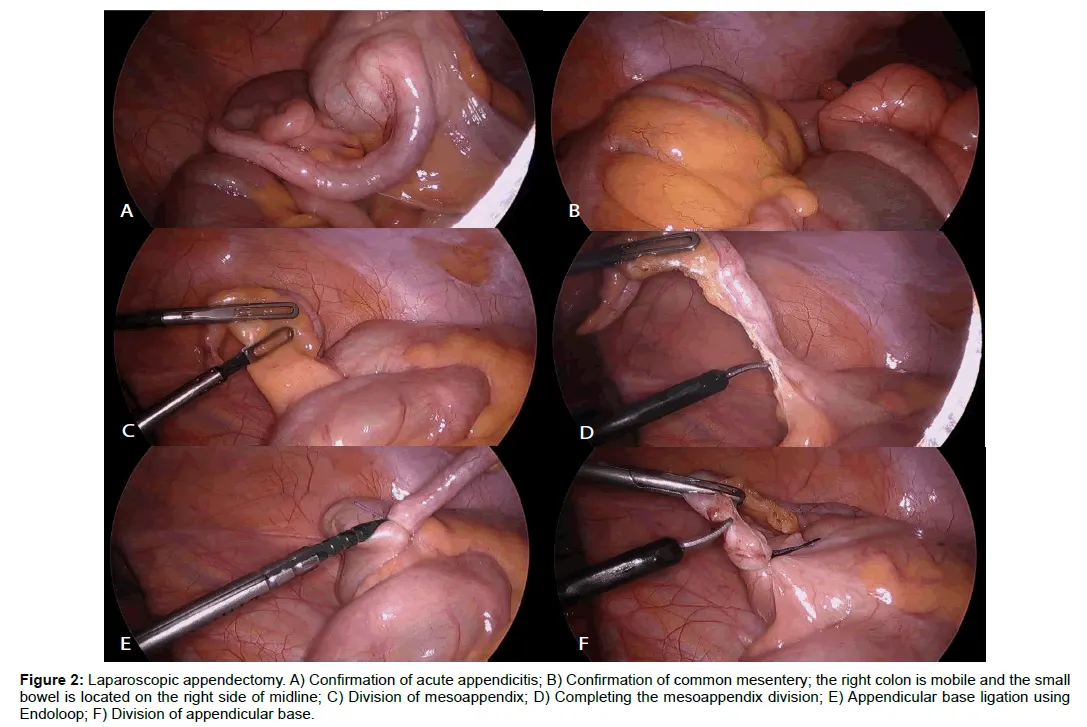
laparoscopic appendetomy:
- For once there are several dozens of trials available from which repetitive information can be extracted. The largest and most recent of the prospective randomized trials included 523 patients . It showed that the operation time for laparoscopy was longer (+ 25 minutes) but patients had less pain and recovered quicker (- 1 week).
- There was no statistical difference for hospital stay or sick leave. The latter reflect administrative decision that may not be affected by the operative method.
- There are at least six structured reviews (meta-analysis) which included information from up to 28 prospective trials . There is universal finding of the longer operation time, but most also establish less pain and shorter recovery. Hospital stay is slightly shorter and wound infections are fewer.
- The conversion rate was about 10% in the studies. Information that was not systematically analyzed includes the number of laparoscopic surgeons available, whether the same surgeons did all the open and laparoscopic operations, and the missed inclusions and exclusions. The cost for laparoscopy has also not been systematically studied.
- There is overwhelming evidence of some gains with laparoscopy but its limitation has not been given the same attention. Hence it remains a policy decision to balance the gains against a more expensive and more difficult method that cannot manage all cases of appendicitis.
- It is this reviewers impression that laparoscopy for appendicitis is currently loosing that trial in favor of the open operation. The penetration of laparoscopic appendectomy varies widely from one hospital to another but the overall penetration is probably no more than around 15% for western countries.
comments on diagnosis technique:
- Considering the evidence available it is reasonable that new foundations are laid for the treatment of suspected appendicitis. Its essence would be to find out in new trials what proportion of those with appendicitis that need an operation. The equivalent of the cost of the unnecessary operations avoided will pay for much better diagnostic work-up to increase both the diagnostic accuracy and the adequacy of an operation. Because the helical CT scan promises real change in this respect it is now important that this means of diagnosis is further investigated.
- Laparoscopic diagnosis has been proposed with claims that if there is appendicitis it can be removed simultaneously. However it is not the appendectomy that is the issue but the unnecessary operation. Unless the laparoscopic diagnosis can be obtained on the awake patient at a price comparable with a CT scan there seems to be little place for it.
- Laparoscopic appendectomy is sufficiently investigated. Under the current principles of treatment for appendicitis, it is an administrative decision whether laparoscopy should be pressed further.
- There should be simple ligation of the appendix stump in the future, primary closure of the wound, and no drain. Antibiotic treatment should be of short duration, usually single dose unless there is perforation and abscess
after surgery to do following things:
- Take proper rest and sound sleep
- Keep your incision(s) clean and dry to avoid infection
- For some time eat soft foods
- Drink a lot of water daily to avoid constipation
- Eat healthy and ensure you get enough sleep as these may aid in speedy recovery
- Try to go for a daily stroll
- Avoid lifting anything that would cause you to strain for around two weeks
- For the first several days, you might want to take a moderate laxative.
- Don’t stand for long as your abdominal muscles may hurt after prolonged standing if you underwent open surgery
- Stay away from intense activities like bicycling, running, weight lifting, or aerobic activities for around two weeks or till the time your doctor tells you to do so
- After a few days, gradually get back to your regular routine. Consistently engage in little exercise
- If you have any odd symptoms, contact your doctor
Following surgery, physicians assist the patient in their recovery, and recommend a diet that promotes energy and speeds up the healing process. Immediately following surgery, you could notice that your bowel motions are irregular. To prevent constipation and maintain stable blood sugar levels one should follow soft foods and drink a lot of water (unless your doctor tells you not to). Your typical diet post surgery should include;
- Fibrous foods: Many patients may experience constipation following surgery. Increased fiber intake will make it simpler to pass stools and prevent the patient from having to exert any effort on the muscles used to push the stool out of the body.
- Easily digestible foods: Go for lighter options like fruits, vegetables, dishes made with rice, or meals made with wheat. Foods like soups, milk, or light breakfasts are simple to digest and provide the body with energy. Rice contains carbohydrates that not only provide the body with energy but also aid in blood vessel creation, tissue growth, and repair.
- Foods that help boost immunity: Including foods that strengthen the immune system always aids in warding off any illnesses. The patient will undoubtedly be given antibiotics until they are well, but consuming foods high in vitamin A and C, such as eggs, carrots, fresh vegetables, lemon, oranges, and amla (Indian Gooseberry), aids in digestion and the formation of antibodies. Consuming vitamin E helps protect cells from free radical damage. Zinc-rich foods and supplements, such as dry fruits, eggs, whole grains, and zinc supplements, can also aid in the prevention of infection and are essential for the production of white blood cells.
- Return to your regular diet: The patient should return to their regular diet if they are not experiencing any nausea, vomiting, or indigestion, according to our specialists. This may promote faster healing and improved energy in the body. Include extra fresh fruits, vegetables, whole grains, low-fat dairy, poultry, and seafood in your diet if at all feasible. Inflammation can be decreased by eating more dry fruits and unsaturated fats like ghee.
side effect of surgery:
Over the following several days of appendicitis surgery, you can suffer certain operation-related side effects. Typical side-effects of appendicitis surgery include:
- Nausea
- Constipation for short period of time after surgery
- Gas problem due to the carbon dioxide that was pumped into the abdomen during the laparoscopy surgery
- Stomach bloating which will lessen over the next few weeks
aftercare of surgery:
You must keep the incision clean and dry at home. You'll receive bathing advice from your doctor.
- Any surgical staples or stitches will be taken out during a subsequent appointment visit. If adhesive strips were applied, it is best to keep them dry.
- They mostly disappear after a few days
- Your abdominal muscles and the incision may hurt, especially after prolonged
- standing. As advised by your doctor, take a painkiller
- Your risk of bleeding may increase if you use aspirin or other painkillers. Take only medications that have been prescribed by your doctor
- Walk and move about a little but stay away from anything strenuous.
If you experience any of the following symptoms, get in touch with your doctor right away:
- Cold or fever
- Bruising, bleeding, or other discharge at the site of the incision
- More discomfort near the incision site
- Vomiting
- Lack of appetite or inability to consume any food or liquids
- Persistent coughing, breathing difficulties, or shortness of breath
- Stomach cramps, discomfort, or swelling
- No bowel movement for at least two days
- Diarrhea for more than three days
result:-
The safest and most successful approach to treat appendicitis, as well as to stop it from coming back and spreading the infection, an immediate surgical procedure should be performed. The minor condition might turn deadly if the operation is postponed as the appendix could rupture, spilling germs into the bloodstream and creating a potentially fatal infection.
2) pathalogical condition : kidney stones in urinary system
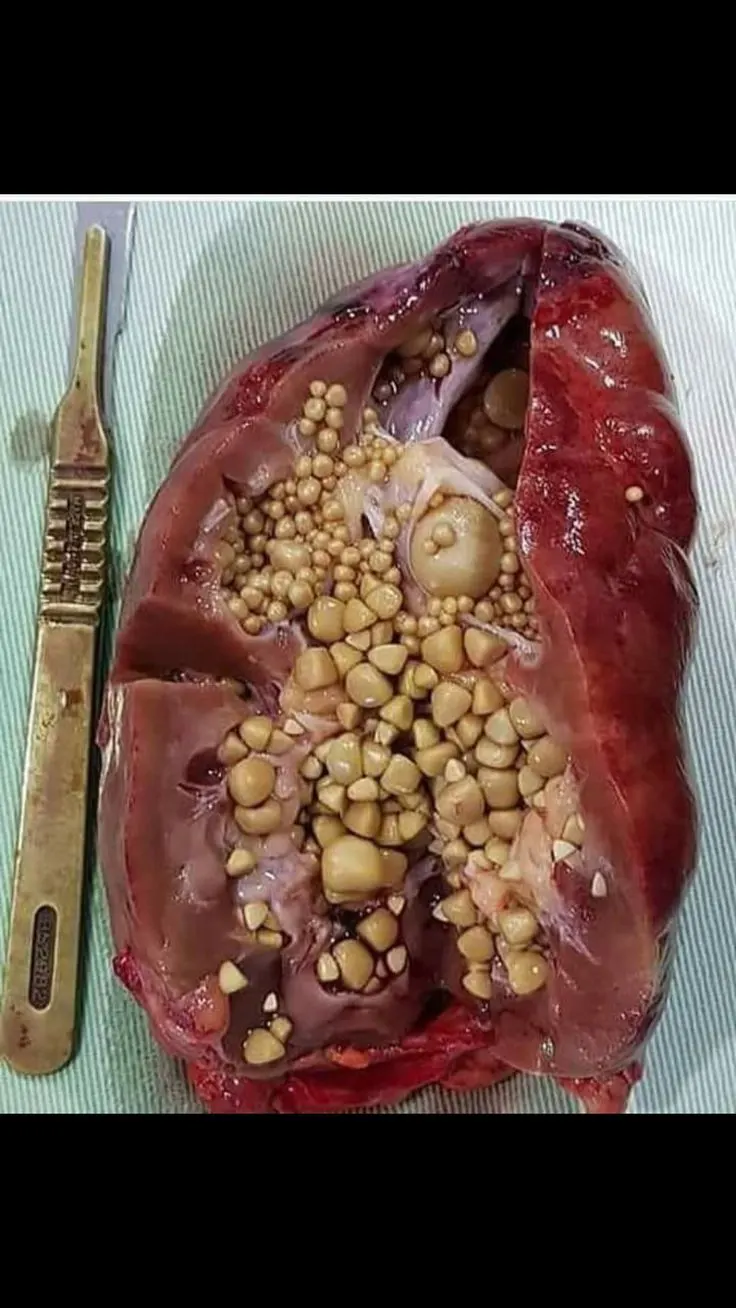
- Kidney stones (also called renal calculi, nephrolithiasis or urolithiasis) are hard deposits made of minerals and salts that form inside your kidneys.
- Diet, excess body weight, some medical conditions, and certain supplements and medications are among the many causes of kidney stones. Kidney stones can affect any part of your urinary tract — from your kidneys to your bladder. Often, stones form when the urine becomes concentrated, allowing minerals to crystallize and stick together.
- Passing kidney stones can be quite painful, but the stones usually cause no permanent damage if they're recognized in a timely fashion. Depending on your situation, you may need nothing more than to take pain medication and drink lots of water to pass a kidney stone. In other instances — for example, if stones become lodged in the urinary tract, are associated with a urinary infection or cause complications — surgery may be needed.
- Your doctor may recommend preventive treatment to reduce your risk of recurrent kidney stones if you're at increased risk of developing them again.
symptoms:
kidney stone usually will not cause symptoms until it moves around within the kidney or passes into one of the ureters.The ureters are the tubes that connect the kidneys and bladder.
If a kidney stone becomes lodged in the ureters, it may block the flow of urine and cause the kidney to swell and the ureter to spasm,which can be very painful. At that point, you may experience these symptoms:
- Severe, sharp pain in the side and back, below the ribs
- Pain that radiates to the lower abdomen and groin
- Pain that comes in waves and fluctuates in intensity
- Pain or burning sensation while urinating
Other signs and symptoms may include:
- Pink, red or brown urine
- Cloudy or foul-smelling urine
- A persistent need to urinate, urinating more often than usual or urinating in small amounts
- Nausea and vomiting
- Fever and chills if an infection is present
Pain caused by a kidney stone may change — for instance, shifting to a different location or increasing in intensity — as the stone moves through your urinary tract.
When to see a doctor
Make an appointment with your doctor if you have any signs and symptoms that worry you.
Seek immediate medical attention if you experience:
- Pain so severe that you can't sit still or find a comfortable position
- Pain accompanied by nausea and vomiting
- Pain accompanied by fever and chills
- Blood in your urine
- Difficulty passing urine
Causes:-
- Kidney stones often have no definite, single cause, although several factors may increase your risk.
- Kidney stones form when your urine contains more crystal-forming substances — such as calcium, oxalate and uric acid — than the fluid in your urine can dilute. At the same time, your urine may lack substances that prevent crystals from sticking together, creating an ideal environment for kidney stones to form.
Types of kidney stones
Knowing the type of kidney stone you have helps determine its cause, and may give clues on how to reduce your risk of getting more kidney stones. If possible, try to save your kidney stone if you pass one so that you can bring it to your doctor for analysis.
Types of kidney stones include:
-
Calcium stones. Most kidney stones are calcium stones, usually in the form of calcium oxalate. Oxalate is a substance made daily by your liver or absorbed from your diet. Certain fruits and vegetables, as well as nuts and chocolate, have high oxalate content.
Dietary factors, high doses of vitamin D, intestinal bypass surgery and several metabolic disorders can increase the concentration of calcium or oxalate in urine.
Calcium stones may also occur in the form of calcium phosphate. This type of stone is more common in metabolic conditions, such as renal tubular acidosis. It may also be associated with certain medications used to treat migraines or seizures, such as topiramate (Topamax, Trokendi XR, Qudexy XR).
- Struvite stones. Struvite stones form in response to a urinary tract infection. These stones can grow quickly and become quite large, sometimes with few symptoms or little warning.
- Uric acid stones. Uric acid stones can form in people who lose too much fluid because of chronic diarrhea or malabsorption, those who eat a high-protein diet, and those with diabetes or metabolic syndrome. Certain genetic factors also may increase your risk of uric acid stones.
- Cystine stones. These stones form in people with a hereditary disorder called cystinuria that causes the kidneys to excrete too much of a specific amino acid.
risks factors are:-
Factors that increase your risk of developing kidney stones include:
- Family or personal history. If someone in your family has had kidney stones, you're more likely to develop stones, too. If you've already had one or more kidney stones, you're at increased risk of developing another.
- Dehydration. Not drinking enough water each day can increase your risk of kidney stones. People who live in warm, dry climates and those who sweat a lot may be at higher risk than others.
- Certain diets. Eating a diet that's high in protein, sodium (salt) and sugar may increase your risk of some types of kidney stones. This is especially true with a high-sodium diet. Too much salt in your diet increases the amount of calcium your kidneys must filter and significantly increases your risk of kidney stones.
- Obesity. High body mass index (BMI), large waist size and weight gain have been linked to an increased risk of kidney stones.
- Digestive diseases and surgery. Gastric bypass surgery, inflammatory bowel disease or chronic diarrhea can cause changes in the digestive process that affect your absorption of calcium and water, increasing the amounts of stone-forming substances in your urine.
- Other medical conditions such as renal tubular acidosis, cystinuria, hyperparathyroidism and repeated urinary tract infections also can increase your risk of kidney stones.
- Certain supplements and medications, such as vitamin C, dietary supplements, laxatives (when used excessively), calcium-based antacids, and certain medications used to treat migraines or depression, can increase your risk of kidney stones.
DIAGNOSIS:-
If your doctor suspects that you have a kidney stone, you may have diagnostic tests and procedures, such as:
- Blood testing. Blood tests may reveal too much calcium or uric acid in your blood. Blood test results help monitor the health of your kidneys and may lead your doctor to check for other medical conditions.
- Urine testing. The 24-hour urine collection test may show that you're excreting too many stone-forming minerals or too few stone-preventing substances. For this test, your doctor may request that you perform two urine collections over two consecutive days.
-
Imaging. Imaging tests may show kidney stones in your urinary tract. High-speed or dual energy computerized tomography (CT) may reveal even tiny stones. Simple abdominal X-rays are used less frequently because this kind of imaging test can miss small kidney stones.
-
Ultrasound, a noninvasive test that is quick and easy to perform, is another imaging option to diagnose kidney stones.
- Analysis of passed stones. You may be asked to urinate through a strainer to catch stones that you pass. Lab analysis will reveal the makeup of your kidney stones. Your doctor uses this information to determine what's causing your kidney stones and to form a plan to prevent more kidney stones.
Treatment :
Small stones with minimal symptoms:
Most small kidney stones won't require invasive treatment. You may be able to pass a small stone by:
- Drinking water. Drinking as much as 2 to 3 quarts (1.8 to 3.6 liters) a day will keep your urine dilute and may prevent stones from forming. Unless your doctor tells you otherwise, drink enough fluid — ideally mostly water — to produce clear or nearly clear urine.
- Pain relievers. Passing a small stone can cause some discomfort. To relieve mild pain, your doctor may recommend pain relievers such as ibuprofen (Advil, Motrin IB, others) or naproxen sodium (Aleve).
- Medical therapy. Your doctor may give you a medication to help pass your kidney stone. This type of medication, known as an alpha blocker, relaxes the muscles in your ureter, helping you pass the kidney stone more quickly and with less pain. Examples of alpha blockers include tamsulosin (Flomax) and the drug combination dutasteride and tamsulosin (Jalyn).
Large stones and those that cause symptoms
Kidney stones that are too large to pass on their own or cause bleeding, kidney damage or ongoing urinary tract infections may require more-extensive treatment. Procedures may include:
-
Using sound waves to break up stones. For certain kidney stones — depending on size and location — your doctor may recommend a procedure called extracorporeal shock wave lithotripsy (ESWL).
ESWL uses sound waves to create strong vibrations (shock waves) that break the stones into tiny pieces that can be passed in your urine. The procedure lasts about 45 to 60 minutes and can cause moderate pain, so you may be under sedation or light anesthesia to make you comfortable.
ESWL can cause blood in the urine, bruising on the back or abdomen, bleeding around the kidney and other adjacent organs, and discomfort as the stone fragments pass through the urinary tract.
-
Surgery to remove very large stones in the kidney. A procedure called percutaneous nephrolithotomy (nef-row-lih-THOT-uh-me) involves surgically removing a kidney stone using small telescopes and instruments inserted through a small incision in your back.
You will receive general anesthesia during the surgery and be in the hospital for one to two days while you recover. Your doctor may recommend this surgery if ESWL is unsuccessful.
-
Using a scope to remove stones. To remove a smaller stone in your ureter or kidney, your doctor may pass a thin lighted tube (ureteroscope) equipped with a camera through your urethra and bladder to your ureter.
Once the stone is located, special tools can snare the stone or break it into pieces that will pass in your urine. Your doctor may then place a small tube (stent) in the ureter to relieve swelling and promote healing. You may need general or local anesthesia during this procedure.
-
Parathyroid gland surgery. Some calcium phosphate stones are caused by overactive parathyroid glands, which are located on the four corners of your thyroid gland, just below your Adam's apple. When these glands produce too much parathyroid hormone (hyperparathyroidism), your calcium levels can become too high and kidney stones may form as a result.
Hyperparathyroidism sometimes occurs when a small, benign tumor forms in one of your parathyroid glands or you develop another condition that leads these glands to produce more parathyroid hormone. Removing the growth from the gland stops the formation of kidney stones. Or your doctor may recommend treatment of the condition that's causing your parathyroid gland to overproduce the hormone.
Prevention:
Prevention of kidney stones may include a combination of lifestyle changes and medications.
Lifestyle changes
You may reduce your risk of kidney stones if you:
-
Drink water throughout the day. For people with a history of kidney stones, doctors usually recommend drinking enough fluids to pass about 2.1 quarts (2 liters) of urine a day. Your doctor may ask that you measure your urine output to make sure that you're drinking enough water.
If you live in a hot, dry climate or you exercise frequently, you may need to drink even more water to produce enough urine. If your urine is light and clear, you're likely drinking enough water.
- Eat fewer oxalate-rich foods. If you tend to form calcium oxalate stones, your doctor may recommend restricting foods rich in oxalates. These include rhubarb, beets, okra, spinach, Swiss chard, sweet potatoes, nuts, tea, chocolate, black pepper and soy products.
- Choose a diet low in salt and animal protein. Reduce the amount of salt you eat and choose nonanimal protein sources, such as legumes. Consider using a salt substitute, such as Mrs. Dash.
-
Continue eating calcium-rich foods, but use caution with calcium supplements. Calcium in food doesn't have an effect on your risk of kidney stones. Continue eating calcium-rich foods unless your doctor advises otherwise.
Ask your doctor before taking calcium supplements, as these have been linked to increased risk of kidney stones. You may reduce the risk by taking supplements with meals. Diets low in calcium can increase kidney stone formation in some people.
Ask your doctor for a referral to a dietitian who can help you develop an eating plan that reduces your risk of kidney stones.
Medications:
Medications can control the amount of minerals and salts in the urine and may be helpful in people who form certain kinds of stones. The type of medication your doctor prescribes will depend on the kind of kidney stones you have. Here are some examples:
- Calcium stones. To help prevent calcium stones from forming, your doctor may prescribe a thiazide diuretic or a phosphate-containing preparation.
- Uric acid stones. Your doctor may prescribe allopurinol (Zyloprim, Aloprim) to reduce uric acid levels in your blood and urine and a medicine to keep your urine alkaline. In some cases, allopurinol and an alkalizing agent may dissolve the uric acid stones.
- Struvite stones. To prevent struvite stones, your doctor may recommend strategies to keep your urine free of bacteria that cause infection, including drinking fluids to maintain good urine flow and frequent voiding. In rare cases long-term use of antibiotics in small or intermittent doses may help achieve this goal. For instance, your doctor may recommend an antibiotic before and for a while after surgery to treat your kidney stones.
- Cystine stones. Along with suggesting a diet lower in salt and protein, your doctor may recommend that you drink more fluids so that you produce a lot more urine,. If that alone doesn't help, your doctor may also prescribe a medication that increases the solubility of cystine in your urine.
PROJECT B:
1) pathalogical condition :- Tuberculosis of respiratory system
- It is caused by a bacteria called Mycobacterium tuberculosis. These bacteria usually attack or infect the lungs, but sometimes other parts of the body, too, like the spine or brain.
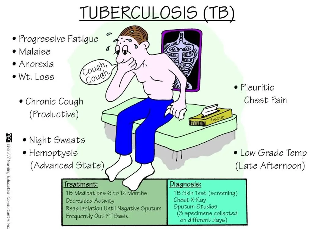
Causes:
1. Bacteria Mycobacterium tuberculosis
2. Contact with a contagious person
3. Spreads through air
4. Weak immune system
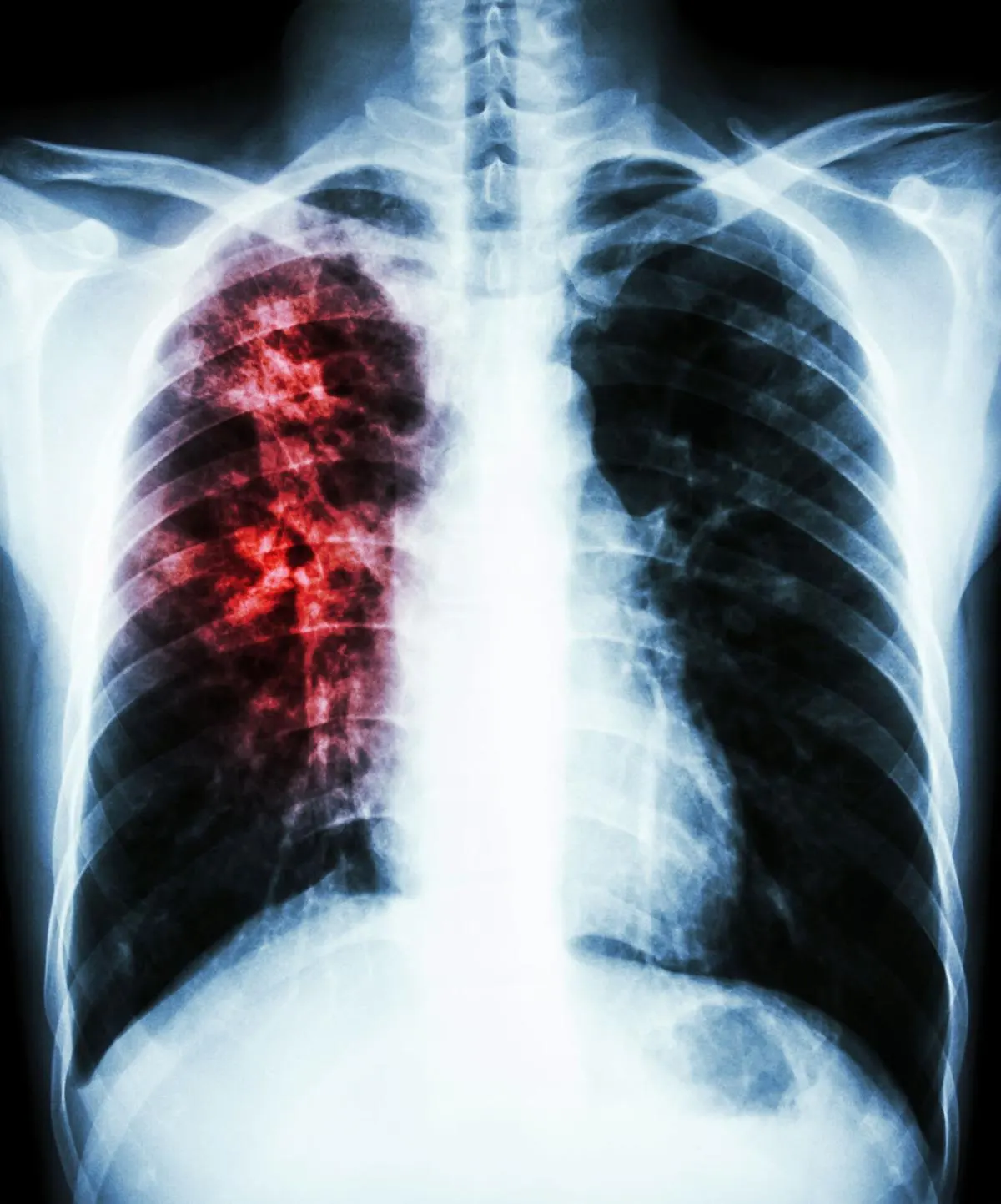
ABOVE fig shows xray of tuberculosis
Symptoms:
1. A persistent cough that can last for more than 3 weeks
2. Coughing blood
3. Chest pain
4. Weight loss
5. Fever
6. Chills
Prevention:
1. Wearing a surgical mask in public places.
2. Avoid going to crowded places.
3. Medications to infected people do not let them spread disease.
4. Vaccine
DIAGNOSIS:
- During the physical exam, your doctor will check your lymph nodes for swelling and use a stethoscope to listen to the sounds your lungs make when you breathe.
- The most commonly used diagnostic tool for tuberculosis is a skin test, though blood tests are becoming more commonplace. A small amount of a substance called tuberculin is injected just below the skin on the inside of your forearm. You should feel only a slight needle prick.
- Within 48 to 72 hours, a health care professional will check your arm for swelling at the injection site. A hard, raised red bump means you're likely to have TB infection. The size of the bump determines whether the test results are significant.
Results can be wrong
- The TB skin test isn't perfect. Sometimes, it suggests that people have TB when they don't. It can also indicate that people don't have TB when they do.
- You can have a false-positive result if you've been vaccinated recently with the bacille Calmette-Guerin (BCG) vaccine. This tuberculosis vaccine is seldom used in the United States but is widely used in countries with high TB infection rates.
- False-negative results also can occur.
Blood tests
- Blood tests can confirm or rule out latent or active tuberculosis. These tests measure your immune system's reaction to TB bacteria.
- These tests require only one office visit. A blood test might be useful if you're at high risk of TB infection but have a negative response to the skin test, or if you've recently received the BCG vaccine.
Imaging tests
- If you've had a positive skin test, your doctor is likely to order a chest X-ray or a CT scan. This might show white spots in your lungs where your immune system has walled off TB bacteria, or it might reveal changes in your lungs caused by active tuberculosis.
Sputum tests
- If your chest X-ray shows signs of tuberculosis, your doctor might take samples of your sputum — the mucus that comes up when you cough. The samples are tested for TB bacteria.
- Sputum samples can also be used to test for drug-resistant strains of TB. This helps your doctor choose the medications that are most likely to work. Getting results of these tests can take four to eight weeks.
Treatment:-
- If you have latent TB, your doctor might recommend treatment with medication if you're at high risk of developing active TB. For active tuberculosis, you must take antibiotics for at least six to nine months.
- The exact drugs and length of treatment depend on your age, overall health, possible drug resistance and where the infection is in your body.
Most common TB drugs
If you have latent tuberculosis, you might need to take only one or two types of TB drugs. Active tuberculosis, particularly if it's a drug-resistant strain, will require several drugs at once. The most common medications used to treat tuberculosis include:
- Isoniazid
- Rifampin (Rifadin, Rimactane)
- Ethambutol (Myambutol)
- Pyrazinamide
If you have drug-resistant TB, a combination of antibiotics called fluoroquinolones and injectable medications, such as amikacin or capreomycin (Capastat), are generally used for 20 to 30 months. Some types of TB are developing resistance to these medications as well.
Some drugs might be added to therapy to counter drug resistance, including:
- Bedaquiline (Sirturo)
- Linezolid (Zyvox)
Medication side effects
Serious side effects of TB drugs aren't common but can be dangerous when they do occur. All tuberculosis medications can be toxic to your liver. When taking these medications, call your doctor immediately if you have any of the following:
- Nausea or vomiting
- Loss of appetite
- A yellow color to your skin (jaundice)
- Dark urine
- Easy bruising or bleeding
- Blurred vision
Completing treatment is essential
- After a few weeks, you won't be contagious and you might start to feel better. Don't stop taking your TB drugs — you must finish the full course of therapy and take the medications exactly as prescribed by your doctor.
- Stopping treatment too soon or skipping doses can allow the bacteria that are still alive to become resistant to those drugs, leading to TB that is much more dangerous and difficult to treat.
- A program called directly observed therapy (DOT) can help people stick to their treatment regimen. A health care worker gives you your medication so that you don't have to remember to take it on your own.