Project 2
1.Stenosis in Gastrointestinal tract
a)Normal function of the organ
Each part of the digestive system helps to move food and liquid through our GI tract,breaks it into smaller parts and its beneficial parts are absorbed giving back energy and nutrients
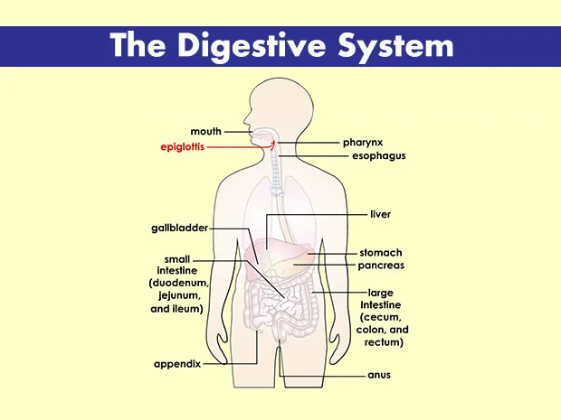
b)Current Pathological state in detail
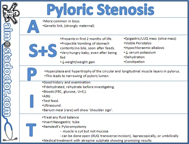
Partial obstruction that results in a narrowing of the opening(pylorus) of the intestine
c)Reason for the Pathological condition
- Family history
- Premature birth
- Smoking during Pregnancy
- Adhesions in abdomen that form after surgery
d)Effect of Homeostasis imbalance on human body
- Organ Homeostasis is essential for Organ Physiology and disease Prevention
- Decreased tolerance towards commensal bacteria as well as concomitant reduction in microbial diversity
2)Diagnostic method assisted by surgeon
a)Diagnostic method
- Abdominal X-ray
- CT scan
- Ultrasound in children
b)Data Acquisition method
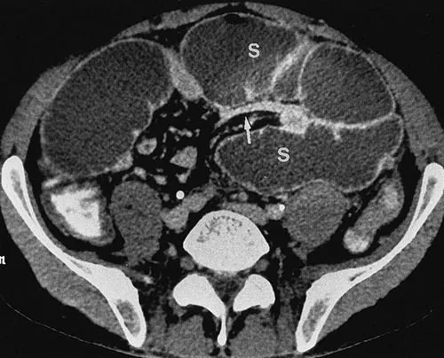
Data Obtained from the above CT scan image
c)How doctor identifies the problem from data ,Parameters he considered
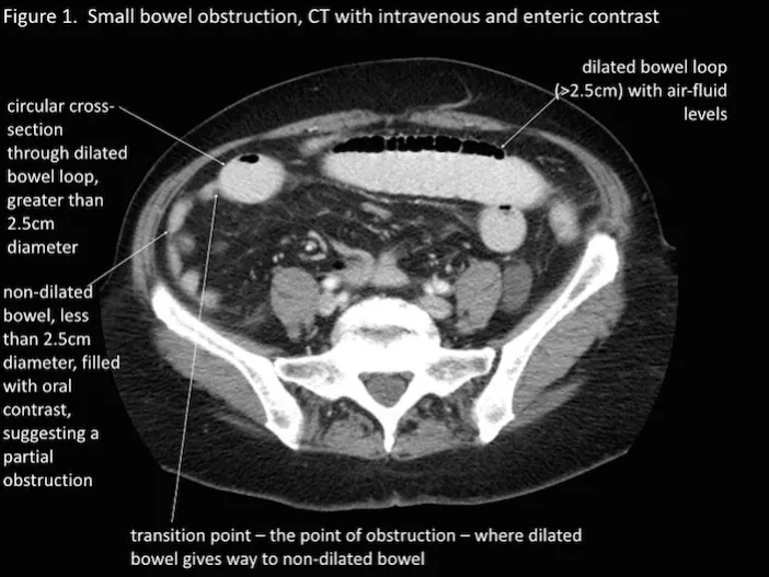
d)Result to the patient
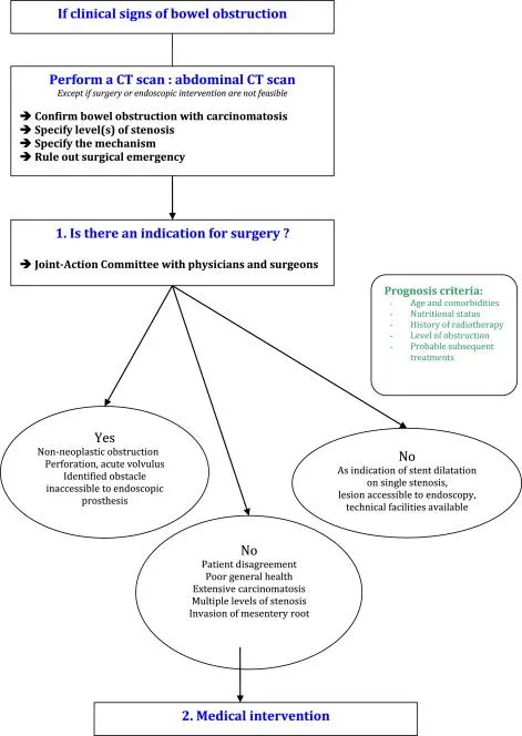
3.Medication
Pyloric stenosis is always treated with surgery, which almost always cures the condition permanently. The operation, called a pyloromyotomy, divides the thickened outer muscle, while leaving the internal layers of the pylorus intact. This opens a wider channel to allow the contents of the stomach to pass more easily into the intestines.
- A minimally invasive approach to abdominal surgery, called laparoscopy is generally the first choice of surgery for pyloric stenosis.
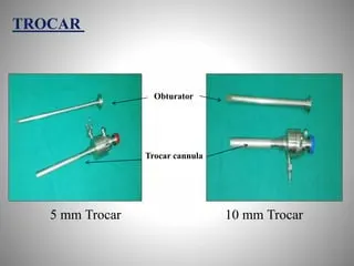
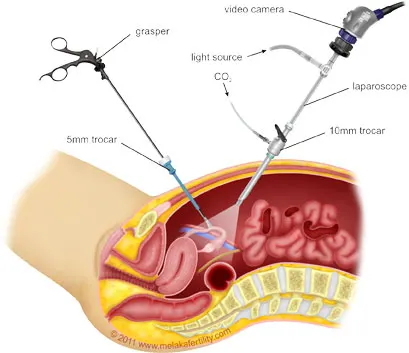
- To perform laparoscopic surgery, the surgeon inserts a rigid tube (called a trocar) into the abdominal cavity through a small incision (cut).
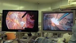
- The tube allows the surgeon to place a small camera into the abdomen and observe the structures within on an external monitor.
- The abdomen is inflated with carbon dioxide gas, which creates room to view the contents of the abdomen and to perform the operation.
- Additional rigid tubes are placed through small incisions and used to insert small surgical instruments into the abdomen.
- These instruments are used together with the camera to perform the operation. Tubes and instruments are removed when the operation is finished and the incisions are closed with sutures (stitches) that are absorbed by the body over time.
Laparoscopic pyloromyotomy generally involves the use of two or three trocars, and therefore usually requires two or three small incisions. If the surgeon decides that a laparoscopic operation is not the best way to treat the problems that are found in the operating room, then the operation will be changed (converted) to use an older surgical technique. Conversion to a nonlaparoscopic operation (called an “open procedure”) is rare and requires a larger incision, which may take longer to heal.
d)Instruments for Laparoscopy
- Gas delivery system
- light source
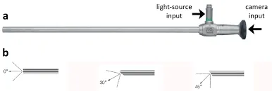
- Camera system
- monitor
- Trocars
e)Link of Youtube videos
https://www.youtube.com/watch?v=sPyZRkkxqNs
5)Post surgical care
- For some days food will be given through the intravenous technique and for internal feeding Frederick miller feeding tube is very effective.
- When the amount of fluid decreases and normalizes, mom can start feeding baby orally.
- Tylenol and ibuprofen may be required.
- Keep in touch with the doctor after the baby’s discharge.
6)Medical devices
a)Insufflators: . Endoscopic insufflators are used in laparoscopic surgeries to create space for the surgeon to work. A lapraroscopic insufflator inflates a patient's body using pressurized gas – commonly carbon dioxide (CO2) rather than ambient air or oxygen (O2) because CO2 has fewer negative effects on the body.
b)Trocars:Trocars are used during laparoscopic procedures and other minimally invasive surgery (MIS) to make small, puncturelike incisions in outer tissue layers. These incisions allow surgeons to insert cannulas through which surgical instruments can be introduced
c)light source :Conventional endoscopes utilize white light from a xenon arc lamp to illuminate tissue while performing endoscopic procedures.
d)Camera system : relays images of the inside of the abdomen or pelvis to a television monitor.
Project B
Chronic obstructive pulmonary disease (COPD) in Respiratory system
It is a chronic inflammatory lung disease that causes obstructed airflow from the lungs. Symptoms include breathing difficulty, cough, mucus (sputum) production and wheezing.
Emphysema and chronic bronchitis are the two most common conditions that contribute to COPD. These two conditions usually occur together and can vary in severity among individuals with COPD.
Chronic bronchitis is inflammation of the lining of the bronchial tubes, which carry air to and from the air sacs (alveoli) of the lungs. It's characterized by daily cough and mucus (sputum) production.
Emphysema is a condition in which the alveoli at the end of the smallest air passages (bronchioles) of the lungs are destroyed as a result of damaging exposure to cigarette smoke and other irritating gases and particulate matter.
1.
a).Normal function of the organ
The main function of the lungs is the process of gas exchange called respiration (or breathing). In respiration, oxygen from incoming air enters the blood, and carbon dioxide, a waste gas from the metabolism, leaves the blood. A reduced lung function means that the ability of lungs to exchange gases is reduced.
b)Current pathological state
c)Reason for COPD
Over time, exposure to irritants that damage your lungs and airways can cause chronic obstructive pulmonary disease (COPD), which includes chronic bronchitis and emphysema. The main cause of COPD is smoking, but nonsmokers can get COPD too.
d)Effect of Homeostasis imbalance on human body
In COPD patients, chronically elevated carbon dioxide shifts the normal acid-base balance toward acidic. There is the retention of carbon dioxide, which is hydrated to form carbonic acid.
In smoking COPD patients, long-term smoking can lead to exhaustion of the compensatory anti-inflammatory compacity, which leads to the imbalance between pro- and anti-inflammatory mechanisms, and eventually disrupts the immune homeostasis.
2)
a)Diagnostic methods
- Lung /pulmonary function tests
- Chest X-ray
- Ct scan
- Arterial blood gas analysis
b)Data aquisition method
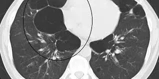
Dark region shown in Ct image shows Emphysema condition in COPD patient
c)Parameters to identify COPD
- In COPD, CT can identify key morphological features, including emphysema, bronchial wall thickening and gas trapping. These pathologies contribute directly to airflow obstruction and, therefore, CT has the potential to provide vital insights into the underlying pathophysiological changes of COPD.
- CT images can identify emphysema better and at an earlier stage than a chest x-ray. They can also identify other changes of COPD such as enlarged arteries in the lungs.
d)Result to the Patient
3)a)Minor condition
For people with COPD, short-acting bronchodilator inhalers are the first treatment used. Bronchodilators are medicines that make breathing easier by relaxing and widening the airways. There are 2 types of short-acting bronchodilator inhaler: beta-2 agonist inhalers – such as salbutamol and terbutaline.
b)Major condition
- Lung volume reduction surgery
- Lung transplant
- Bullectomy
4)surgical solution
a)Name of surgery :Lung Transplant
b)surgical position :For a single lung transplant, we will be positioned on the side opposite the side of the transplant. For a bilateral sequential lung transplant, we will be lying on the back with the arms above our head
c) surgical procedure
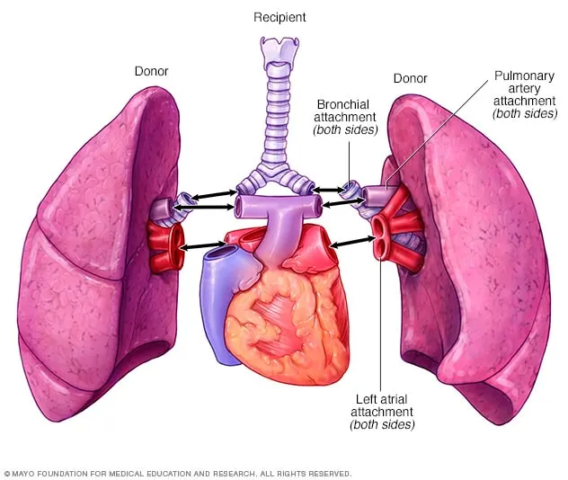
- During a lung transplant, surgeons remove our diseased lung and then attach a donor lung to our airway and to the blood vessels that lead to and from our heart. A lung transplant is a surgical procedure to replace a diseased or failing lung with a healthy lung, usually from a deceased donor.
- An intravenous (IV) line will be put in our arm or hand.
- Thin, flexible tubes (catheters) will be put in our neck, wrist, under the collarbone (subclavian), or the groin. These are used to monitor our heart and blood pressure, and to take blood samples.
- A breathing tube will be put into our throat and hooked up to a breathing machine (ventilator
- A catheter will be put into our bladder to drain urine during the surgery.
- Make an incision in our chest.
- Cut off the airways and blood vessels to our diseased lung.
- Remove the diseased lung and replace it with the healthy donor lung.
-
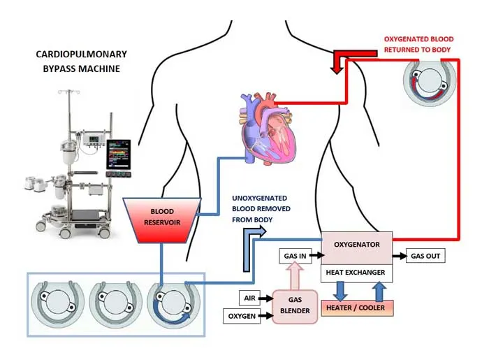
Depending on your lung condition and the type of transplant being done, you may be put on a cardiopulmonary bypass machine (heart-lung machine). This machine sends blood and oxygen to our body during the procedure.
- Reattach the airways and blood vessels.
- Close up the incision.
-
One or more tubes will be put in our chest. These are to remove air, fluid, and blood from the chest and to allow the new lung(s) to expand fully.
-
A thin tube (epidural catheter) may be put in. This is done to send pain medicine into your back. It may be done in the operating room or in the recovery room.
- During the procedure — most often with a double-lung transplant — we may connect it to a heart-lung machine.
- This machine supports our heart and lungs during surgery. It oxygenates our blood outside the body and pumps it back into our body.
d)Instruments during transplant
- Endotracheal tube
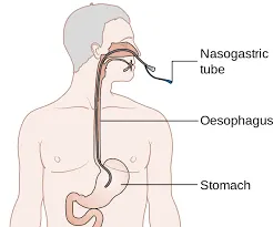
- nasogastric tube
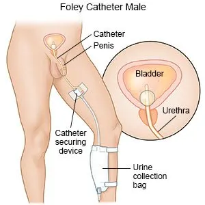
- Urinary catheter
- Heart lung machine
- Tracheostomy
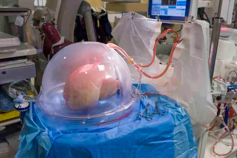
- Ex vivo lung perfusion
e)Lung Transplant
https://www.youtube.com/watch?v=0-X3J1biPZA
5)Post surgical care
- After Surgery, the patient will be taken to the transplant intensive care unit (ICU) where they will stay for several days. Here, they will remain on a ventilator until they can breathe on their own. This may take a few hours to a few days.
- pain medicine and anti-rejection drugs will be given
- When the body detects new lung or lungs, the immune system begins working to fight what it views as a threat. Anti-rejection drugs suppress the immune system's natural response to allow the body to accept the new lungs.
- When they become stronger, will move to a patient room within the transplant unit, can expect to stay in the hospital for 3 to 4 weeks.
- The lung transplant team will monitor progress to make sure the body isn't rejecting the lungs and that aren't developing an infection or other problems.
- They will begin rehabilitation, including physical therapy and breathing and speech exercises.
- we may be kept at NPO status (nothing by mouth) for days to months post transplant to ensure that our swallowing function is working correctly and also to prevent aspiration of food or drink into our new lungs
6)Medical devices
a)Ex vivo lung perfusion
Lungs can be damaged when the brain dies, before they're removed for donation.
Ex vivo lung perfusion is a technique designed to overcome this problem.
It involves removing the lungs from the body and placing them in a piece of equipment called a perfusion rig.
Blood, protein and nutrients are then pumped into the lungs, which repairs the damage
b)Endotracheal tube
Breathind tube that s inserted through mouth into lungs
c)Nasogastric tube
It goes into stomach through nose
d)Urinary catheter:Drains urine freely into a bag
e)Tracheostomy: helps to breath without ventilator and take out excess mucus