Project 1
PART-A
.Clinical conditions associated with the urinary system
- Neurogenic bladder
1.Gross anatomy of the organ involved-Urinary system
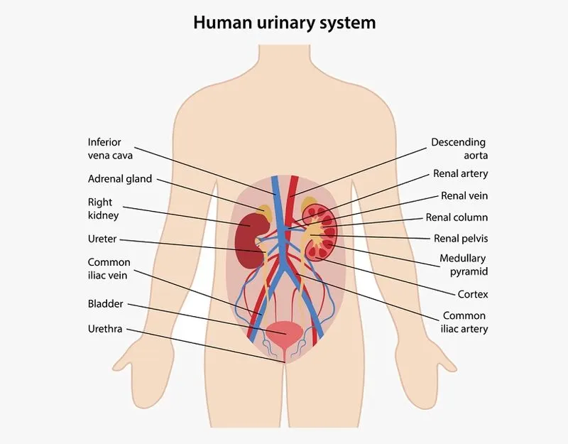
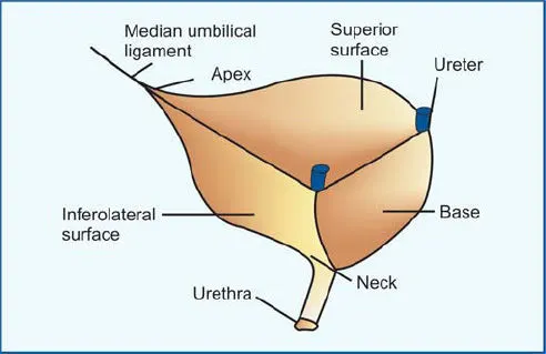
Gross structure of urinary bladder
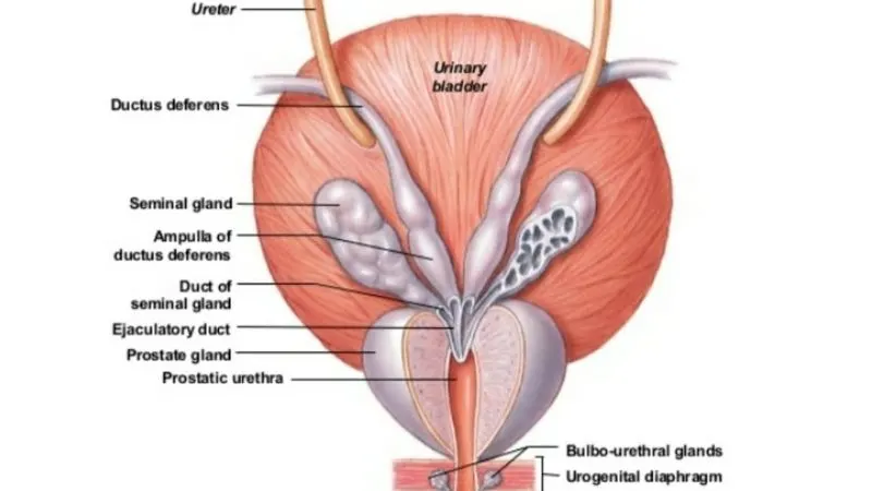
Macroscopic structure of bladder in males
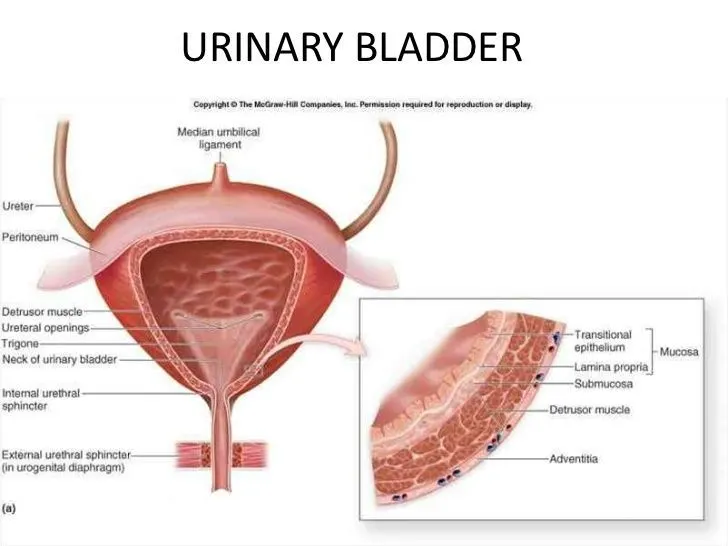
microscopic structure of urinary bladder
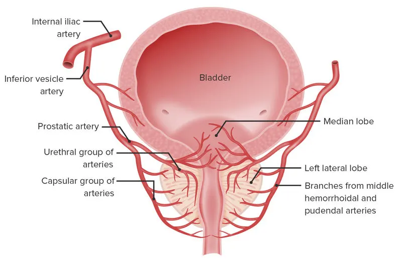
Blood supply of bladder
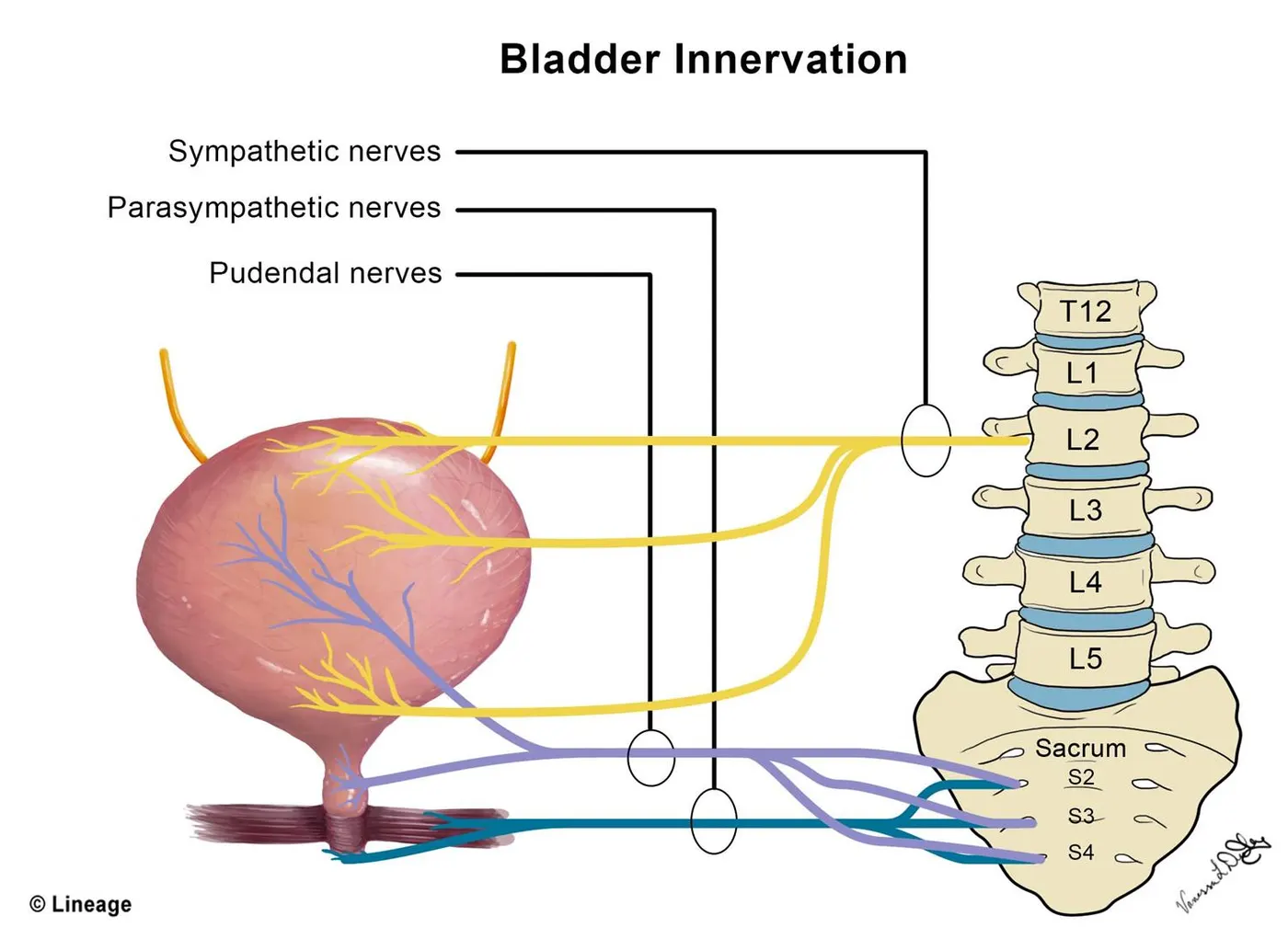
Nerve supply of uriary bladder
2.Common disorders
Clinical presentation
-Characteristic symptoms of neurogenic bladder include recurrent urinary tract infections, urine leakage, overactive bladder, frequent urination, urinary retention, and underactive bladder.
Medical conditions include Stroke,Parkinsons disease,Spinal cord injuries which also leads to this condition
Diagnostic techniques:
-X-rays of the skull and spine:This imaging test uses invisible energy beams to make images of tissues,bones and organs.
-Imaging tests(CT and MRI) of bladder and ureters.
-Ultrasound(sonography):It is an imaging test that uses high frequency sound waves to create images of the organs on a copy screen .
Cystoscopy(detailed )
- It is the examination of the inside of the bladder.A cystoscope is a long thin instrument with an eyepiece on the external end and tiny lens and a light on the end that is inserted into the bladder.
- The cystoscope is inserted into the patients urethra and the small lens magnifies the inner lining of urethra and bladder,allowing to see inside the hollow bladder.Two types of cystoscopy:Rigid and Flexible
It can diagnose
- Bladder cancer/Urethral cancer
- Bladder stones
- Bladder control problems
- Enlarged prostate
- Urethral strictures
Treatments using cystoscopy
- Injects dye for an X-ray Procedure that tracks urine flow.Retrogrde pyelogram is an imaging test that uses X-rays to look at your bladder,Ureters and Kidneys. This test is usually done during a test called Cystoscopy.
- Remove bladder stones,abnormal tissue polyps or tumours
- Take small pieces of bladder or urethral tissue to biopsy.
3.Management and Treatment plan
Medical management
-Lifestyle changes
-Drugs(oxybutynin, tolterodine,etc.)
-Continuous catheterization
- Artificial sphincter.
- Electrical device implanted near the bladder nerves to stimulate the bladder muscles.
- Sling surgery.(sling surgery involves making a cut in the lower tummy (abdomen) and vagina so a sling can be placed around the neck of the bladder to support it and prevent urine leaking.
- Creation of an opening (stoma) in which urine flows into a special pouch (this is called urinary diversion)
4.Surgical management
Eg:SLING SURGERY
Anatomical basis of surgery
Vaginal sling procedures are types of surgeries that help control stress urinary incontinence. There is urine leakage that happens when we laugh, cough, sneeze, lift things, or exercise. The procedure helps close the urethra and bladder neck. The urethra is the tube that carries urine from the bladder to the outside
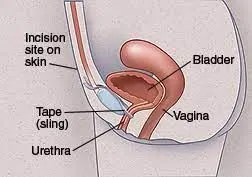
Placement for sling supporting the bladder neck,secured behind the pubic bone
Organ structure and anatomical relation of the bladder during the surgery
Vaginal sling procedures use different materials:
- Tissue from our body
- Man-made (synthetic) material known as mesh
We have either general anesthesia or spinal anesthesia before the surgery starts.
- With general anesthesia, we are asleep and feel no pain.
- With spinal anesthesia, we are awake, but from the waist down we are numb and feel no pain.
A catheter (tube) is placed in the bladder to drain urine from our bladder.
The doctor makes one small surgical cut (incision) inside the vagina. Another small cut is made just above the pubic hair line or in the groin. Most of the procedure is done through the cut inside the vagina.
The doctor creates a sling from the tissue or synthetic material. The sling is passed under the urethra and bladder neck and is attached to the strong tissues in the lower belly, or left in place to let the body heal around and incorporate it into the tissue.
Instruments and Equipments used for Surgery
- Cutting and dissecting instruments(scalpels,scissors...)
- Grasping or holding instruments(forceps,clamps,.....)
- Hemostatic instruments(to stop bleeding)
- Retractors(to expose body cavities)
- tissue unifying instruments and material
Post operative care after sling surgery
Light meal with clear liquid
Do lighter activity for 4 weeks after surgery
Notice a slower stream or mild difficulty voiding.
Incision is within the vagina and thus will not be seen. The surgeon may leave “vaginal
packing,” which is a long strip of gauze saturated with estrogen cream, which is packed into the
vagina to aid in healing.
Post operative monitoring system
- Cardiac Monitor.
- ICU Monitor.
- Multipara Monitor.
- Bedside Monitor & Portable Patient Monitor.
- Vital Signs Monitor.
- Capnography Etco2 Monitor.
- Fetal Doppler Monitor.
- Central Patient Monitoring System.
5.Recent advances in the field
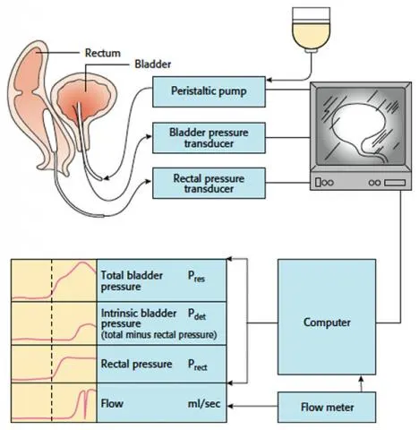
Videourodynamics
when cystometry is carried out using a contrast filling medium and the procedure is visualised radiographically
Useful to see reflux into the ureters
thickening of the bladder wall and bladder diverticula
The figure shows videourodynamics
B.KIDNEY STONE
1.Gross anatomy of kidney
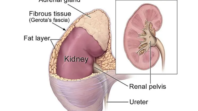
Macroscopic structure of kidney
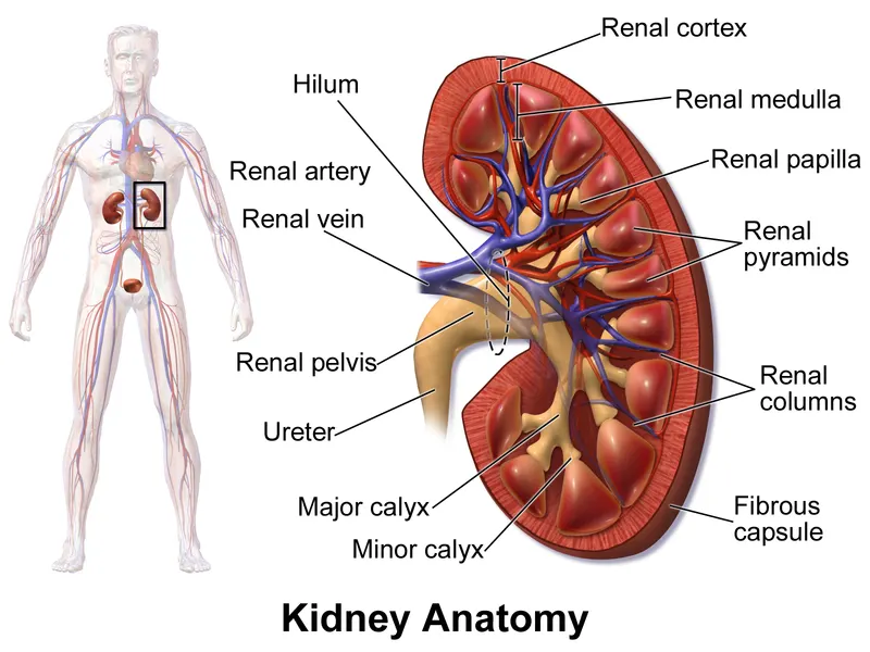
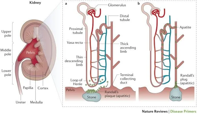
Microscopic structure of kidney
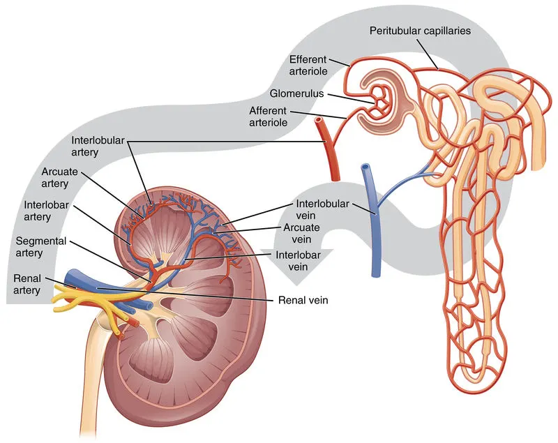
Blood supply of kidney
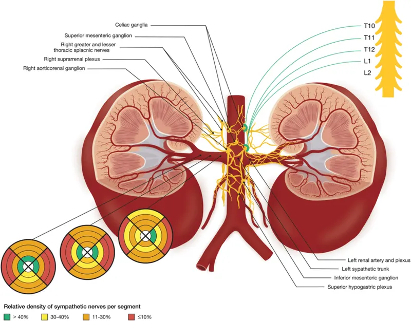
Nerve supply of kidney
2.Common Disorders
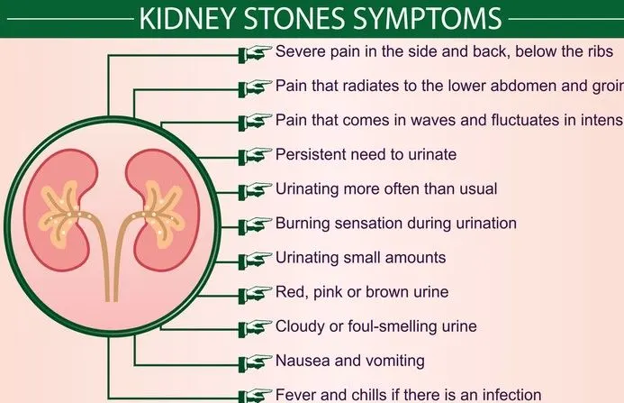
-Clinical presentation of kidney stone
Diagnostic Techniques of Kidney stones:
- Blood testing.Blood tests may reveal too much calcium or uric acid in the blood.
- Urine testing. The 24-hour urine collection test may show that we are excreting too many stone-forming minerals or too few stone-preventing substances.
- Imaging tests may show kidney stones in the urinary tract.
- High-speed or dual energy computerized tomography (CT) may reveal even tiny stones. Simple abdominal X-rays are used less frequently because this kind of imaging test can miss small kidney stones.
- Ultrasound, a noninvasive test that is quick and easy to perform, is another imaging option to diagnose kidney stones.
Computerised Tomography -A Diagnostic Technique
- (CT) scans are the most commonly used imaging test for detecting kidney stones. CT scans of your abdomen and pelvis combine multiple x-ray images to provide a 3D picture of your urinary tract. This is generally considered to be the most accurate and useful imaging test for visualizing kidney stones.
- Computed tomography is a noninvasive diagnostic imaging procedure that uses a combination of X-rays and computer technology to produce horizontal, or axial images of the body. A CT scan shows detailed images of any part of the body, including the bones, muscles, fat, and organs. CT scans are more detailed than standard X-rays.
- In standard X-ray, a lot of detail about internal organs and other structures is not available.CT scans show the exact size and location of the stone.
- CT scans may be done with or without "contrast." Contrast refers to a substance taken by mouth or injected into an intravenous (IV) line that causes the particular organ or tissue under study to be seen more clearly.
- CT scans of the kidneys can provide more detailed information about the kidneys than standard Kidney,Ureter and bladder X-rays, thus providing more information related to injuries and/or diseases of the kidneys
- . CT scans of the kidneys are useful in the examination of one or both of the kidneys to detect conditions such as tumors or other lesions, obstructive conditions, such as kidney stones, congenital anomalies, polycystic kidney disease, accumulation of fluid around the kidneys
- While this is considered safe for most people, CT scans may not be recommended for some patients such as pregnant people and children and it has low risk of radiation.
3.Medical Management and Treatment Plan
Medical management
The type of medication, known as an alpha blocker, relaxes the muscles in the ureter, helping to pass the kidney stone more quickly and with less pain. Examples of alpha blockers include tamsulosin (Flomax) and the drug combination dutasteride and tamsulosin .Drink plenty of water.Small kidney stones can be managed by these methods
Surgical Procedures
4.Surgical Management for Kidney stones
Eg:Percutaneous Lithotripsy
Anatomical basis for surgery
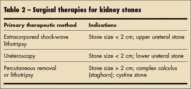
Percutaneous nephrolithotomy is typically recommended when:
- Large kidney stones block more than one branch of the collecting system of the kidney. These are known as staghorn kidney stones.
- Kidney stones are larger than 0.8 inch (2 centimeters) in diameter.
- Large stones are in the tube connecting a kidney and the bladder (ureter).
- Other therapies have failed.
Organ Structure and Anatomical relation of the organ during the Procedure
A specialized needle is inserted into a urine-collecting chamber of the kidney (calyx). The path of this needle becomes the passage for performing the rest of the procedure.
A surgeon or a radiologist with specialized training in this procedure uses X-ray, CT or ultrasound images to guide the placement of the needle.
There may have a flexible tube (catheter) passed through the urethra, bladder and ureter into the kidneys. . Through this catheter, the doctor can put a specialized tracer substance into the kidney that outlines structures inside the kidney so that they're more visible during imaging. Or, a tiny camera may be threaded through the catheter, which allows the doctor to see the needle as it's placed in the kidney and other work during surgery.
Next, the surgeon place a tube (sheath) along the path of the needle. Using specialized instruments that pass through the sheath, the surgeon breaks up the stones and removes them.
The surgeon may then place a different tube, called a nephrostomy tube, in this same passageway. The nephrostomy tube allows urine to drain directly from the kidney into a bag worn outside the body during recovery.
Instruments needed during surgery
- Intra Corporeal Pneumatic Lithotripter ( To Break the Stone).
- I.P. Needle.
- PTFE Guide Wire.
- Alkine Canulla.
- Facial Dilator SET.
- Alkine Telescopic Dilator SET.
- PCNL Forceps – Alligator / Tripronge / Peanut Jaw / U Handle.
- Ureteric Catheter.
- Double J Stent.
- Flow Pump for continuous irrigation.
- LED Light Source.
- Fiber Optic Cable.
- Postoperative Care
- Dressing
It is common to leak fluid from the incision site for a day or two after removal of the
nephrostomy tube. If this occurs it is best to keep the site covered with clean gauze. The gauze
should be changed on a daily basis and more frequently if needed. Once the incision has
stopped draining it may be left uncovered. - Ureteral Stent: A ureteral stent is a small flexible plastic internal tube that is placed to promote drainage of your kidney down to the bladder. This will be removed in your surgeon’s office in typically 1-2 weeks following surgery.
Post operative monitoring system
- Cardiac Monitor.
- ICU Monitor.
- Multipara Monitor.
- Bedside Monitor & Portable Patient Monitor.
- Vital Signs Monitor.
- Capnography Etco2 Monitor.
- Fetal Doppler Monitor.
- Central Patient Monitoring System.
5.Recent advances in the field
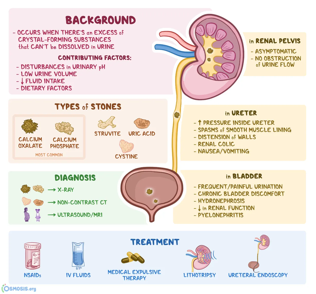
Researchers have applied Nobel prize-winning microscope technology to uncover an ion channel structure that could lead to new treatments for kidney stones. The study published in Nature Structural and Molecular Biology, revealed atomic-level details of the protein that serves as a passageway for calcium across kidney cell membranes.
Approximately 80 percent of kidney stones are comprised of calcium salts. They are extremely painful to pass, and depending on size and location can require surgery to remove. Ion channels that span kidney cell membranes help reabsorb calcium from the urine before it can form kidney stones. The new study is the first to show molecular details of the essential kidney calcium channel, called TRPV5, in its closed form. The study also reveals how inhibitor molecules attach to and close the channel, leaving calcium stranded in the urine where it can form kidney stones.
Now that we know what the protein looks like in its inhibited state, drugs can be made with the intention of modulating TRPV5 activity and potentially treating kidney stones directly
Laser removal
surgery is minimally invasive
requires no incisions
Through ureteroscope,access ureter and kidney.
Uses holmium energy to break up kidney stones
removes some pieces through urethra and later passes the remaining though urine
C.Chronic kidney disease
Chronic kidney disease, also called chronic kidney failure, involves a gradual loss of kidney function. our kidneys filter wastes and excess fluids from the blood, which are then removed through the urine. Advanced chronic kidney disease can cause dangerous levels of fluid, electrolytes and wastes to build up in the body.
1.Gross anatomy,Micro and macro structure,blood and nerve supply remains same as in Kidney stone
2.Clinical conditions
Diseases and conditions that cause chronic kidney disease include:
- Type 1 or type 2 diabetes
- High blood pressure
- Glomerulonephritis
- Interstitial nephritis , an inflammation of the kidney's tubules and surrounding structures.
- Polycystic kidney disease or other inherited kidney diseases
- Prolonged obstruction of the urinary tract, from conditions such as enlarged prostate, kidney stones and some cancers
- Vesicoureteral reflux, a condition that causes urine to back up into your kidneys
- Recurrent kidney infection, also called pyelonephritis
Various Diagnostic Techniques
- Blood tests. Kidney function tests look for the level of waste products, such as creatinine and urea, in our blood.A creatinine level of greater than 1.2 for women and greater than 1.4 for men may be an early sign that the kidneys are not working properly. As kidney disease progresses, the level of creatinine in the blood rises.
- Urine Tests:check the urine for albumin. Albumin is a protein found in our blood. A healthy kidney doesn't let albumin pass into the urine. A damaged kidney lets some albumin pass into the urine.
A diagnostic Technique
- GFR - A blood test measures how much blood our kidneys filter each minute, which is known as the glomerular filtration rate (GFR).
- GFR=125 ML/Min or 180 L/day.
- Plasma Volume(70 kg young adult man)=about 3L,the kidneys filter the plasma some 60 times in a day.
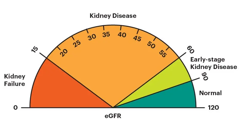
What is a normal eGFR number?
In adults, the normal eGFR number is more than 90. eGFR declines with age, even in people without kidney disease.
Factors affecting GFR:
- -change in renal blood flow
- Glomerular capillary hydrostatic pressure
- Glomerular capillary permeability
- Effective filtration surface area
- Size ,shape and electrical charge of macromolecules
3.Medical management
The main treatments are: lifestyle changes – to help you stay as healthy as possible and medicine – to control associated problems, such as high blood pressure and high cholesterol.
People with chronic kidney failure have 3 treatment choices
- Dialysis
- Renal transplant
- conservative treatment
Dialysis
For a small proportion of people with CKD, the kidneys will eventually stop working.
This usually hapens gradually, so there should be time to plan the next stage of our treatment.
One of the options when CKD reaches this stage is dialysis. This is a method of removing waste products and excess fluid from the blood.
There are 2 main types of dialysis:
- haemodialysis – this involves diverting blood into an external machine, where it's filtered before being returned to the body
- peritoneal dialysis – this involves pumping dialysis fluid into a space inside our tummy to draw out waste products from the blood as they pass through vessels lining the inside of our tummy
Haemodialysis is usually done about 3 times a week, either at hospital or at home. Peritoneal dialysis is normally done at home several times a day, or overnight.
If we don't have a kidney transplant, treatment with dialysis will usually need to be lifelong.
4.Surgical procedures
Anatomical basis of surgery
Wet ischemic time(time from cessation of circulation to removal of organ and its placement in cold storage)should not exceed 30 min.Living donor transplants function immediately after transplant.
Anatomical relation of organ to be noted
Blood relative
HLA identical or haploidentical
with negative cross match
Excellent medical condition with normal renal function
Instruments used for surgery
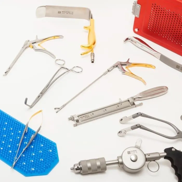
- Clamp Tube, Clamp Dissection, Clamp Artery, Forceps Tissue, Clamp Gall-Duct, Jaw inserts, Clip applier, Clamp Ring.
- Dilators, Punch Tissue, Punch Vascular, Stilet, Probes.
- Elevators, Hook Phlebectomy.
- Forceps and Forceps HF.
- Hook Vessel, Hook Nerve, Hook Skin, Hook Dura, etc.,Spatula, Retractor Wound, Retractor Epicardial.
Post operative care
- Cardiac Monitor.
- ICU Monitor.
- Multipara Monitor.
- Bedside Monitor & Portable Patient Monitor.
- Vital Signs Monitor.
- Capnography Etco2 Monitor.
- Fetal Doppler Monitor.
- Central Patient Monitoring System.
Post operative monitoring system
- Cardiac Monitor.
- ICU Monitor.
- Multipara Monitor.
- Bedside Monitor & Portable Patient Monitor.
- Vital Signs Monitor.
- Capnography Etco2 Monitor.
- Fetal Doppler Monitor.
- Central Patient Monitoring System.
5.Recent advances
Researchers and firms hoping to transform medicine with 3D-printed organs have made strides in recent years, bolstered by increased funding and interest.
They’re working to develop customized 3D-printed organs, including hearts and kidneys, from patients’ own cells — a process they hope will reduce the likelihood of patients’ bodies rejecting them and speed up delivery of the organs to those who need them. We're talking about a technology that can rebuild different parts of the body as needed.”
The technology often works like traditional 3D printing, which leans on a digital blueprint to form a 3D object by dispensing material through a nozzle. In this case, however, the material is created using “bioink” made of cells instead of materials like plastic or metal, along with employing other techniques. Using patients’ cells could eliminate the need for harmful immunosuppressive medication.
Cell-integrated and wearable artificial kidneys aim to create convenient and efficient systems of filtration and restore elements of immunoregulatory function.
possible newer techniques involved in managing disorder
suggested initial evaluation
- complete urinalysis (U/A)
- urine albumin-to-creatinine ratio (UACR)
- creatinine with estimated GFR, blood urea nitrogen (BUN), electrolytes, glucose, calcium, phosphorus, albumin
- complete blood count (CBC)
For further evaluation ,the following tests are often ordered
- hepatitis B serology
- hepatitis C serology
- antinuclear antibody test (ANA)
- rheumatoid factor (RF)
- complement 3 (C3)
- complement 4 (C4)
- serum protein electrophoresis (SPEP) and urine protein electrophoresis (UPEP) (in patients over the age of 40)
- renal ultrasound to measure kidney size and to check for echogenicity and hydronephrosis
- dilated retinal exam
If a patient with diabetes has retinopathy, albuminuria, and negative screening tests listed above, it is reasonable to assume the diagnosis is diabetic kidney disease. Patients who do not conform to these criteria should be discussed with a nephrologist
Part-B
1.Cranial bone
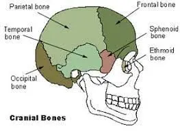
There are eight cranial bones, each with a unique shape:
- Frontal bone. This is the flat bone that makes up your forehead. It also forms the upper portion of your eye sockets.
- Parietal bones. This a pair of flat bones located on either side of your head, behind the frontal bone.
- Temporal bones. This is a pair of irregular bones located under each of the parietal bones.
- Occipital bone. This is a flat bone located in the very back of your skull. It has an opening that allows your spinal cord to connect to your brain.
- Sphenoid bone. This is an irregular bone that sits below the frontal bone. It spans the width of your skull and forms a large part of the base of your skull.
- Ethmoid bone. This is an irregular bone located in front of the sphenoid bone. It makes up part of your nasal cavity.
The cranial bones are held together by unique joints called sutures, which are made of thick connective tissue. They’re irregularly shaped, allowing them to tightly join all the uniquely shaped cranial bones.
2.Microcopic anatomy of bone
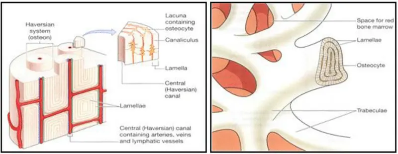
- The bone consists of several cylindrical microscopic units called Haversian systems(osteon)
- It contains a central canal known as Haversian canal.
- It carries blood vessels and nerves.The Haversian canals of two adjacent haversian systems are interconnected by transverse canal.known as volkmans canal.
- The canal surrounde by concentric layers of bone matrix called lamellae
- The bony lamellae contain fluid filled spaces called lacunae.
- Each lacunae enclose bone cell called osteocyte
- The cytoplasmic processes of a bone cell are attached to the membranes of adjacent cells.
- Materials can move rapidly between blood vessels and bone cells
3. Blood supply of bone
The external carotid is the main blood supply to the skull bones and meninges. It travels up the side of the neck with eight main branches feeding the superficial structures of the skull and face. Of these branches, the maxillary artery is the most prominent and clinically relevant.
.
4.Bone remodelling
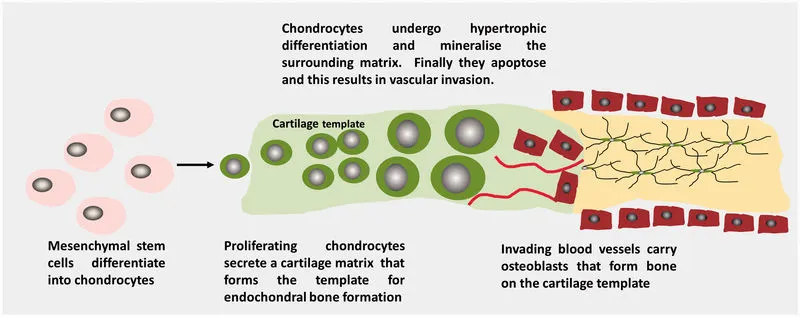
Bone remodeling is a lifelong process where mature bone tissue is removed from the skeleton and new bone tissue is formed. These processes also control the reshaping or replacement of bone following injuries like fractures but also micro-damage, which occurs during normal activity.
5.Anatomical basis of bone implant surgery
- Diseases of the bone, like osteonecrosis or cancer
- Spinal fusion surgery( if we have an unstable spine)
- Dental implant surgery( if we want to replace missing teeth)
- Surgically implanted devices, like in total knee replacement, to help promote bone growth around the structure
These bone grafts can provide a framework for the growth of new, living bone. Hips, knees, and spine are common locations for bone grafting, but we might need bone grafting for a different bone in our body.
6.Indications of bone implant
Bone grafting is done for numerous reasons, including injury and disease. There are four main reasons bone grafts are used:
- A bone graft may be used in the case of multiple or complex fractures or those that don’t heal well after initial treatment.
- Fusion helps two bones heal together across a diseased joint. Fusion is most often done on the spine.
- Regeneration is used for bone lost to disease, infection, or injury. This can involve using small amounts of bone in bone cavities or large sections of bones.
- A graft can be used to help bone heal around surgically implanted devices, like joint replacements, plates, or screws.
Contraindications of bone implant
All surgical procedures involve risks of bleeding, infection, and reactions to anesthesia. Bone grafts carry these risks and others, including:
- pain
- swelling
- nerve injury
- rejection of the bone graft
- inflammation
- reabsorption of the graft