Project 2
Respiratory system:
- Pathological condition:
Lungs: Pleural effusion
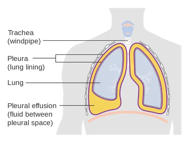
- It is the buildup of excess fluid between the tissues that line the lungs and the chest.
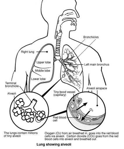
- The normal function of the lungs is the process of gas exchange called respiration.
- Pathology state: Because of the fluid accumulation difficulty in breathing (fast breath), including cough, chest pain and shortness of breath.
- Reasons: It can occur due to poor pumping of the heart, inflammation, infection, lung injury or tumors
- Homeostasis change: Pleural effusion can result in chronic heart failure because the heart cannot pump fluid away from the lungs, fluid that seep from the lungs places additional stress on the dysfunctioning heart. Large pleural effusions can cause disabling shortness of breath
- Diagnostic method:
- X- ray, CT or Ultrasound
- Data: The data acquisition is done either by X-ray, CT or ultrasound
And in the above image the data acquisition is done based on the waves off of the body structures. The strength of the sound signal and the time it takes for the wave to travel through the body provide the information necessary to produce an image.
- The clinicians assess the data by observing the anechoic area that denotes the presence of fluid accumulation
- Result: the treatments include the antibiotics, water pills, and removal of the fluid by draining
- Clinical solutions:
- Minor severity:
- If the pathological condition is less severe, the clinicians suggest for antibiotics or diuretic.
- Antibiotics stops the growth of bacteria. Diuretic helps in eliminating the extra water from the body. It’s also prescribed to treat high blood pressure.
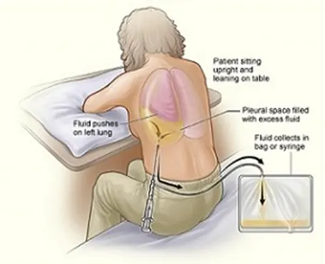
- Draining of fluid from the chest cavity either with a needle or by inserting a small tube into the chest
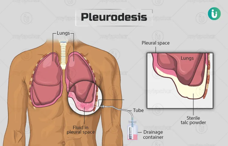
- Pleurodesis is a treatment that creates mild inflammation between the lung and chest cavity pleura. After drawing the fluid, they inject a drug into the area, which causes the two layers of the pleura to stick together, which prevents the future buildup of fluid between them.
- Major severity:
- If the condition persists even after draining the fluid, the doctor may recommend for thoracoscopic decortication or thoracoscopic debridement or pleurectomy
- Surgical solution:
- Thoracoscopic decortication:
- The patient is positioned as for a posterolateral thoracotomy
- If the symptoms don’t improve with drainage and antibiotics, the doctor may recommend thoracoscopic decortication or thoracoscopic debridement.
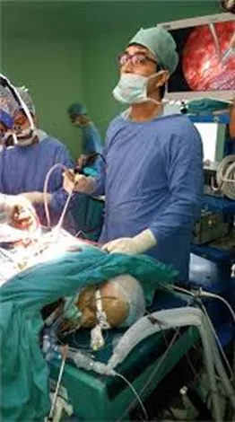
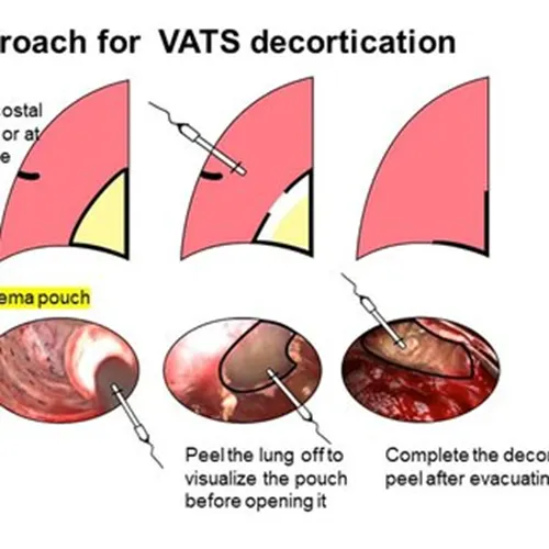
- They will insert a thoracoscope into the pleural space then either remove any tissue that is causing a problem or surgically clean a wound to enable it heal.
- The video assisted thoracoscopic surgery (VATS) is usually performed via an anterior approach. Three ports can be inserted as per the surgeon’s preference.
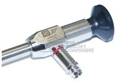
- A 30-degree camera is used for visualization during the procedure
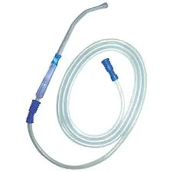
- The cautery hook and suction cannula are effective instruments for dissection
- Limits of the dissection are the same as in one open surgery
- The camera port can be switched to perform adhesiolysis at different portions of the pleural cavity
- The chest tube can be inserted in the port sites
- Instruments required for the surgery:
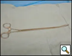
- A curved ring forceps
- 30-degree thoracoscope
- Yankauer sucker
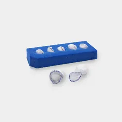
- Peanut dissecton
- Long curettes
- post-surgical care:
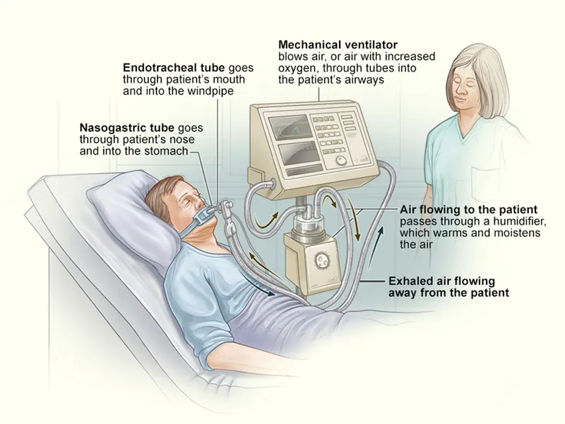
- The patient often requires mechanical ventilation
- Intensive monitoring must be ensured during the initial postoperative period in these patients
- Apart from serial chest radiographs, periodic arterial blood gas analysis might be required in these patients
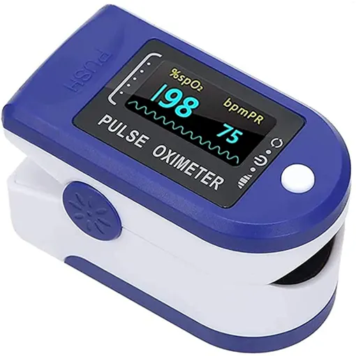
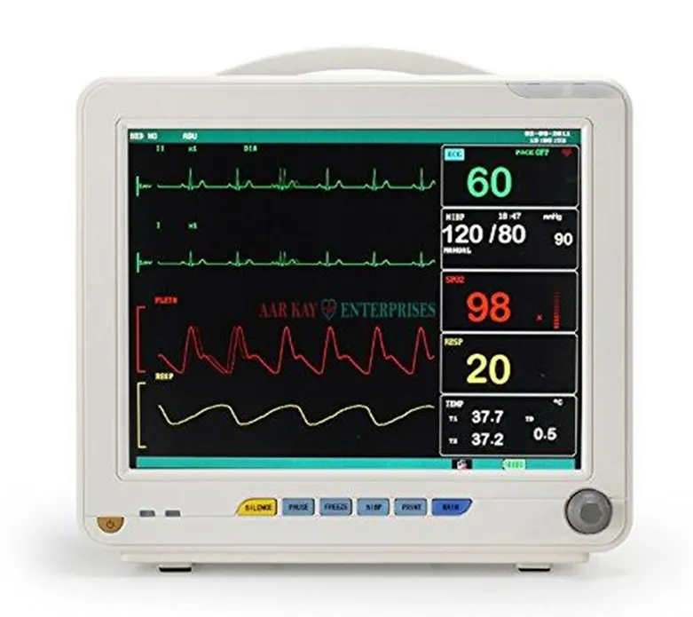
- The oxygen saturation (spO2), blood pressure, heart rate, is monitored using multipara monitor
- Devices used:
- Multipara monitor: used to monitor the oxygen saturation (spO2), blood pressure (BP), and heart rate
- Ventilator: that provides mechanical ventilation by moving breathable air and out of the lungs
- Pulse oximeter: the saturation of oxygen in the blood is monitored