Project 1
Part A
- Clinical disorders of digestive system:
- The pathological disorders related to digestive system are
- GERD (Gastrointestinal reflux disease)
- Hemorrhoids
- Tumor or cancer
- Esophageal varices
- Cholelithiasis
GERD (Gastro esophageal reflux disease):
- It is the disease associated with the digestive system caused by the problem in esophageal sphincter muscle associated with the symptoms such as heart burn, discomfort in the chest, and difficulty in swallowing.
- Usually diagnosed with the upper GI Scopy or Endoscopy.
- The organ involved in this pathological condition is the stomach.
Anatomy of Stomach:
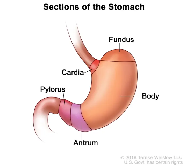
- The digestive system starts with the oral cavity and ends at the anal canal
- The stomach is the organ where the food is digested and it has three parts:
- The cardia
- The fundus
- The body
- The antrum
- The pylorus
- The food ingested is digested with the help of the enzyme gastric and pepsin and thus the absorption of nutrients takes place.
- The GERD occurs when the acid in the stomach backwash to the esophagus and cause irritation in the linings of esophagus.
Macroscopic and microscopic structure of stomach:
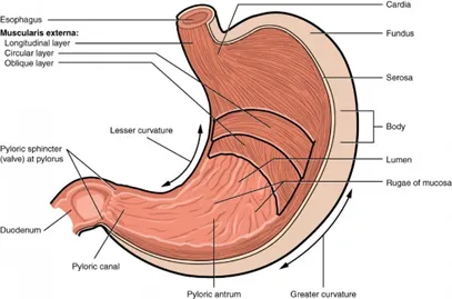
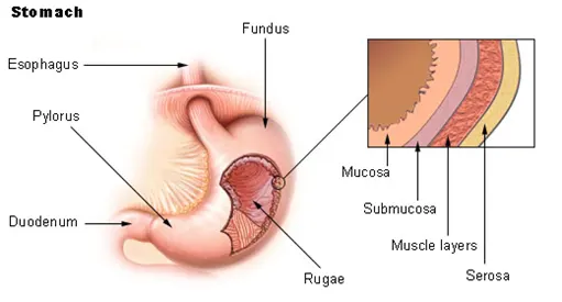
- The stomach consists of a mucosa, submucosa, muscularis externa, subserosa and serosa
- The muscular layer of the layers is the inner oblique layer, the middle circular layer and myenteric plexus (Auerbach’s plexus)
- Mucosa lining of the stomach has several pits wherein the gastric juices are secreted from the gastric glands
- The sphincter muscles help in the movement of food from stomach to the intestines.
Blood supply and nerve supply of the stomach:
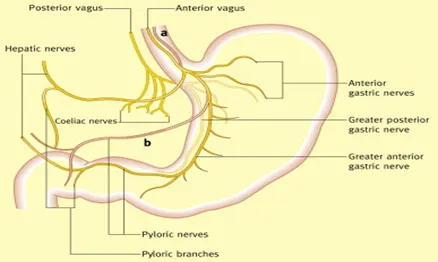
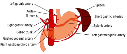
- The main arteries of the stomach are the common hepatic artery, splenic artery, gastroduodenal artery, supra duodenal artery,
- The major nerve supplies are parasympathetic and sympathetic nerves.
- The vagus nerve supplies parasympathetic via the right posterior and left vagal trunks
- Atherosclerosis:
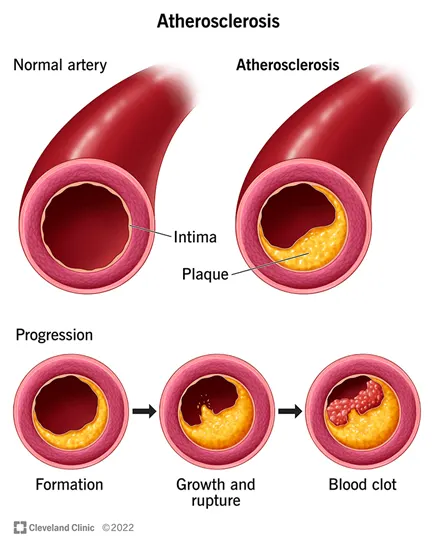
- It is the pathological disorder associated with the cardiovascular system.
- Caused due to the deposition of fats or blood clot which further results in the narrowing of blood vessels.
- Thus, the blood flow to the heart decreases and may lead to heart attack, mini strokes and poor wound healing
- And usually have no symptoms until the plaque ruptures
Diagnostic technique:
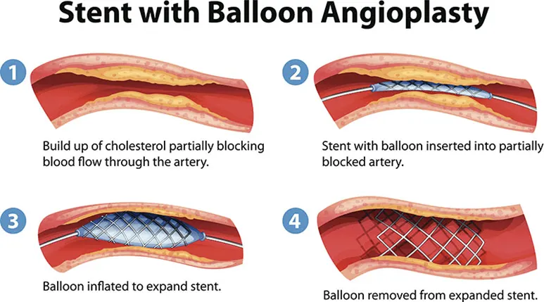
- The diagnostic technique used to treat atherosclerosis is the angioplasty and stent is placed in the site of block
- It is also called as the percutaneous coronary intervention (PCI)
- By inserting catheter along with the dye into the blood vessel and X-Ray are taken to find the location of blocked arterial wall
- And the stent is placed at the site of plaque via another catheter with balloon
- Once they inflate the balloon and the plaque is moved aside and the way for blood supply is made and the stent is locked at the site
- Kidney stone/Renal calculus/urolithiasis:
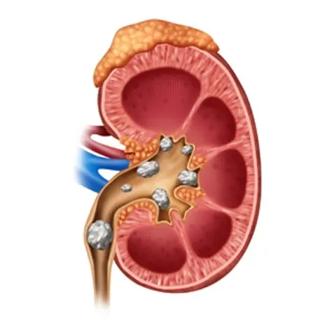
- The renal calculus is related to the excretory system
- It is the stone formation in the kidney due to the deposition of mineral and salts.
- Often associated by abdominal pain and groin pain
- The treatment includes Extracorporeal shockwave therapy, Ureteroscopic stone removal and laser lithotripsy
Medical management:
- The supportive care initially is drinking lots of water by dissolving the stone by medication
- If the condition is severe due to hard deposition, then through ultrasound technique the calculus is examined whether it is multiple calculus or cortical calculus
- Depending on the severity of the calculus the procedural plans are made by the clinicians
Extracorporeal shockwave therapy
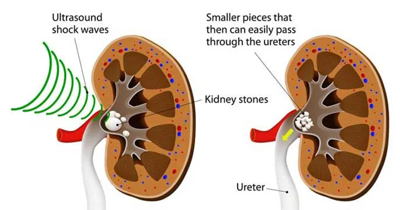
- It is a non-invasive procedure which sends acoustic shockwaves into the site of calculus
- ESWL is generally a painful procedure, done by breaking up the stones with minimal collateral damage by using an externally applied, focused, high intensity acoustic pulse
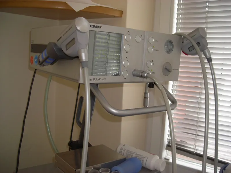
- During the procedure the patient is sedated or anaesthetized to reduce the discomfort
- The controlled reinjuring of tissues helps in regenerating the blood vessels and bone
- Lung cancer:
- Lung cancer can occur mostly in person who smokes often
- There are two types of lung cancer non-small cell cancer and small cell cancer
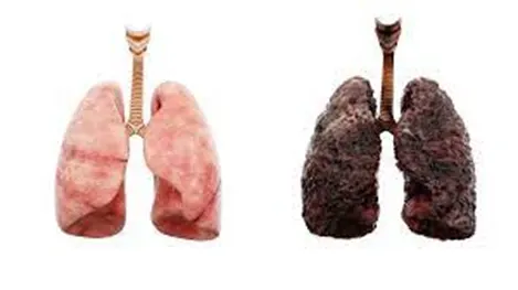
- Most common symptoms are cough, chest pain, wheezing and weight loss
- There are various types of treatments for lung cancer such as surgery, chemotherapy, radiation therapy, targeted drug therapy and immunotherapy
Structure of lung:
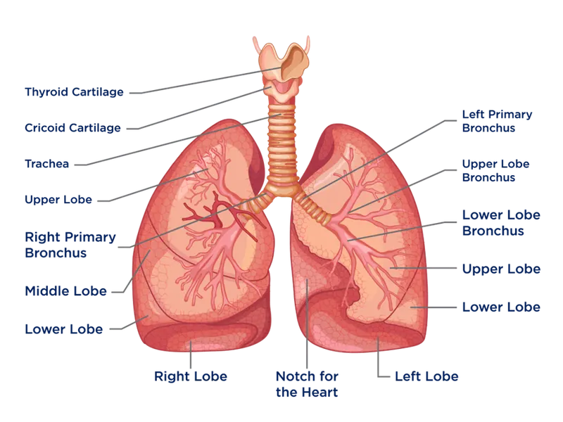
- The lung has an apex, three boarders and three surfaces
- It is located in the intrathoracic space, which is filled with the mediastinum
- The right lung has three lobes – inferior, middle and superior lobe
- The left lung has two lobes separated by an oblique fissure
- Both the lungs rest on the diaphragm
- There are 10 segments in the right lung and 8-10 segments in the left lung
Diagnostic methods
- The diagnostic methods include pulmonary lobectomy, wedge resection and thoracoscopic surgery, thoracotomy, and radiation therapy
- Lobectomy is the surgical removal of a part of the lung
- Wedge resection is the removal of a triangular piece of tissue from the lung
- Thoracoscopy is a surgery using a tiny camera inserted in the chest through a small cut, which allows the doctor to see inside the lungs without making a big opening
Thoracoscopy
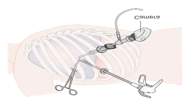
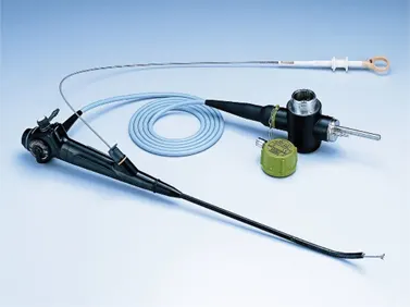
- Video assisted thoracoscopy is a type of surgery for diagnosing and treating a variety of conditions involving the chest area
- It uses a video camera called thoracoscope
- They instrument is used for biopsy part of the lung, the lymph nodes, the tissue around the lung, the tissue around the heart or the esophagus
- There also risk factors associated with the procedure such as air leak, partial collapsed lung, pneumonia, blood clot and complications with the anesthesia
Post operative care
- The pulmonologists treat the respiratory tract diseases and the cardiothoracic surgeon treat diseases affecting organs inside the chest
- Oncologist specializes in cancer
- One of the post operative cares is the palliative care wherein the interdisciplinary medical caregiving approach aimed at optimizing quality of life for terminal illness
Equipment’s used in the post operative care:
- Blood pressure monitors
- Kidney machines
- Bi-pap and C-Pap
- Pulse oximeter
- Oxygen concentrators
- Patient monitors
- Nebulizer
Bi-Pap and C-Pap machine:
- Bi-Pap (Bilevel positive airway pressure) is a type of ventilator which helps with breathing
- It delivers higher air pressure when you breathe in
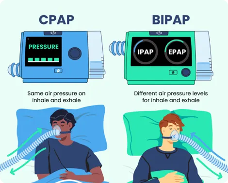
- On the other hand the C-Pap (Continous positive airway pressure) delivers the same amount of pressure at all times
- It is used to treat sleep-related breathing disorders including sleep apnea
- Recent advances in the medical field:
- Ultrasound is the technique which is applicable in various diagnostic and therapeutic purpose
- The various departments it is applicable is radiology, obstetrics and gynaecology and cardiology
- It is the easiest and less time consuming diagnostic technique unlike the MRI and CT
- It helps the clinicians to detect several diseases such as canerous cells to showing real-time images of mother’s womb
- The emerging technologies of are ultra-compact ultrasound, 3D and 4D ultrasound, artificial in telligence, tissue harmonic imaging and volumentric ultrasound
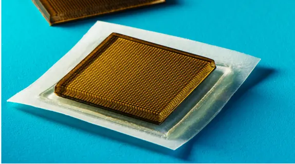
Stamp-sized Ultrasound Patch Can Image Internal Organ:
- One of the emerging technologies is the stamp-sized ultrasound patch which is an ultra-compact ultrasound technique which helps in viewing the lesions, cyst, fibroids, etc.
- Wherein the large, bulky and complicated circuitary is reduced into chip sized compact ultrasound
- Beacause that the normal ultrasound equiment is large and requires a lot of space, the image capabilities are often short term
- To tackle this problem the scientists designed a patch – approximately 1 square inch in size and a few millimeters thick – that can be placed anywhere on the body and can image the anatomy
- It is a multi-layered, like a candy wafer, with two main components- an ultrasound probe which is stacked on top of a couplant, the material which helps to transmit the acoustic waves into the body
- It helps in recording the EKG, which records the electrical activity of the heart but not images
Part B
- Flat Bone:
- Flat in appearance
- They are expanded, elongated plates of bones found to outline the cavities of the body
- Each bone consists of two plates of compact bone tissue with spongy bone and marrow
- The intervening spongy tissue in the bone of the vault of the skull is known as diploe which is numerous veins
- Eg; skull, scapula, breast bone, sternum
- Microsopis anatomy of bone:
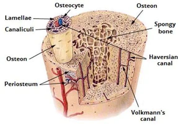
- Osteon is the unit of bone
- It consists of a central canal which is called as the haversian canal surrounded by osteocytes or bone cells which is present in a circular fasion
- Volkmans canal communicate between the two haversian canal which consists of blood vessels and nerves
- Canalculi are the projections from the osteocytes
- Lacunae are the cavities containing the bone cells or the osteocytes
- They are arranged in concentric circles lamallae
- Blood supply of the bone:
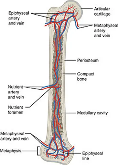
- It is done mainly by the nutrient artery
- The nutrient artery enters the long bone through the nutrient foramen
- Entering into the bone the arteries soon divides into two branches one for each end of bone
- Radiant branches enters the haversian canal and the volkmans canal and supply the cortex
- Epiphyseal arteries are derived from arterial anastomosis around the joint. They are penerated into the epiphysis
- Periosteal artery lie within the periosteum and end by supplying the superficial portion of the cortex the short bones only have epiphysis and metaphyseal branches.
- Bone remodelling and bone formation:
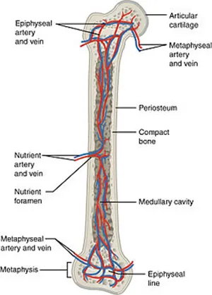
- The area which is very important for the ossification is metaphysis and the epiphyseal cartilage
- So the epiphyseal plates allow for the growth of the bones during childhood
- Bone growth is by increasing thickness and length
- Bone thickness is grown by multiplication of cells in the periosteum
- Bone length is grown by multiplication of cells in the epiphyseal cartilage
- Osteoblasts cells are the progenetor cells or immature bone cells
- These cells are deposited to form few bone on the surface and at the ends of the long bone
- Anatomical basis of bone implant:
- There are different kinds of cranioplasties, but most involve lifting the scalp and restoring the contour of the skull with the original skull piece or a custom contoured graft made from material such as titanium (plate or mesh) and synthetic bone substitute and 3D printing
- The recostruction of cranial defects can be divided into four main phases:
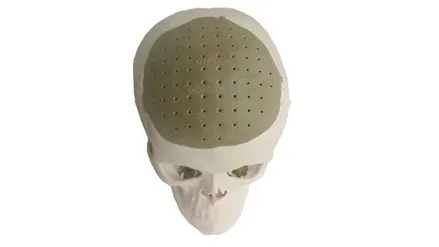
- Medical imaging based on radiological data
- Image processing
- Computer aided design
- Rapid prototyping
- 3D printing allows physicians to carefully craft replacement bones that directly match the sockets and joints with which they’ll fit
- It has been used to create jawbones, spinal discs and vertebrae and biological building materials
- Indications of bone implant:
- Intraarticular fractures
- Diaphyseal fractures
- Fractures of more than one region of the same femur
- Shortening osteotomies
- Tumor resections
- Contraindications of the bone implant:
- Acute or chronic, local or systemic infections
- Blood supply limitations
- Inadequate skin or musculoskeletal system
- Alcohol addict
- Pregnancy