Project 1
PART A: Ans 1)
Respiratory system:
The respiratory system comprises of the nose, pharynx, larynx, trachea, bronchi and the lungs. The primary function of this systems is to ensure the supply of oxygen to the individual tissue, cells and to take away the waste products and carbon dioxide from the cells. The external opening of nose is the nostrils and the dividing partition between the nostrils that forms two nasal cavities. Moreover, the palatine bone separates the nasal cavity from the mouth.
Pharynx on the other hand has three divisions namely, nasopharynx that is behind the nose to soft palate, the oropharynx that sits behind the mouth and laryngopharynx that is associated with the hyoid bone to the esophagus regions. The larynx is the voice box, that is made of cartilage. Next, we have the trachea which is a non collapsible tube that is responsible for conducting air to and from the lungs for respiration. On the other hand, the trachea and the two primary bronchi make up the bronchial tree. Trachea is a long tube like structure that connect the larynx to the bronchi. Trachea is a key component of the respiratory system and plays a key role when we breathe. For instance, during respiration, the air travels from the nose to the mouth through the larynx. Next, it passes through the trachea to reach the bronchi, where the bronchi are responsible for carrying air to the lungs.
Lungs are spongy air filled respiratory organs. That are located on each side of the chest which are covered by a thin tissue known as the pleura. The lung in conical in shape and the hilum of lung is the large depressed region that is located near the center of the medial surface. In addition, there are various structures that enter and leave the lung with the help of its roots. Fissures and lobes of the right lung is further subdivided into two fissures and into three lobes. But the left lung is divided by single oblique fissure into two lobes.
The bronchial artery is the primary source of blood supply for the bronchi. The lungs consist of dual and parallel blood supply provisions also known as pulmonary and systemic circuits. The Pulmonary circuits begins from the heart and brings the deoxygenated blood to the lungs for the gaseous exchange. Next, the circuit also brings back the oxygenated blood back to the heart to be redistributed to the rest of parts of the body. On the other hand, systemic circuit takes the oxygenated blood from the heart and passes to the lung parenchyma. When focusing on the arterial supply, the right ventricle passes the deoxygenated blood to the lungs via pulmonary arteries.
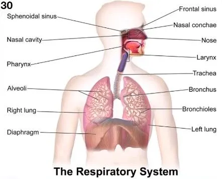
Figure.1: Respiratory system. Source: Skilllync class notes.
2) Common disorders of the organ:
The common disorders of the respiratory system or lung diseases include asthma, fibrosis, lung cancer, and tuberculosis.
For a more specific overview, we will focus on lung cancer as it is one of the most commonly occurring disease worldwide leading a high percentage of mortality rates. Lung cancer is a type of cancer that occurs in the lungs. This disease has a high risk of occurrence in smokers, but lung cancer can also occur in people who are non-smokers. However, the most challenging factor is that lung cancer doesn’t necessarily show symptoms at an early stage due to which it is challenging for early detection of lung cancer. As symptoms develop, they are usually observed in the form of a prolonged cough, coughing up blood, shortness of breath, chest pain, headache, weight loss without attempt, and hoarseness.
Lung cancer can also occur due to genetic factors or through prolonged exposure to secondhand smoke. In addition, certain lifestyle habits are also found to be causes of cancer such as specific consumption of cancer causing substances found in some packaged foods.
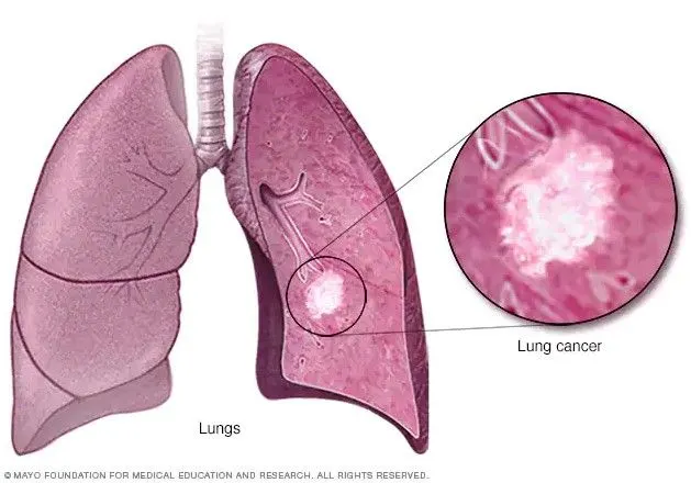
Figure.2: Lung cancer representation. Source
The diagnostic techniques for lung cancer is performed to identify if the prognosis of the disease and also to understand if they are cancerous cells. The diagnostic techniques include:
Imaging procedures: X-ray and CT scans are most commonly performed to reveal an abnormal mass nodule in the lung. However, with CT scan, there is a possibility to identify various lesions as compared to x-ray. Next, we have sputum cytology that is performed when a patient has a cough and producing sputum. It is then studied under the microscope to identify lung cancer cells. Furthermore, biopsy is another common technique that is performed by taking samples of the abnormal cells are studies to reveal if it is cancerous. Biopsy can also be performed in different ways such as bronchoscopy, where doctors examine abnormal or suspected areas of the lung by inserting a lighted tube down the throat and into the lungs. Similarly, mediastinoscopy is also performed by making incision at the base of the neck with surgical procedure is performed to inset the concerned tool behind the breastbone to collect tissue samples from the lymph node.
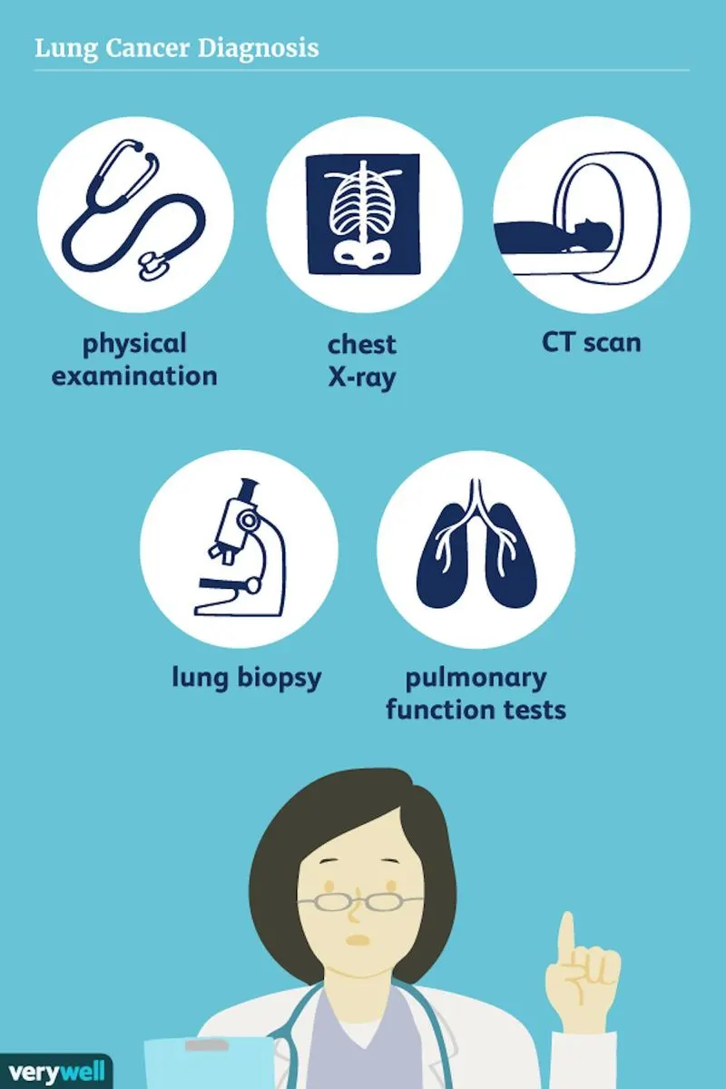
Figure.3: Lung cancer diagnostic techniques. Source
CT Scan:
A CT scan is performed for the chest and abdomen to identify if there is metastasis as part of the initial diagnosis of small cell lung cancer. In particular, CT scan comprises of a multiple cross sectional x-ray imaging of the chest, which is then produced as images in the computer for further analysis. In addition, a 3-dimensonal picture is generated of the internal parts of the body. CT scan is conducted with the help of a contrast agent, which is injected into the veins for making the scans easier to interpret. Moreover, low-dose CT scans are also performed as part of lung cancer screening process to segment the patients as high risk or low risk patient profiles as per specific parameters.
3) At the same time, when lung cancer staging (TNM staging) is performed, a PET/CT scan is also performed to gain the functional and anatomical information of the patient. Staging occurs after the initial investigation is complete. The staging is performed for treatment planning as per the stage of the disease. For instance, if the lung tumor is in the localized area, a surgery may be effective in such cases. However, if there is any possibility of metastasis, then other treatments such as radiotherapy and chemotherapy may be advised as per the stages. The TNM staging stands for:
T for Tumor, which is considered as T1 stating that tumors are less than or equal to 3 centimeters in diameter. Similarly, T2, T3 and T4 stages exists that signify larger sizes of the tumor or that it has spread to other areas such as diaphragm, heart, trachea, or different lung lobes.
N stands for lymph nodes, which signifies that there is lymphatic spread of the cancer and identifying the location of the lymph nodes that is relevant to the original tumor. Again, there are specific number assigned which is N1, N2, N3 that describes which part of lymph nodes that the cancer has spread to such as peribronchial lymph node, subcarinal lymph node etc.
Finally, we have M that stands for metastasis. This stage would signify the extent of the spread. M0 states that the cancer has not spread to distant regions such as brain, bones or liver. Whereas M1 signifies that cancer has distant metastasis.
For treatment plan, if the non-small cell lung cancer is only in one lung, then the prescribed treatment would most likely be surgery to remove the cancerous cells. However, it may be followed up and suggested for chemotherapy to eliminate any remaining cancer cells.
Similarly, if the cancer has not yet spread, but surgery is not the ideal approach due to several patient related complications such as age, then the patient may be suggested to undergo radiotherapy. However, if the cancer has spread significantly, then chemotherapy and immunotherapy is the usual recommendation. But cancer cells occur due to mutation and there is always a chance of regrowth of the cancer cells. In this regard, patients will be specific targeted therapies will be recommended as per the diagnosis.
4)
In general, there are three types of lung cancer surgery:
Lobectomy is performed when one or more significant part of the lung is removed depending on if the cancer is in just one section of the lung.
Pneumonectomy is performed when the entire lung has to be removed. This is generally performed when the cancer is identified in the middle of the lung or has spread entirely in the lung.
Wedge resection is performed when smaller pieces of lung is removed. However, it may not be suitable for all kinds of lung cancer patients. This is generally performed if the cancer is in a smaller area of the lung and is particularly in the very early stages of lung caner.
Post-operative care: Lung cancer surgery is a major surgery and the recovery time may require weeks or even a few months. The length of the recovery depends on the surgery type and what portion of lung was removed. Further, extreme care has to be taken for resuming physical activities and lifting weights.
Patients also tend to face issues like physical pain that may last for few days. Typically, the regions where it may hurt is around the chest, arm and the area of incision. Furthermore, after recovery the patients will be advised for a systematic follow after a period of 3-6 months over a specific period. Besides, doctors also take into consideration the 5-year survival rates as it is lower for lung cancer patients. If the patient is healthy and no new regrowth of cancer has occurred up to 5 years, then the patient may be regarded as cancer free.
5)
New techniques in lung cancer diagnosis and treatment include, new anti-cancer drugs, biopsy techniques and new ways of drug delivery techniques. In terms of new and advanced imaging techniques, nuclear medicine has become an integral part of cancer diagnosis. Various techniques such as PET/CT fusion, PET/MRI, 4DCT, 4DPETCT has come up in recent years with greater resolution and improved ability with fusion approaches to gain the advantages of combining two modalities. Nowadays, it has become possible to gather specific information about the suspected area and also for monitoring possible signs for regrowth of cancer cells after treatment.
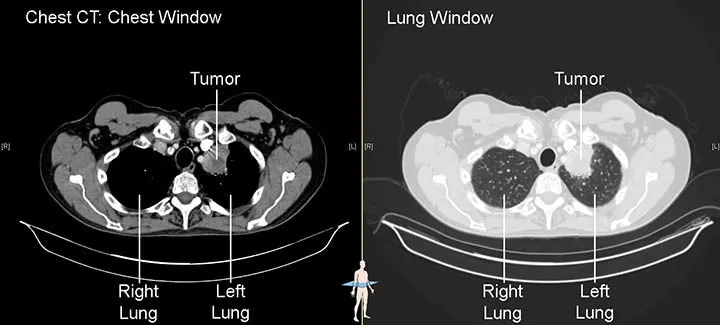
Figure.4: CT SCAN. Source
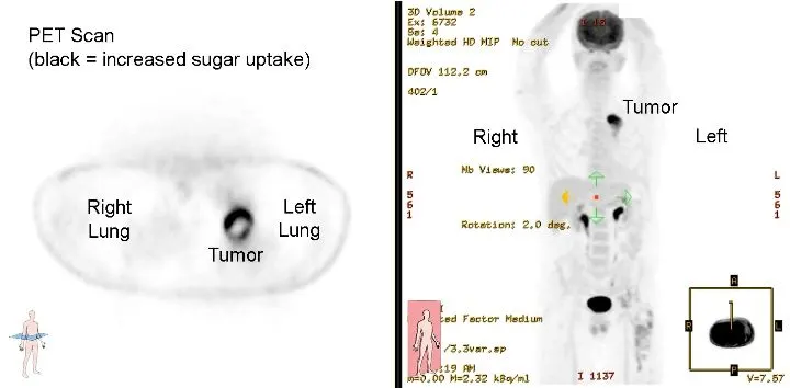
Figure.5: PET SCAN. Source
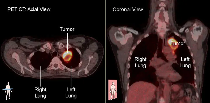
Figure.6: PET/CT Fusion. Source
PART B: Case Study
A 25-year-old biker indulged in illegal racing met with a road traffic accident and severed head injury and skull fracture. After resuscitation and operation done for the hematoma it was resorted to do skull implant for his fracture by the orthopaedician
Discuss the case under the following headings
- Describe the type of bone the skull belongs to
The skull belongs to cranial bone type.
- Describe the microscopic anatomy of bone
In general, there are five major types of bone cells such as osteogenic cells, osteoblasts, osteocytes, bone lining cells and osteoclasts. Each of these bone cells are responsible for bone growth as they mature. However, the osteocytes bone cell occupies small chambers which are responsible for forming concentric circles around canals in the bone. Similarly, bone tissues are made up of collagen and has organic and inorganic components such as calcium phosphate.
- Explain the blood supply of bone
There are some bones that receive tenuous blood supply such as scaphoid, talus, femoral head and odontoid respectively. Similarly, blood supply to long bones are acquired from different sources such as the nutrient artery system, metaphyseal-epiphyseal system and the periosteal system.
- Elaborate on bone remodeling and new bone formation
Bone remodeling consists of three key phases such as resorption when osteoclasts digests old bones, reversal when mononuclear cells start appearing on the bone surface and formation when new bone is produced and replaces the resorbed bone.
- Describe the anatomical basis of bone implant
Cranioplasty is the surgical procedure that is performed to repair any damage related to the bone in the skull. In general, it includes surgically lifting the scalp and restoring the skull shape and contour with the help of original skull parts or with custom grafted materials. These materials include titanium plates, synthetic bone substitutes in a liquid form and other relevant biomaterials.
2nd organ system: Cardiovascular system: Surgical procedures for cardiovascular disease
Coronary angioplasty is a procedure performed for improving the blood flow to the heart. Although, it is not a cure for heart diseases, it is mainly a corrective procedure where a small balloon like formation is inflated surgically in the coronary arteries to open up the clogged area.
Similarly, we have thrombolytic therapy which is a treatment that is infused in patients through the drip to dissolve some of the blood clots that is possibly blocking the coronary artery. In addition, it improves the blood flow to the heart muscle.
Next, we have the bypass surgery that is performed by surgically taking a blood vessel from either the chest, arm or leg and grafting it to the coronary artery. This allows a bypass route for the blood around the blocked artery. Bypass surgery is performed to improve the blood flow to the heart.
Pacemaker surgery is performed by fitting in a small device under the skin of the chest area with wires connected to the heart chambers to create a connection with the pacemaker. This device sends electrical impulse to stimulate the heart muscle to pump regularly.
Defibrillation is yet another intervention that is performed to restore the normal heart rhythm in scenarios where the heart stops beating in the event of a cardiac arrest.
Heart valve surgery is another critical surgery that is performed to fix the damaged heart valve such that the heart is able to pump blood properly.