Project 1
PART-A
Ans1: CARDIOVASCULAR SYSTEM:
- Clinical Condition: Heart attack
-
Heart Attack: A heart attack is also known as myocardial infarction is a acute condition and needs immediate medical attention. It happens when heart muscle doesn't receive enough blood. It happens when the arteries are blocked by atherosclerosis or plaque. The major reason for heart attack is CAD.
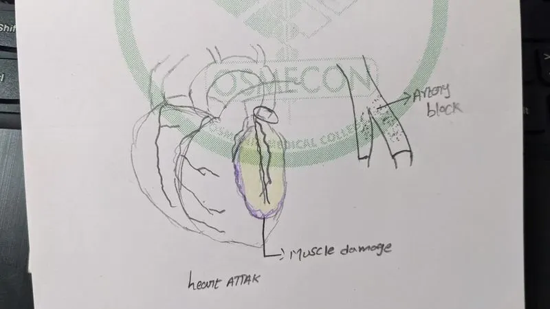
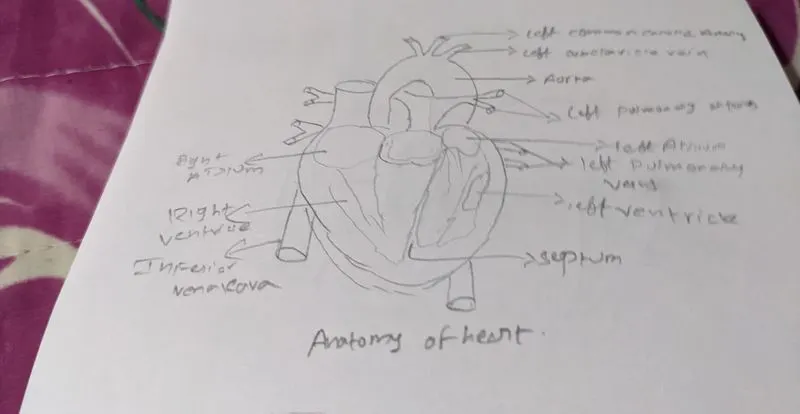
Macroscopic structure & Anotomy of organ:
- The heart is a 4 chambered organ that is situated on the ventral side and on the left side of the body.
- The heart is the organ that pumps blood to various parts of the body.
- The right atrium and ventricle are sepated by valve called tricuspid valve.
- The left atrium and ventricle are separated by valve called Mitral valve or bicuspid valve.
- The right atrium receives 2 major venacavas the superior at upper end and inferior at lower end.
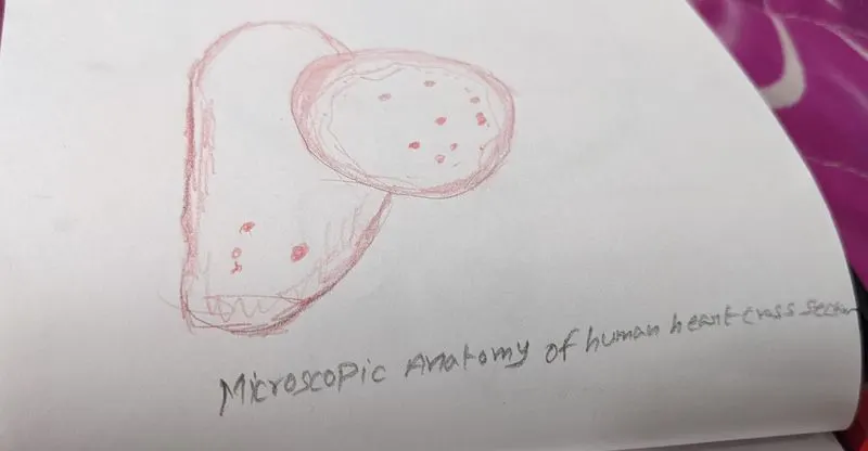
Microscopic structure of heart:
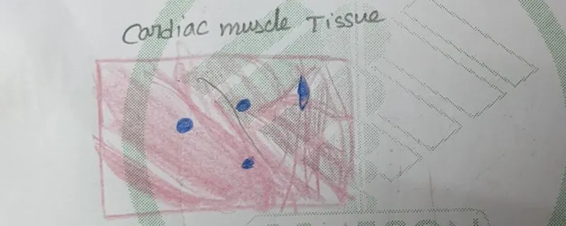
- When we see the cross section of heart in a microscopic tissue we see a muscular tissue.
- The muscular tissue is a special type of tissue it is known as the cardiac muscle and present only in the heart. These can be observed in the microscope.
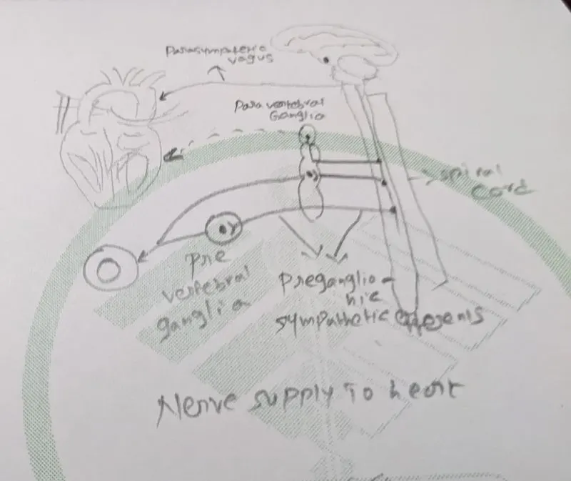
Nerve supply to the heart:
- The innervation of heart refers to the network of nerves responsible for the functioning of the heart.
- The heart is innervated by parasympathetic and sympathetic fibers from autonomic branch of the peripheral nervous system.
- The network of nerves supplying the heart are called cardiac plexus.
- The 10th cranial nerve known as vagus plays an important role in the nerve supply to the heart.
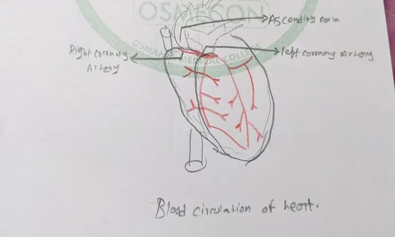
Blood supply to heart:
- Blood supply or circulation with in the heart is called coronary circulation.
- The blood is supplied with right and left coronary arteries majorly contributes the coronary circulation.
Ans2: Common disorders of heart:
- There are many common disorders of heart as
- Arrythmias
- Valve disorders
- heart failure
- Cad
- Heart Attack (MI)
- The common type of disorder i took is Myocardial Infarction
Myocardial Infarction:
- A myocardial infarction is a medical emergency.
- The blockage of blood flow in the heart muscle.
- Myocardial Infarction usually occurs when a blood clot blocks blood flow to the heart. Without blood, tissue loses oxygen and dies.
- CAD is the major issue that causes MI. If Cad is ignored for long time leads to MI.
DIAGNOSTIC PROCEDURES:
- To enumerate the procedures and tests that help to identify the heart attack are:
- Upon arrival to ED one will take Physical exam and a group of tests to analyse the severity of affect.
- The various lab tests are taken such as:
- Troponin level
- Creatinine kinase Test
- Myoglobin test.
- Other diagnostic procedures include:
- ECG
- ECHO cardiogram
- Coronary catheterization (angiogram)
- Various radiology tests can also be done such as
- Cardiac CT
- Cardiac MRI
- X-Ray.
Coronary Catheterization (Angiogram):
- When a person enters the ED with the symptoms of heart attack and the labs show cardiac tissue damage then the person is taken to the cardiac catheterization.
- The angiogram will show the blockage in the heart and if there is any narrowing in the vessels these can be diagnosed by catheterization.
- Procedure:
- The procedure takes upto 20-30 minutes.
- the person for whom the angiogram has to be taken is laid on the ot table .
- A long, thin tube (catheter) is inserted into an artery, usually in the leg, and guided to the heart then inject a contrast dye trough the tube
- This is a clear liquid that shows up under the imaging procedure. A picture of your arteries appears on a screen and the camera is moved around to create a 3D image showing where the narrowings are and how bad they are.
- This helps in treatment of the narrowing.
Ans3: Medical Management:
- The treatment should be given at the earliest as its an acute condition.
- Treatment is needed to fix blood flow and restore oxygen levels.
- Oxygen is given immediately.
- The heart attack treatment depends on whether there's a partial or complete blockage of blood flow.
- the immediate priority in managing acute myocardial infarction is thrombolysis and reperfusion of the myocardium
- The medications will include:
- Vasodilaters
- Antiarrthymatic drugs
- Anticoagulants
SURGICAL PROCEDURES:
- The removal of blockage is necessary for the heart muscle to stop from damaging and to have the blood flow into the heart.
- These surgeries are done to unblock a blocked artery.
- The surgical procedures include:
- Coronary angioplasty and stenting
- Coronary artery bypass Surgery(CABG)
Ans4: Surgical Management:
- Coronary Angioplasty:
- Coronary angioplasty is a surgical procedure to treat the narrowing of blood vessels.
- we have to know where there is blockage in the artery of the heart that causes the narrowing this will be done during the angiography procedure as the dye that is sent through the tube hilights the blockages.
- The heart is located on the ventral side slightly to the left side of the body it is covered by a bilayer called as pericardium.
- As the coronary angioplasty is an invasive surgery we have to have good knowledge on anatomical positions.
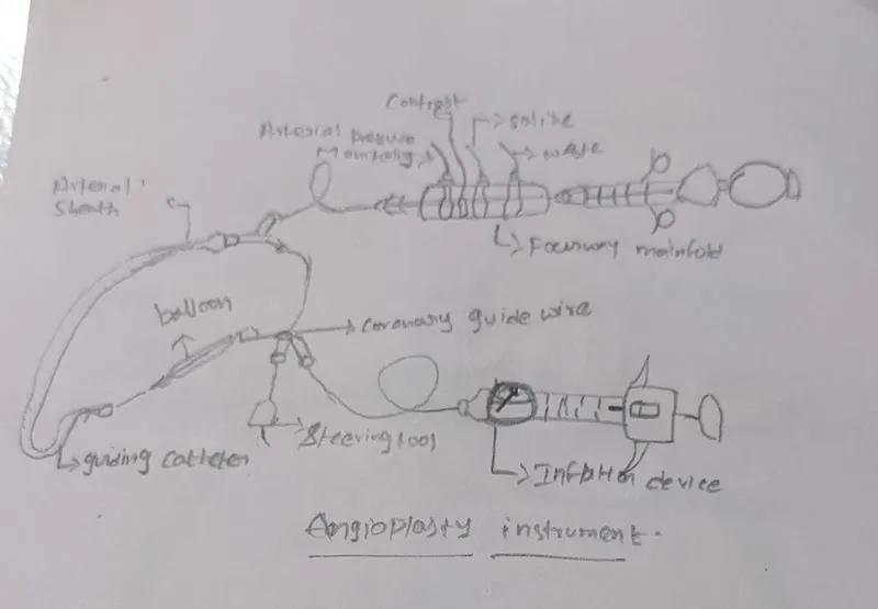
- the angioplasty catheter is entered into the body through the femoral artery mostly and then a dye is sent then the doctor will send a small tube with balloon inflated at the end and then after reaching the artery the balloon is fully inflated then a mesh is kept at the ends of artery to stop the narrowing and then the balloon is removed hence keeping the artery open.
Devices in the Coronary angioplasty:
- The angioplasty procedure will have x-ray imaging equipment, a balloon catheter, sheath, stent and guide wire as the dye is given it can be seen through the x-ray imaging and can be correctly guided and open the narrowed artery.
Post operative care:
- The post operative care is given by doctors to the patients.
- Cardiac rehabilitation is a exercise and education technique that teaches ways to improve heart health after heart surgery. It focuses on exercise, a heart-healthy diet, stress management and a gradual return to usual activities
- Medications will be prescribed that are to be used in a long run.
- Basic monitoring for a post-operative cardiac surgical care patient should include ECG, pulse oximetry.
- The Holter monitoring, echocardiogram
- the echocardiogram uses doppler effect as a result we can see the flow of blood in the heart and ecg waveforms can determine the signals of the heart helping in the postoperative care.
Ans5: Advancements in the treatments:
Recently the reperfusion therapy in the form of Primary Percutaneous Coronary intervention (PPCI) has become the gold standard for the treatment of Acute Myocardial Infarction but still there are large number of deaths due to MI.
Ans1: Gastrointestinal system:Clinical Condition:
- Liver Cirrhosis:
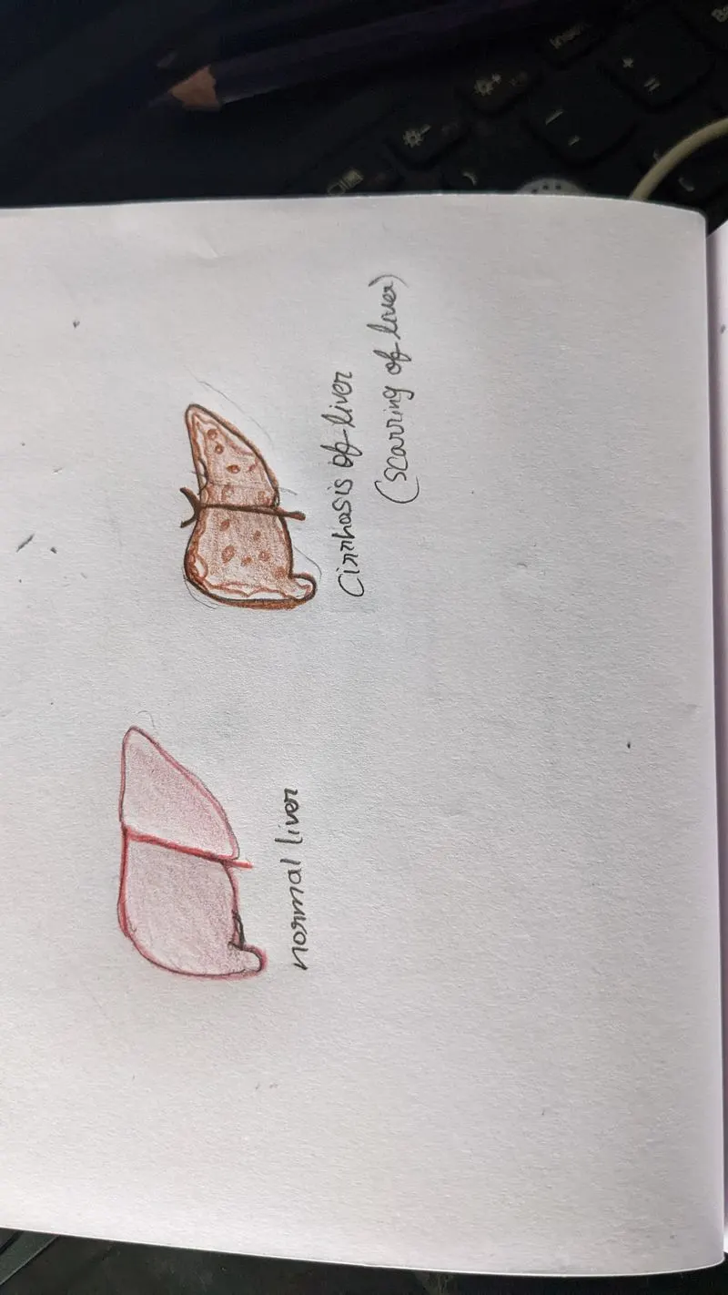
- Cirrhosis is the scarring of liver due to many other chronic diseases.
Macroscopic structure & Anatomy of the Liver:
- It is a wedge shaped organ
- It is the largest gland in the body.
- It is divided into 2 lobes.
- The right and left lobe.
- Porta Hepatis is the deep fissure in the inferior surface of the liver through which neovascular structures and hepatic ducts enter and leave the liver.
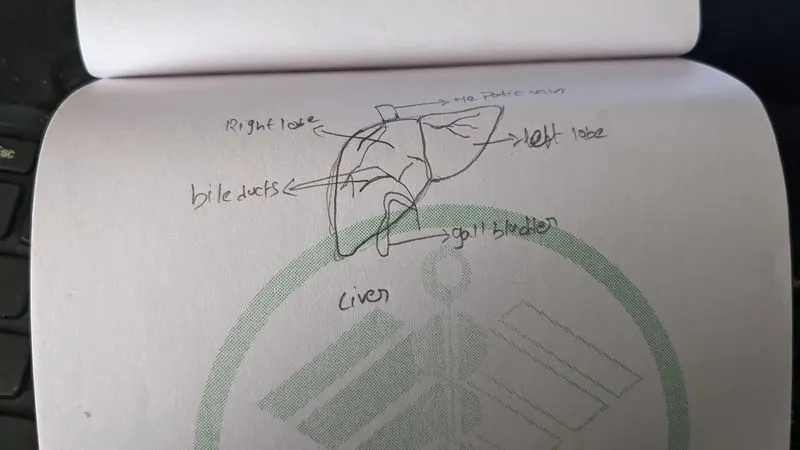
Microscopic anatomy of Liver:
- The liver is a mixed gland surrounded by thin capsule of connective tissue.
- The microscopic anatomy of liver consists of liver lobules with portal and acini conceptios.
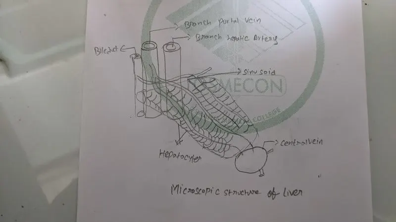
Nerve Supply to the liver:
- The innervation of liver refers to the nerves responsible for the funcioning of liver.
- The liver is innervated by both sympathetic and parasympathetic nervous system
- These nerves are derived from the splanchnic and vagal nerves that surround the portal vein, hepatic artery, and bile duct.
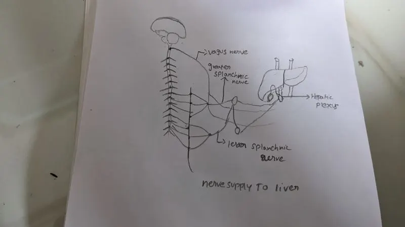
Blood Supply to the liver:
- The blood supply to liver is done with 2 sources.
- the hepatic artery carries oxygenated blood into the liver.
- the hepatic portal vein delivers the deoxygenated blood from small intestine containing nutrients.
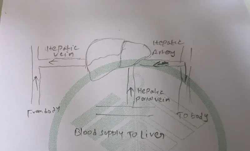
Ans2: Common disorders of liver:
- Fatty liver
- Hepatitis
- cirrhosis of liver
- hemochromatosis
- Liver cancer
- The common liver disease i took is cirrhosis
Cirrhosis of Liver:
- The cirrhosis of liver is the scarring of the liver
- The scarring is also known as fibrosis
- Chronic liver damage from a variety of causes leading to scarring and liver failure.
- The cirrhosis of liver can not be undone.
- treatment can only help but the condition cannot be cured.
Diagnostic Procedures for Cirrhosis:
- To enumerate the diagnostic procedures there are:
- Abdominal CT Scan
- Abdominal Ultrasound
- Body MRI
- Mangetic resonance cholangiopancretography
- Biopsy
- Liver function test.
- Magnetic Resonance elastography
Magnetic Resonance Elastography:
- The magnetic resonance elastography is a new way of imaging process.
- When the patient is presented with the symptoms of liver cirrhosis the doctor wll write for an imaging process.
- The MRE is a non-invasive procedure.
- In this advanced imaging technique the stiffness or hardening of the liver
- The MRI uses high range magnetic resonance and create a image hence providing the light required for the diagnosis of the cirrhosis.
Ans3: Medical management of cirrhosis:
- There is no cure till date for the cirrhosis.
- The treatment can only be given to prevent the further scarring of the tissuue.
- The treatment include to:
- Cut salf off from the food.
- To stop alcohol drinking
- Medictions includes:
- Diuretics
- Ammonia Reducers
- Beta blockers
- Antibiotics
Surgical Procedures for Cirrhosis of liver:
- There are lot of surgical procedures such as:
- Band ligation
- Transjugular Intrahepatic Portosystemic Shunting
- Paracentesis
- Liver Transplantation.
Ans4: Surgical Management of Cirrhosis:
- The surgical procedures are invasive and it needs good anatomical knowledge.
- Transjugular Intrahepatic porosystemic shunt:
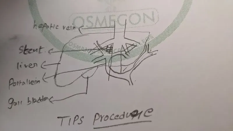
- It is called as TIPS.
- A shunt is an artificial passage which allows fluid to move from one part of your body to another.
- The Tips connects the vein which brings blood from your gastrointestinal tract and intra-abdominal organs to your liver, and the vein from your liver to the right part of your heart.
- while this procedure is performed the patient is sedated.
- The radiologist with the help of ultrasound and fluroscopy help in the guidance.
- Puncture the portal vein through the liver with a special needle and position a wire between the two veins. After the area has been dilated, a stent or stent graft will be placed between the portal and hepatic vein to create a lasting connection.
- When cirrhosis is present the blood flow is blocked
- so when is shunt is placed the block is removed and it reduces ascites.
TIPS DEVICE:
- It is a tract created within the liver using Fluroscopy guidance to connect two veins within the liver. The shunt is kept open by the placement of a small, tubular metal device commonly called a stent
Post operative Care:
- As post operative care patient has to stop drinking alcohol
- should check on getting inflammation and fluid accumalations
- If liver transplant has done patient has been kept on immunosuppresents for lifetime and to keep on checking that the transplanted organ is not getting rejected by the body.
Ans5: Advancements:
- Infusion of anti fibrotic cells to cure the cirrhosis.
- A new therapy that uses blood-vessel-lining cells to regenerate damaged tissue has the potential to treat liver cirrhosis
Ans1: System Involved:
- Nervous system
- Clinical Condition: Epilepsy
- Epilepsy is a condition where there will be abnormal electrical activity in the brain
Macroscopic structure & Anatomy of Brain:
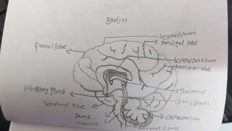
- Brain is the organ involved in the seizure disorder or epilepsy.
- brain is the master organ that regulates the whole body.
- The brain is covered by 3 layers they are:
- Outer duramater.
- Middle Arachnoid membrane
- Inner Pia mater.
- These layers protect the brain
- The brain has 3 parts
- They are:
- Cerebrum
- Cerebellum
- Medulla oblongata
Microscopic Structure of brain:
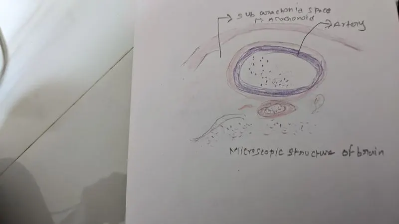
- The human brain is mainly composed of neurons, glial cells neural stem cells and blood vessels.
- These are seen in the microscopic structure when seen through the microscope of brain.
Nerve Supply to the brain:
- The brain is itself a organ that consists of nerves that go through all the other body parts.
- The brain has nerve cells and glial cells.
- The brain has 12 pairs of cranial nerves these play a major role in the nervous system.
Blood supply to the brain:
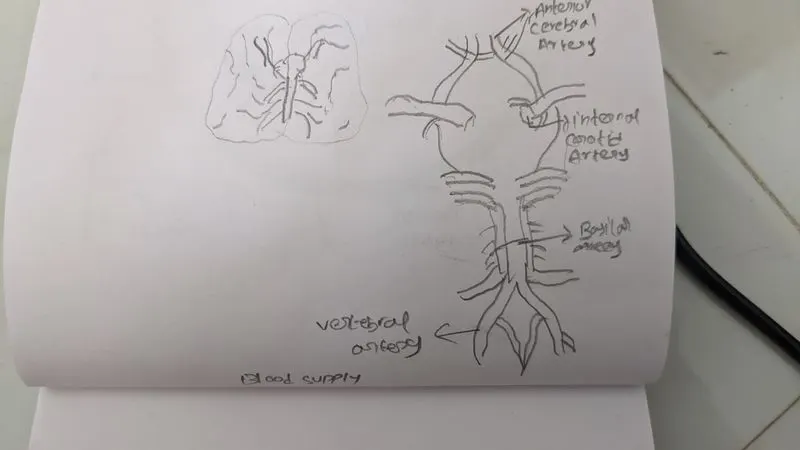
- The brain receives blood from 2 sources
- they are the internal carotid arteries, which arise at the point in the neck where the common carotid arteries bifurcate, and the vertebral arterie.
- The right and left vertebral arteries come together at the level of the pons on the ventral surface of the brainstem to form the midline basilar artery. The basilar artery joins the blood supply from the internal carotids in an arterial ring at the base of the brain.
Ans2: Common conditions of brain:
- Alzheimers disease
- dementia
- Epilepsy
- Brain tumor
- Brain malformations
- Concussions
The common condition of brain i have taken is epilepsy.
Epilepsy:
- Epilepsy is a neurological condition in which the activity of brain is disturbed due to abnormal electrical impulses.
- It causes seizures and sometimes loss of consiousness
- It can be genitic condition or due to the inury of the brain can cause the condition.
- people ust blankly stare or sometimes twich both hands and legs while having a seizure.
- A single seizure cannot be considered as epilepsy.
Diagnostic procedures for epilepsy:
- To eneumarate the diagnostic procedure there are:
- Neurological exam
- Electroencephalogram
- High density EEG
- MRI
- Functional MRI
- PET
- SPECT
- Neuropsychological tests
- Doctors can also use analysing techniques
Electroencephalogram:
- This is the most common type of diagnostic test used to determine the epilepsy or abnormal brain activity.
- The eeg electrodes are attached to the scalp with a gel applied on the scalp
- Then while lying down the doctor will acess the activity of brain by seeing the eeg activity in a video of waves generated
- they calculate the abnormalites while awake, asleep and all.
- So through this procedure we can see the what kind of seizure is affecting us.
Ans3: Medical Management of Epilepsy:
- The medical management of epilepsy is done with treatment options like
- Medications
- preventive care
- diagnising the symptoms
- Taking professional help
- The medications that treat epilepsy are
- Nerve pain medications
- sedative medications
- Anticonvulsant medications
- these help in the medical management.
Surgical Procedures:
- Even though medications are present for the epilepsy.
- many people can catch the side effects of medications or these seizures could not be controlled, in such cases we will perform a surgical procedure.
- There are 2 types of surgical procedures
- they are:
- Lobectomy or resection
- Laser interstitial thermal therapy
Ans4: Surgical Managment:
- The surgical procedures may be little invasive to highly invasive procedures.
- Surgical procedures are performed when there is no change from medications
- The surgical procedure i took is Laser interstitial Thermal therapy.
Laser Interstitial Termal Therapy:
- The laser interstitial termal therapy is a minimally invasive surgery compared to the normal convetional surgery.
- This treatment is given to people who are resistant to drugs and sometime for people with focal seiures.
- During the procedure when the patient is lying down on the table with anesthesia a small hole is made in the skull.
- Then with the help of MRI doctors locate the place where seizure arise and then with the help of laser they will ablate the part that causes the seizure in the brain.
Device LITT Procedure:
The patient will be lying on the table the procedure needs a MRI instrument and a laser probe to make it enter into the brain with the help of MRI the laser light is put on the place required and is the part ablated.
Postoperative care:
- After the surgery patients will have high headaches due to craniotomy or laser surgery. De to this patients are given medications for these pain.
- people after Laser surgery will have less pain in the head and can come back to normal life style early when compared to conventional craniotomy.
Ans5: Advancements:
- Advanements in medical maintainance can not only acheived by medications but also by devoloping the novel drugs that are already being implemented,
- The maintainance apart from medications are : Nerve stimulations, Deep brain stimulations, responsive neurostimulation.
PART-B
Case Study:
TYPE Of Skull Bone
- The skull is as per skeleton is called as axial skeleton.
- The skeleton is divided into axial skeleton and appendicular skeleton.
- The axial skeleton is present in the centre of the body and supports all the vital organs,
- The skull also protects the brain.
- The skull as per the shape of bone is called as flat bone,
- as they are flat in shape and elongated bones.
Microscopic Anatomy Of Bone:
- When we see a section of bone under microscope we can see
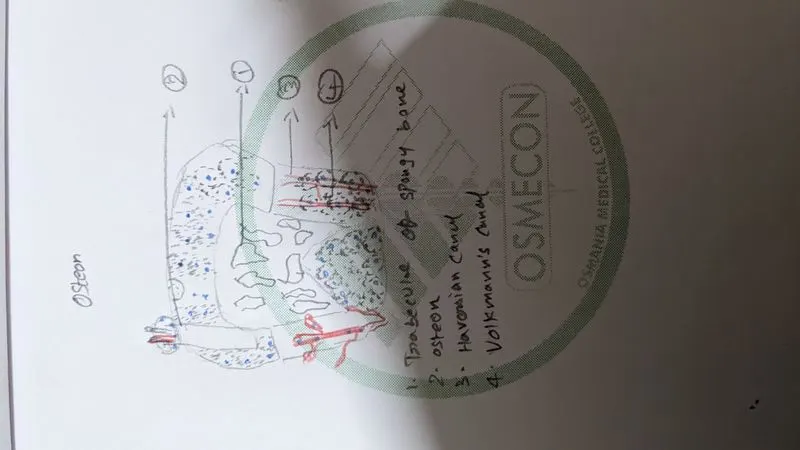
- The haversian system
- The haversian system is called as osteon.
- Osteon is the unit of bone.
- The haversian system consists of:
- Central haversian canal
- The osteocytes are present in circular fashion around the central canal in concentric fashion. these are present in the lamina along the lacunae.
- The haversian canal consists of blood vessels and nerves
- The volkmann's canal is the canal that connects two haversian canals.
- Lacunae: These are the cavities containing osteocytes.
- Canaliculi: These are tiny canals that forms the transport system.
Blood Circulation of bones:
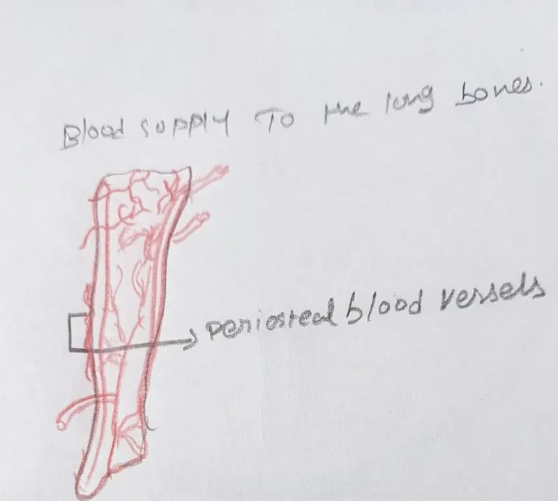
- Blood supply to the bones is mainly by the nutrient artery.
- The nutrient artery enters the long bone through the nutrient foramen
- After entering into the bone it soon divides into two branches, for each end of the bone, each artery again break up into small parallel branches which run into the metaphysis where they freely anastamose into the terminal branches of metaphyseal artery.
- Radial branches enters the haversian canal and the volkmann's canal and supply the cortex.
- Metaphyseal or Juxta epiphyseal arteries are the small arteries that arise from the arterial anastamosis around the joint. Then they enter the bone by peircing the capsular ligament.
- Epiphysea arteries that arise from the arterial anastamosis around the oint they penetrate into the epiphysis.
- Periosteal arteries lie within the periosteum and end by supplying the superficial cortex.
- The short bones have metaphyseal and epiphyseal arteries.
- The flat bones have nutrient artery and periosteal artery.
BONE REMODELLING AND NEW BONE FORMATION:
- Bone remodelling is a life long process.
- In general bone remodelling is described as removal of old mineralised bone by osteoclasts and then formation of bone matrix/bone tissue by osteoblast.
- Osteoclasts are devoloped from HSC (Hemotopoitec Stem cells).
- These HSC give rise to Myeloid progenitor that give rise to monocyte which give rise to osteoclast.
- Osteoblast is devoloped from MSC.
- These MSC give rise to osteoblast progenitor that give rise to osteoblast.
- The osteoclast forms depression in the bone and then it secretes the enzymes that dissolve the bone and the osteoblast synthesize the bone.
- This is a life long process.
- NEW BONE FORMATION:
- Osteoblasts secrete the osteoid to form a spongelike network of bony processes called trabeculae
- The new bone formation radiates outward from ossification centres in the membrane. This process is called intermembranous ossification.
ANATOMICAL BASIS OF BONE IMPLANT:
- The anatomical basis of implant is where the bone should be implanted.
- The length of the bone and the breadth of the bone for the implant purpose.
- We have to have knowledge of the location so that we can use the type of material for the purpose of implantation.
- Most of the bone implants are made up of stainless steel or titanium.
- Even though these are metals while using for a implant alloys will be used.
INDICATIONS AND CONTRAINDICATIONS OF BONE IMPLANTS:
- The orthopedic implants are either the bone or cartilage implants.
- The indications of bone implants are:
- When patient is having congenital bone disease or deformity
- chronic pain in the Joints
- Acute pain due to traumatic injury
- there are different kinds of implants such as:
- Plates
- Screws
- Prosthesis
- Removal of implants:
- The implants provide a sheild and give the bone the normal metabolism.
- but the implants should be removed after particular time when the bone is healed.
- The contraicdications of bone implants are the implants in hip and knee are limited life time and has to be replaced.