Project 1
PART A
Three systyems selected from Module 1 to Module 6:
1.Endocrine System
2.Urinary System
3.Cardiovascular system
Answer 1. One common clinical condition related to Endocrine System: Hypothyroidism.
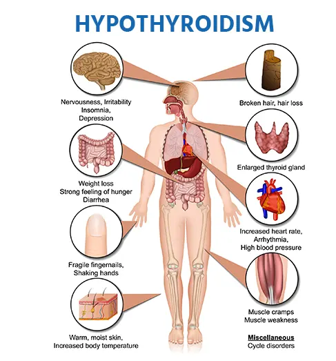
It is a condition where there is decrease in the production of thyroid hormone or what we called as T3 or T4. Symptoms are: fatigue, poor concentration, feeling mentally foggy, dry skin, constipation, feeling cold, fluid retention, muscle and joint ache, depression. Prolonged or excessive menstrual bleeding in case of women.
Other common causes are- Hashy motos thyroiditis. This is autoimmune condition, which causes inflammation of thyroid gland. Thyroid Hormone resistance can also cause Hypothyroidism.
Gross anatomy of the organ involved: Thyroid Gland is situated at the base of the neck and appears to be butterfly in shape. It is wrapped around the lateral sides of the trachea. It is largest and the most highly vascular endocrine gland. It presents two conical lateral lobes and isthmus. Isthmus is located on the anterior aspect of the trachea over 2nd and 4th tracheal rings. Lobes extend sideways upto the oblique line of Thyroid cartilage. Gland is enclosed by true and false capsule. Condensation of connective tissue is the true capsule. False capsule is provided by the paratracheal layer of deep cervical fascia of the neck.
Macroscopic and microscopic structure of organ involved: Diagram is attached. The Thyroid gland is enveloped by fascia consisting of anterior and posterior parts of the deep cervical fascia. The gland weighs approx. 10-20g, and each lobe measure an average of 5cm in length, 2.5cm in width and 1.5cm in depth. The gland is slightly heavier and bigger in size during menstruation and pregnancy. Thyroid lobes are located lateral to the trachea and esophagus, anteromedial to the carotid sheath and posteromedial to the strap muscles, overlying from the level of 5th cervical vertebra down to the first thoracic vertebra. The shape of a gland varies from a H to a U form, consisted of two elongated lateral lobes with superior and inferior poles that are joined at the midline of an isthmus. The lenght of isthmus is in between 12 and 15 high, connecting the two lobes. Ocassionally, the isthmus may be absent, and gland exists as two separate lobes.
Blood Supply of the organ involved: The Thyroid gland is a highly vascular organ among other endocrine organs, in a sense that there is a rich blood flow with large amounts of anastomosis in the gland. Arterial supply is bilateral from both the external carotid system and superior thyroid artery and subclavian system with the lower thyroid branchof the thyrocervical trunk. It may be a single thyroid ima artery arising from the branchiocephalic artery. The superior thyroid arteries originate from the ipsilateral external carotid arteries and are divided into anterior and posterior branches in the apex of the thyroid lobes. Inferior thyroid arteries originate from the thyrocervical shortly after the origin of thr subclavian arteries. The inferior thyroid arteries extend from the neck to the back of the carotid sheath and enter the thyroid lobes at the midpoints. The inferior thyroid artery passes through the recurrent laringeal nerve(RLN) and requires the identification of RLN before the arterial branches are ligated. The inferior thyroid artery provides an arterial supply of the cervical esophagus with subclavian artery and branches directly from the aorta, intercostal arteries and tracheobronchial arteries.
Nerve Supply of the organ involved: The external branch of superior laringeal nerve innervates the inferior constrictor and cricothyroid muscles. The external branch travels with the superior thyroid artery until approximately 1cm before the artery enters the superior thyroid pole and then divides into branches that enter the lateral inferior pharyngeal constrictor muscle and the cricothyroid muscle. A few smaller branches may be seen entering into the superior thyroid. The RLN is associated with the inferior thyroid artery at approximately the junction of the lower and middle thirds of the thyroid gland. On the left the RLN ascends in the tracheoesophageal grove and crosses deep to the inferior thyroid artery, on the right the RLN crosses more obliquely and is oriented more laterally than caudally. While the nerve most often crosses the deep to the inferior thyroid artery, documented variations include passing anterior to the artery as well as passing between branches of inferior thyroid artery. Another variation of nerve is a non-recurrent laryngeal nerve, in which the laryngeal nerve branches directly from the vagus nerve. This variation occurs more commonly on the right side.
One common clinical condition related to Urinary System: Urinary Incontinence-the loss of bladder control
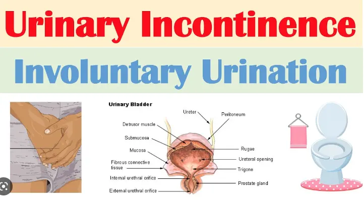
https://www.youtube.com/watch?v=Vli_6spE8HY for more details.
It is a condition where bladder control issues arise when the sphincter muscles of the urethra are too weak or too active. If the sphincter muscles are too weak, a cough or sneeze can cause urination. The sphincter muscles that are too active can trigger a sudden strong urge to urinate with little urine in the bladder. These disorders are diagnosed as urinary incontinence(UI). Women experience UI twice as often as men. It becomes more common with age.
UI can be caused by everyday habits, underlying medical conditions or physical problems. Ui is of two types-Temporary and Persistent. Temporary UI-certain drinks may act as diuretics-stimulating your bladder and increasing your volume of urine. Ex-alcohol, caffeine, carbonated drinks, artificial sweeteners, large doses of vit c, certain heart and blood pressure medications, etc. UI is also caused by easily treatable conditions such as Urinary Tract Infections(UTI) and constipation. Persistent UI- Can be due to Pregnancy, child birth, changes with age, menopause, enlarged prostate, Prostate Cancer, Obstruction and Neurological Disorders.
Gross Anatomy of the Organ Involved: The appearance of the bladder varies depending on the amount of urine present in it. When full it exhibits an oval shape and when it is empty it is flattened by overlying bowel. The external features of the bladder:
Apex- Located superiorly pointing towards the pubic symphysis. It is connected to the umbilicus by the median umbilical ligament.
Body- main part of the bladder located between the apex and the fundus.
Fundus(or base)-located posteriorly. It is triangular-shaped with the tip of the triangle pointing backwards.
Neck- formed by the convergence of the fundus and the two inferolateral surfaces. It is continuous with the urethra.
Urine enters the bladder through the left and the right ureters and exits via the urethra. Internally these orifices are marked by the trigone- the triangular area located within the fundus. In contrast with the rest of the internal bladder, trigone has smooth walls(this is explained by the different embryological origin, the trigone is developed by the integration of two mesonephric ducts at the base of the bladder). The musculature of the bladder plays a key role in the storage and emptying of the urine. In order to contract during micturition, the bladder wall contains specialised smooth muscles-known as detrusor muscle. Its fibres are oriented in multiple directions, thus retaining structural integrity when stretched. It receives innervations from both sympathetic and parasympathetic nervous system. The fibers of detrusor muscle often become hypertrophic(presenting as prominent trabeculae) in order to compensate for increased workload of the bladder emptying. This very common in conditions that obstruct urine outflow such as benign prostatic hyperplasia. There are two muscular sphincters located in urethra:
Internal Urethral Sphincter:
Male- consists of circular smooth fibres, which are under autonomic control. It is thought to prevent seminal regurgitation during ejaculation.
Female- thought to be functional sphincter(i.e. no sphincteric muscle present). It is formed by the anatomy of the bladder neck and proximal urethra.
External Urethral Sphincter: It has the same structure in both the sexes. It is skeletal muscle, and under voluntary control. However in males, external sphincteric mechanism is more complex as it correlates with fibes of the rectourethralis muscle and the levator ani muscle.
Macroscopic and Microscopic Structure of the organ involved:
Blood of the Organ Involved: The vasculature of bladder is primarily derived from internal iliac vessels. Arterial Supply is via the superior vesicle branch of the internal iliac artery. In males this is supplemented by the inferior vesicle artery and in females by the vaginal arteries. In both sexes, the obturator and inferior gluteal arteries may also contribute small branches. Venous drainage is achieved by the vesicle venous plexus, which empties into the internal iliac veins. The vesicle plexus in males is in continuity at the retropubic space with the prostate venous plexus(plexus of Santorini), which also receives blood from the dorsal vein of the penis. The superolateral aspect of the bladder drains into the external iliac lymph nodes. The neck and fundus drain into the internal iliac, sacral and common iliac nodes.
Nerve Supply of the organ involved: Neurological control is complex with the bladders receiving input from both the autonomic(sympathetic and parasympathetic) and somatic arms of the nervous system:
Sympathetic: Hypogastric Nerve(T12-L2). It causes relaxation of the detrusor muscle, promoting urine retention.
Parasympathetic: Pelvic Nerve(S2-S4). Increased signal from this nerve causes contraction of the detrusor muscle, stimulating micturition.
Somatic: Pudendal Nerve(S2-4). It innervates the external urethral sphincter, providing volunary control over micturition.
In addition to efferent nerves supplying the bladder, there are sensory(afferent) nerves that report to the brain. They are found in the bladder wall and signal the need to urinate when the bladder becomes full.
One common clinical condition related to Cardiovascular System: Heart Attack
A Heart Attack or Myocardial Infarction, usually occurs when the flow of blood to the heart is severely reduced or blocked. Blockage is usually due to a buildup of fat, cholesterol and other substances in the heart(coronary) arteries. The fatty cholesterol-containing deposits are called plaques. The process of plaque build up is called artherosclerosis. Sometimes a plaque can rupture and form a clot that blocks blood flow. A lack of blood flow can damage or destroy part of heart muscle. Prompt treatment is needed for heart attack to prevent death.
Symptoms include: Chest pain that may feel like pressure, tightness, pain, squeezing or aching; Pain or discomfort that spreads to the shoulder, arm, back, neck, jaw, teeth or sometimes the upper belly; Cold sweat; Fatigue; Heartburn or indigestion; Lightheadedness or sudden dizziness; Nausea; Shortness of breath. Women may have typical symptoms such as sharp pain felt in the neck, arm or back. Sometimes, the first symptom sign of a heart attack is sudden cardiac arrest.
Causes: Heart Muscle need constant supply of oxygen-rich blood. Coronary arteries give this critical blood supply to the heart. If a person has coronary artey disease, those arteries become narrow and blood can't flow as well as it should. When blood supply is blocked then a person has a heart attack. Fat, calcium, proteins and inflammatory cells build up in an individuals arteries to form plaque. These plaque deposits are hard on the outside and soft and mushy in the inside. When the plaque is hard, the outer shell cracks. This is called a rupture. Platelets(disc shaped-things in your plaque, that help it clot and )come to the area, and blood clots form around the plaque. If a blood clot blocks an artery, a persons heart muscles become starved for oxygen. The muscle cells soon die, causing permanent damage. Rarely, a spasm in a coronary artery can also cause heart attack. During this coronary spasm, arteries restrict or spam on and off, cutting off blood supply to heart muscles. It can happen while you are are at rest and if you don't have serious coronary artery disease. Each coronary artery sends blood to different part of your heart muscle. How much the muscle is damaged depends on the size of the area that the blocked artery supplies and the amount of time between the attack and the treatment. Heart muscle start to heal soon after heart attack. This takes about 8 weeks. Just like askin wound, a scar forms in the damaged area. But the new scar tissue doesn't move the way it should. So the heart can't pump as much after heart attack. How much that ability to pump is affected depends on the size and location of the scar.
Gross Anatomy of the organ involved: The heart is a four chambered hollow muscular organ approximately the size of human fist. It is located in the mediastenum which is present on the superior surface of the diaphragm. Left to the midline. Anterior to the vertebral column and posterior to the sternum. Somewhat pyramidal or conical in shape and placed obliquely behind the sternum & adjoining parts of the costal cartilage. So that, 1/3rd of the heart is to the right of the median plane and 2/3rd of the heart is to the left of the median plane. Length is 12cm, width is 9cm, weighs 250g in females and 300g in males. Apex of the heart is formed by the left ventricle. It is present half inch medial to the mid-clavicular line. Apex beat can be felt here. Heart has three surfaces. 1st is the sternocostal surface, which lies just behind sternum or anterior surface. The diaphragmatic surface on which it rests on the diaphragm and left surface. It has 4 borders, right border, left border, upper border and inferior border. Apex of the heart is a conical area formed by the left ventricle. It is directed downwards and forwards to the left. It lies at level of 5th left intercostal space. 3.5 inches to the midline and just medial to the mid-clavicular line. Apex beat is the outermost and the lowermost thrust of the cardiac contraction during ventricular systole felt on the front of the chest or at the point of maximum cardiac impulse. It is felt in usually left 5th intercostal space. In the infants, the apex beat may be raised and may lie in the 3rd or 4th left intercostal space. The base of the heart or the posterior portion of the heart is formed mainly by the two chambers, i.e., the left and the right atrium and 2/3rd of the space is formed by posterior surface of the right atrium.
Macroscopic and Microscopic Structure of the organ involved:
Blood Supply and Nerve Supply to the organ involved: Blood supply on the right side of the heart:-Heart supply is mainly by the coronary arteries. Coronary arteries and their branches run on the surface of the heart lying within the subpericardial fibro fatty tissue. Right coronary artery-it arises from the anterior aortic sinus of the ascending aorta just above the anterior aortic valve. After arising from the ascending aorta, the right coronary artery first runs forwards between the pulmonary trunk and the right auricle and then it descends almost vertically in the right atrioventricular grove upto the junction of the right and the inferior border of the heart. At the inferior border of the heart it turns posteriorly and runs in the posterior atrioventricular grove upto the posterior interventricular grove where it terminates by anastomosing with the left coronary artery.
Blood supply on the left side of the heart:- Left coronary artery arises from the left posterior sinus of the ascending aorta immediately above the aortic valve. After arising from the ascending aorta, the left coronary artery, the left coronary artery runs forwards and to the left between the pulmonary trunk and the left auricle. It divides into the anterior interventricular and circumflex artery. These are the main two branches of the left coronary. Other branches of left coronary include:Diagonal branches, Atrial branches, Conus Artery.
Answer 2. Common Disorder associated with the organ(Thyroid): Hypothyroidism, Hyperthyroidism, Thyroid Nodules, Grave's Disease, Goitre, Hashimoto's Thyroiditis
Hyperthyroidism- In this condition, Thyroid Gland is overactive. It produces too much thyroid hormone. This can cause many of your body's function's to speed up. Grave's disease is the most common cause of hyperthyroidism, affecting about 70% of people with an overactive thyroid. Nodules on your thyroid-a condition called toxic nodular goiter or multinodular goitre can also cause the gland to overproduce its hormones.
Hypothyroidism- In this condition there is a decrease in production of thyroid hormone or what we called as T3 OR T4. Common causes of Hypothyroidism are Hashimoto's Thyroiditis. This is autoimmune condition whic causes inflammation of thyroid gland. Thyroid Hormone resistance can also cause hypothyroidism.
Thyroid Nodules are solid or fluid filled lumps that form within your thyroid. Most thyroid nodules are not serious and don't cause symptoms. Only small percentage of thyroid nodules are cancerous. Several conditions can cause nodules to develop in your thyroid-overgrowth of normal thyroid tissue, thyroid cyst, chronic inflammation of the thyroid, multinodular goitre, thyroid cancer, iodine deficiency.
Grave's Disease is an immune system disorder that results in overproduction of thyroid hormone. This disease is caused by malfunction in the body's disease fighting immune system. It is unknown why this happens. The immune system produces antibodies designed to target a specific virus, bacterium, or other foreign substance. In Grave's disease-for reasons that aren't well understood- the immune system produces an antibody to one part of the cells in the hormone producing gland in the neck(thyroid gland). Thyroid function is regulated by a pituitary gland. The antibody associated with Grave's Disease-thyrotropin receptor antibody(TRAb) acts like a regulatory pituitary hormone. That means TRAb overrides the normal regulation of thyroid , causing an overproduction of thyroid hormones.
Goitre happens when the thyroid gland grows larger. It has several possible causes and may or may not be associated with abnormal thyroid hormone levels. It is treatable. Entire thyroid can grow larger or it can develop one or more small lumps called thyroid nodules. Goitre is an adaptive reaction of the cells in your thyroid to any process that blocks thyroid hormone production. While most common cause of Goitre worldwide is iodine deficiency.
Hashimoto's Thyroiditis is an autoimmune disease in which immune system turn's against body's own tissues. In people with Hashimoto's, the immune system attacks the thyroid. This can lead to hypothyroidism. The exact cause of Hashimoto's is not known, but many factors play a role- Genes, Hormones, Excessive iodine, radiation exposure, etc.
Common Disorder associated with the organ(Urinary Bladder): Overactive bladder, Urinary Incontinence, Interstitial Cystitis, bladder cancer
Overactive bladder called as OAB, causes a frequent and sudden urge to urinate that may be difficult to control. The person may feel like need to pass urine many times during the day and night, also may experience unintentional loss of urine(urgency incontinence).
Urinary incontinence is the loss of bladder control. The severity ranges from ocassionally leaking urine when you cough or sneeze to having an urge to urinate that is so sudden and strong a person don't get to a toilet in time. It occurs more often as people get older. It can be caused by everyday habits, underlying medical conditions or physical problems.
Interstitial Cystitis is a condition causing bladder pressure, bladder pain and sometimes pelvic pain. The pain ranges from mild discomfort to severe pain. The condition is a part of a spectrum of diseases known as painful bladder syndrome. The bladder is a hollow muscular organ that stores urine. The bladder expands until it is full and then signals your brain that it is time to urinate, communicating through the pelvic nerves. This creates the urge to urinate for most people. With interstitial cystitis these signals get mixed- the person feels the need to urinate more often and with smaller volumes of urine than most people. Interstitial cystitis most often affects women and can have long lasting impact on quality of life. Thexact cause of interstitial cystitis is not known, but it is likely that many factors contribute. For instance, people with interstitial cystitis may also have a defect in the protective lining(epithelium) of the bladder. A leak in the epithelium may allow toxic subsances in urine to irritate the persons bladder wall. Other possible but unproven possible factors include an autoimmune reaction, heredity, infection or allergy.
Bladder cancer is a common type of cancer that begins n the cells of bladder. Bladder is a hollow muscular organ in your lower abdomen that stores urine. Bladder cancer most often begins in the cells(urothelial cells) that line the inside of the bladder. Urothelial cells are also found in the idneys and ureters. Most bladder cancers are diagnosed at an early stage, when the cancer is highly treatable. But even early stage bladder cancers can come back after successful treatment. Bladder cancer begins when the cell in the urinary bladder develop changes(mutations) in their DNA. A cell's DNA contains instructions that tell the cell what to do. The changes tell the cell to multiply rapidly and to go on living when healthy cells would die. The abnormal cells form a tumor that can invade and destroy normal body tissue. In time the abnormal cells can break away and spread through the body.
Common Disorder associated with the organ(Heart):Coronary Artery Disease(CAD), Heart Arrhythmias, Hear Failure, Heart Valve Disease
CAD is the most common heart problem. With CAD you may get blockage in your coronary arteries. That can lead to flow of blood to your heart muscles, keeping it from getting the oxygen it needs. The disease usuall starts as a result of artherosclerosis, a condition sometimes called hardening of arteries.
Heart Arrhythmia is an irregular beating pattern. Severe arrhythmias often develop from other heart problem but also may happen on their own.
Heart Failure-In this condition, the heart doesn't pump blood as well as it should to meet the body's needs. It is usually caused by Coronary artery disease, but it can also happen because you have Thyroid disease, high blood pressure, heart muscle disease, or certain other conditions.
Heart Valve Disease- The heart has four valves that open and close to direct blood flow between heart's four chambers, lungs and blood vessels. An abnormality could make it hard for a valve to open and close the right way. When that happens, blood flow could be blocked or blood can leak. The valve may not open and close right. The causes of heart valve problems include infections such as rheumatic fever, congenital heart disease, high blood pressure, coronary artery disease, or as a result of heart attack.
Various Diagnostic Techniques and investigations used in diagnosis of disorders
The diagnostic process incorporates a number of different types of examinations, including clinical evaluation, blood tests, imaging tests, biopsies and other tests:
For thyroid diseases:- Physical Exam, Hormone and Antibody blood tests, thyroid ultrasound, CT or MRI Scans, Radioactive iodine uptake and Fine needle aspiration biopsy.
Fine Needle aspiration biopsy: A needle biopsy, also known as a fine needle aspiration (FNA) biopsy, is used to help evaluate suspicious thyroid lumps and nodules. During a needle biopsy, a thin needle is inserted directly into the nodule, and cells are withdrawn and evaluated in a laboratory for signs of cancer. Some healthcare providers use ultrasound while conducting a biopsy to guide the needle position. While 95% of thyroid nodules are not cancerous, FNA, along with some additional tests such as the Veracyte Afirma test, can make the results of your biopsy more accurate and may spare you unnecessary surgery for nodules that turn out to be benign. Fine needle aspiration is generally considered a safe procedure. Complications are infrequent.
A fine needle aspiration is most often done on swellings or lumps located just under the skin. A lump may be felt during a doctor's examination. Or it may be discovered on an imaging test such as:CT scan and ultrasound.
Doctors may recommend fine needle aspiration for areas such as:
- cysts (fluid-filled lumps)
- nodules or masses (solid lumps)
- enlarged lymph nodes
Without a biopsy, it's usually hard for a doctor to confirm what these abnormal areas contain. And you may not know if they are a threat to your health. A fine needle aspiration biopsy may also be performed during an endoscopy or bronchoscopy. An endoscopy is a procedure that allows the doctor to view the inside of a person's body, and a bronchoscopy is a procedure that allows the doctor to view the inside of a person’s lungs. A fine needle biopsy is an effective tool in evaluating and diagnosing suspect lumps or masses. A quick diagnosis can mean that cancer is detected early, giving more options for treatment, or that benign lumps are diagnosed without the need for surgery. It is non-invasive and only slightly uncomfortable, compared to a surgical biopsy which requires a general anaesthetic, involves pain and the possibility of infection or scarring
For Heart:- ECG(Electrocardiogram), chest x-ray, stress test, tilt table test, echocardiogram, cardiac catherisation, electrophysiology test, CT Heart scan, Myocardial Biopsy, Heart MRI, Pericardiocentasis and Cardiac Perfusion scan.
Cardiac Catherisation: also called cardiac cath or coronary angiogram is an invasive imaging procedure that allows your doctor to evaluate your heart function. Cardiac catheterization is used to:
- Evaluate or confirm the presence of coronary artey disease or valve disease or disease of the aorta
- Evaluate heart muscle function
- Determine the need for further treatment (such as an interventional procedure or coronary artey bypass graft or CABG, surgery)
During a cardiac catherisation, a long, narrow tube called a catheter is inserted through a plastic introducer sheath (a short, hollow tube that is inserted into a blood vessel in your arm or leg). The catheter is guided through the blood vessel to the coronary arteries with the aid of a special x-ray machine. Contrast material is injected through the catheter and x-ray movies are created as the contrast material moves through the heart’s chambers, valves and major vessels. This part of the procedure is called a coronary angiogram (or coronary angiography). The digital photographs of the contrast material are used to identify the site of the narrowing or blockage in the coronary artery. Additional imaging procedures, called intra vascular ultrasound(IVUS) and fractional flow reserve (FFR), may be performed along with cardiac catheterization in some cases to obtain detailed images of the walls of the blood vessels. Both of these imaging procedures are currently only available in specialized hospitals and research centers.
With IVUS, a miniature sound-probe (transducer) is positioned on the tip of a coronary catheter. The catheter is threaded through the coronary arteries and, using high-frequency sound waves, produces detailed images of the inside walls of the arteries. IVUS produces an accurate picture of the location and extent of plaque.
With FFR, a special wire is threaded through the artery and a vasodilator medication is given. This test is functionally performing a very high quality stress test for a short segment of the artery.
For Urinary Bladder:-Cystometry, uroflowmetry, urethral pressure profile and leak point pressure, transurethral resection of bladder tumor(TURBT), Renal Function Test, Urine analysis, urine dipstick and renal biopsy, CT Scan, MRI, Bone scan, ultrasound.
Magnetic resonance imaging (MRI) is a medical imaging technique that uses a magnetic field and computer-generated radio waves to create detailed images of the organs and tissues in human body. Most MRI machines are large, tube-shaped magnets. When you lie inside an MRI machine, the magnetic field temporarily realigns water molecules in your body. Radio waves cause these aligned atoms to produce faint signals, which are used to create cross-sectional MRI images — like slices in a loaf of bread.The MRI machine can also produce 3D images that can be viewed from different angles.An MRI (magnetic resonance imaging) scan is a painless test that produces very clear images of the organs and structures inside your body. MRI uses a large magnet, radio waves and a computer to produce these detailed images. It doesn’t use X-Rays(radiation).
Because MRI uses powerful magnets, the presence of metal in your body can be a safety hazard if attracted to the magnet. Even if not attracted to the magnet, metal objects can distort the MRI image. Before having an MRI, you'll likely complete a questionnaire that includes whether you have metal or electronic devices in your body.
Unless the device you have is certified as MRI safe, you might not be able to have an MRI. Devices include:
- Metallic joint prostheses
- Artificial heart valves
- An implantable heart defibrillator
- Implanted drug infusion pumps
- Implanted nerve stimulators
- A pacemaker
- Metal clips
- Metal pins, screws, plates, stents or surgical staples
- Cochlear implants
- A bullet, shrapnel or any other type of metal fragment
- Intrauterine device
Answer 3. Management and treatment plan utilized in the disorder.
Thyroid Nodules: Treatment depends on the type of thyroid nodule. Treatment options include:
- No treatment/watch and wait: If the nodules aren’t cancerous, you and your healthcare provider may decide that you don’t need to be treated at this time. You’ll see your provider regularly so they can check for any changes in the nodules.
- Radioactive Iodine: Your provider may use radioactive iodine to treat hyperfunctioning thyroid nodules and goiters with several nodules. Your thyroid gland absorbs the radioactive iodine, causing the nodules to shrink.
- Surgery: Surgery to take out the nodules is the best treatment for nodules that are cancerous, cause obstructive symptoms like breathing or swallowing issues and are “suspicious” (they can’t be diagnosed without being surgically removed and examined.
Various factors including serum TSH, clinical risk factor assessment, size of the nodule, ultrasound characteristics, patient preferences and results of the FNA biopsy should be considered in management of thyroid nodule. FNA biopsy cytological diagnosis is the most crucial determinant in decision making. For autonomous or hyperfunctioning nodules, if the patient has hyperthyroidism, management options include radioiodine therapy or surgery. If the patient has subclinical hyperthyroidism (low TSH with normal FT4), management depends on clinical risk of complications . Nodules less than 1 cm with some exceptions should not be biopsied and followed up closely.
Also for these patients the frequency and duration of follow up will depend on the additional risk factors present. For nodules selected for FNA, management primarily depends on cytologic results. According to the Bethesda Classification scheme, FNA of the nodules yields six major results with subsequent different management for each category. However the management of indeterminate nodules (FLUS/AUS and FN/SFN) has similar principles and will be discussed together.
The risk of malignancy in nodules reported as benign is 0–3 %. Patients with benign nodules are usually managed conservatively without surgery; immediate further diagnostic studies are not required. Though there is a risk of false negative results associated with cytology reporting, initial benign FNA has negligible mortality risk in long term follow up.
Malignant- This category includes papillary cancer, follicular carcinoma, Hurthle Cell(oncocytic) carcinoma, medullary cancer, thyroid lymphoma, anaplastic cancer and cancer metastatic to the thyroid. Surgery is generally recommended for these patients.
Non-diagnostic- This category includes cytologically inadequate specimen. If no or scant follicular tissue is obtained, the absence of malignant cells does not mean a negative biopsy in patients with nondiagnostic FNA. In such cases, FNA using US-guidance should be repeated and if possible with onsite cytological assessment. If the results are still nondiagnostic, core needle biopsy or close observation or diagnostic surgical excision can be considered depending on the suspicious pattern on sonography, clinical risk factors and growth of the nodule during active surveillance.
Pregnancy- The evaluation of a thyroid nodule in a pregnant woman should be done in same way as one would in non-pregnant state. However, for the pregnant women with nodule and suppressed TSH that persists after first trimester, further evaluation can be delayed after pregnancy and cessation of lactation when the radionuclide scan can be performed.
Heart Attack: Treating a heart attack means restoring blood flow to the affected heart muscle as soon as possible. This can happen in a variety of ways, ranging from medication to surgery. It's extremely likely that treatment will use several of the following methods:
Supplementary Oxygen- People having trouble breathing or with low blood oxygen levels will often receive supplementary oxygen along with other heart attack treatments. You can breathe the oxygen either through a tube that sits just below your nose or a mask that fits over your nose and mouth. This increases the amount of oxygen circulating in the blood and reduces the strain on your heart.
Medications-
- Anti-clotting medications: This includes aspirin and other blood-thinning medicines.
- Nitroglycerin: This medicine is used to relieve chest pain. It also is a powerful vasodilator, meaning it causes blood vessels to widen so blood can pass through more easily.
- Thrombolytic (clot-busting) medications: These intravenous (IV) medications cause blood clots to break down and dissolve. These medications are usually used only within the first 12 hours after a heart attack.
- Anti-arrhythmia medications: Heart attacks can often cause malfunctions in your heart’s normal beating rhythm called arrhythmias. Some arrhythmias can be life-threatening. Anti-arrhythmia medications can stop or prevent these malfunctions.
- Pain medications: The most common pain medication given during heart attack care is morphine. This can help alleviate chest pain.
Percutaneous Coronary Intervention- Restoring circulation to the affected heart muscle is usually done with a procedure called percutaneous coronary intervention. Often called PCI for short, this procedure uses a catheter-based device inserted into a major blood vessel (usually one near your upper thigh). Once the catheter is inserted into the blood vessel through a small incision, the provider threads it up to the blocked artery on your heart. Once it reaches the location of the blockage, the provider will inflate a small balloon on the end of the device to widen the blood vessel and clear the blockage. PCI is a critical tool in restoring blood flow, and the sooner that happens, the better the chance of a good outcome. Hospitals use a metric called “door-to-balloon time” to measure their ability to treat a heart attack. This is the average time it takes for patients to undergo PCI after they first come into the Emergency Room. PCI often includes the placement of a stent at the site of the blockage. Stents are made of metallic mesh and look like tube-shaped scaffolds. They help hold the artery open so another blockage doesn’t happen in the same spot. Some stents are coated with a medication that prevents clotting or tissue growth on the stent itself (either of which could cause another blockage).
Coronary Artery Bypass Grafting- Patients who have severe blockages of their coronary arteries may undergo coronary artery bypass grafting. This surgery is often called open-heart surgery, bypass surgery or CABG (the acronym is pronounced the same as “cabbage”). CABG involves taking a blood vessel from elsewhere in the body (usually your chest, arm or leg) and using it to construct a detour for blood to use. This reroutes blood around the blocked artery section (or more than one artery; a double bypass goes around two arteries, three is a triple, and so on).
We can reduce risk of having heart attack by:
- Schedule a chekup
- Quit Tobacco Products
- Exercise Regularly
- Eat Healthy diet
- Maintain Healthy weight
- Manage your existing health condition
- Reduce your stress
- Take your medications
- Keep all your medical appointments
Urinary Incontinence-loss of bladder control: There are many different factors that your healthcare provider will consider when creating a treatment plan for your incontinence. The type of incontinence and the ways it affects your life are both big considerations. Your provider will also talk to you about the type of treatment you are most comfortable with. There are three main types of treatment you can explore for incontinence-medications, lifestyle changes and surgery. Each option has pros and cons that your provider will discuss with you.
Medications to treat incontinence-There are quite a few medications that can reduce leakage. Some of these drugs stabilize the muscle contractions that cause problems with an overactive bladder. Other medications actually do the opposite thing — relaxing muscles to allow your bladder to empty completely. Hormone replacement therapies can-often involving replacing estrogen that’s decreased during menopause — may also help restore normal bladder function. In many cases, medications can work very well to return normal function to the bladder. Your provider will carefully select a medication that matches your specific needs. Often, your provider will start you on a low dose of the medication and then increase it slowly. This is done to try and reduce your risks of side effects and to keep track of how well the medication is working to treat your incontinence.
Common medications that can be used to treat incontinence include:
- Oxybutynin (Ditropan®), oxybutynin XL (Ditropan XL®), oxybutynin TDDS (Oxytrol®).
- Tolterodine (Detrol®).
- Solifenacin (Vesicare®).
- Fesoterodine (Toviaz®).
- Darifenacin (Enablex®).
- Trospium (Sanctura XR®)
- Antideppressant Medications-Imipramine (Norfranil, Tipramine, Trofranil).
- Mirabegron (Myrbetriq®)
Lifestyle changes to manage incontinence- Sometimes, there are changes to your everyday life that can actually help your incontinence. These changes often include exercises you can do to strengthen your pelvic floor muscles, changes to your normal habits and an improved diet. Some people notice improvements by making these changes at home and don’t need additional treatment. A person can talk to their healthcare provider about these home treatment options for incontinence before starting any of them. You might not be able to treat all types of incontinence with these lifestyle changes. Your provider might also make detailed suggestions to you about the best lifestyle changes to try given your incontinence diagnosis. Lifestyle changes to help improve incontinence can include:
- Emptying your bladder on a regular schedule. This is also called timed voidings-a practice of going to the bathroom on a regular schedule instead of waiting for the urge to go.
- Emptying your bladder before physical activities. If you’re planning to exercise or do a physical activity, plan to empty your bladder before the activity starts to avoid leakage.
- Avoiding lifting heavy objects. If you need to move something large, grab an additional person to help you.
- Doing regular Kegel exercises to help strengthen your pelvic floor muscles.
- Avoiding drinking caffeine or a lot of fluids before starting an activity. If you experience frequent urination and leakage at night, you might also want to avoid drinking beverages right before bed.
- Wearing pads and products that are designed to catch any leaked urine can also be a useful way to manage incontinence. These products can usually be worn under your clothes without anyone noticing and they provide reassurance that you won’t experience urine leaking through your clothes.
- Practicing bladder training to help stretch out the amount of time between each trip to the bathroom. The goal in bladder training is to try and wait a little longer between urinating each time to build up strength.
- Maintaining a healthy body weight. Having excess body weight can be one cause of incontinence. By eating a healthy diet and exercising, you can reduce the risk of incontinence.
There are also devices that you can use at home to help with incontinence. One over-the-counter device that you can use is a:
A Vaginal Insert-There are over-the-counter devices that can be placed in the vagina that can compress the urethra and help reduce stress incontinence in women.
Procedures and surgeries to treat incontinence- If other non-invasive treatment options have failed to treat your incontinence, there are several procedures that your provider might suggest. These procedures range from simple injections to more complicated surgeries. Your provider will discuss the best procedural option for you based on the type of incontinence you have and your symptoms.
Procedures to treat incontinence can include:
- Bulking Agents: This treatment option is an injection that’s typically used in women with stress incontinence. A permanent substance is injected into the lining of your urethra to help increase the size of the urethra’s lining.
- Botulinum toxin injections(BOTOX): You might think of Botox® as a cosmetic treatment, but it can also be used to relax muscles in your body. Your provider might inject Botox® into your bladder to help relax the muscles-helping with urge incontinence. This treatment isn’t permanent and will need to be repeated over time.
- Neuromodulation Devices: Pacemakers that stimulate the nerves to the bladder to improve control can be implanted. Furthermore, a nerve near your ankle can be stimulated to achieve better bladder control.
- Sling Procedures: There are sling procedures to treat incontinence for both men and women. In women, either a synthetic material or strip of your own tissue is typically used to support the urethral channel.
- Artificial Urethral Sphincter: This is a device that’s used in men with stress incontinence that is placed to close the urethra when not urinating. It’s typically used for leakage after prostate cancer surgery.
Answer 4. Surgical Management of the Disorder
According to:
- Anatomical basis of surgery
- Organ Structure and anatomical relation of organ to be noted
- Detailed description of instruments and equipments used for surgery
- Brief note of post-operative care
- Elaborate equipment/ medical devices used to monitor in post operative care.
Thyroidectomy:- A thyroidectomy is the surgical removal of all or part of the thyroid gland. Thyroidectomy is just one of the treatments for thyroid disease and is the main surgical treatment for thyroid cancer. The thyroid, located at the base of your neck, makes a hormone that is sent into your bloodstream. The thyroid hormone controls the speed of your metabolism. The thyroid gland makes this hormone from iodine. Iodine is absorbed from the foods we eat.The pituitary gland (located in your brain) controls how much thyroid hormone to make. It does this by making thyroid stimulating hormone (TSH). TSH tells the thyroid gland to make more or less thyroid hormone.
Surgery might be necessary to remove a large goiter or nodule. Surgery might also be needed to remove a hyperthyroid gland that cannot be treated with medicine, or for thyroid cancer.
Before Thyroidectomy:
- For thyroid nodules, the patient should have a thyroid ultrasound, and a fine-needle aspiration should be performed for large or suspicious nodules. Those with lesions suspicious for papillary or medullary thyroid cancer should also have an ultrasound performed of the lateral neck compartments to be evaluated for metastatic disease.
- CT imaging of the neck is helpful, especially in patients with significant goiters, to rule out substernal extension. Note that contrast-enhanced scans can limit the ability to provide radioactive iodine in patients with iodine-avid tumors.
- Additionally, all patients undergoing thyroidectomy should have their vocal cord function evaluated and documented prior to surgery.
Equipments used in surgery: A basic head and neck set, to include the following, is necessary:
- Electrocautery- Electrocautery, also known as thermal cautery, refers to a process in which a direct or alternating current is passed through a resistant metal wire electrode, generating heat. The heated electrode is then applied to living tissue to achieve hemostasis or varying degrees of tissue destruction. In electrocautery, the current does not pass through the patient; thus, the procedure can be safely used in patients with implanted electrical devices such as cardiac pacemakers, implantable cardioverter-defibrillators, and deep-brain stimulators.
- Intraoperative Neural monitoring of Recurrent Laryngeal Nerve- The Nerve Integrity Monitor is the most widely used device for laryngeal nerve monitoring providing both
audio and visual evoked waveform information when either the recurrent laryngeal or vagus nerve is stimulated. This nerve monitoring device transforms laryngeal muscle activity into audible and visual electromyographic (EMG) signals whenever the RLN or vagus nerve is stimulated intraoperatively. During IONM, a low pressure-cuffed silicone endotracheal tube is used. This tube is similar to a standard endotracheal tube but in addition, it has two integrated stainless steel contact electrodes on each side of the tube that monitor vocal cord EMG activity. It is imperative that these endotracheal surface electrodes make good contact with the luminal surface of the true vocal cords. Correct positioning of the endotracheal contact electrodes can be confirmed by direct laryngoscopy. Following correct positioning of the endotracheal tube, the vocal cord electrodes and ground wires are connected to the NIM 3 monitor via a connector box. Both the recording electrodes and nerve stimulator probe require grounding electrodes (adhesive or subcutaneous) usually placed on the patient’s shoulder closest to the monitor unit. Alternatively, grounding electrodes can be placed on the sternum region. The patient and electrical set-up of the endotracheal tube monitoring system - Transcutaneous Electrical Nerve Stimulator- A transcutaneous electrical nerve stimulator (TENS) sends electrical pulses through the skin to start your body’s own pain killers. The electrical pulses can release endorphins and other substances to stop pain signals in the brain. There are two theories about how transcutaneous electrical nerve stimulation (TENS) works. One theory is that it is a therapy that uses low voltage electrical current to provide pain relief. A TENS unit consists of a battery-powered device that delivers electrical impulses through electrodes placed on the surface of your skin. The electrodes are placed at or near nerves where the pain is located or at trigger points.It cannot be used on patients having: A cardiac pacemaker or defibrillator, Spinal cord stimulator, In-dwelling pumps or monitors, any implanted metallic or electronic device.
Post Operative Care:
Immediate post operative course: patients who have undergone thyroidectomy remain overnight for a 23-hour stay. Some surgeons perform thyroid surgery on an outpatient basis. Patients who undergo a total thyroidectomy have their calcium levels monitored for iatrogenic hypoparathyroidism. Recent studies have compared the use of postoperative parathyroid hormone as a replacement to measuring serum calcium levels in predicting hypoparathyroidism. Al-Dhahri et al describes the use of postoperative PTH 6 hours after surgery with a cutoff of 1.7 pmol/L as being more accurate at predicting which patients are at risk for hypocalcemia. Graff et al, on the other hand, note that a PTH value less than 14 pg/mL was 100% sensitive and 83% specific, while combining this value with a postoperative serum calcium raised specificity to 88%.
Long Term Monitoring: Patients undergoing a total thyroidectomy are iatrogenically hypothyroid following the operation. Medical management of hypothyroidism and continued monitoring are imperative. Those patients with thyroid cancer should also be monitored for disease recurrence.
Equipment/ Medical Devices used to monitor in Post operative Care:
Patient Monitor- It is a multiparameter medical device that provides continuous, real-time evaluations of patient vital signs.Various measuring parameters are: non-invasive blood pressure (NIBP), pulse oximetry and temperature, ECG, respiration and blood oxygen levels or blood saturation or SpO2.
Incentive Spirometer-An incentive spirometer is a handheld medical device that measures the volume of your breath. It helps your lungs recover after surgery or lung illness, keeping them active and free of fluid.A piston rises inside the device to measure your breath volume when you breathe from an incentive spirometer. A healthcare professional can set a target breath volume for you to hit. Breathing slowly with a spirometer allows your lungs to inflate fully. These breaths help break up fluid in the lungs that can lead to pneumonia if it’s not cleared.
Heart Attack Surgical Management and Treatment:- Surgical treatments for heart attack include Coronary Angioplasty & Stenting and coronary artery bypass surgery (CABG).
Coronary Angioplasty & Stenting:- This procedure is done to open clogged heart arteries. It may also be called percutaneous coronary intervention (PCI). If you've had a heart attack, this procedure is often done during a procedure to find blockages (cardiac catheterization). During angioplasty, a cardiologist guides a thin, flexible tube (catheter) to the narrowed part of the heart artery. A tiny balloon is inflated to help widen the blocked artery and improve blood flow. A small wire mesh tube (stent) may be placed in the artery during angioplasty. The stent helps keep the artery open. It lowers the risk of the artery narrowing again. Some stents are coated with a medication that helps keep the arteries open.
The healthcare provider uses fluoroscopy during the surgery. Fluoroscopy is a special type of X-ray that’s like an X-ray "movie." It helps the doctor find the blockages in the heart arteries as a contrast dye moves through the arteries. This is called coronary angiography. The healthcare provider may decide that you need another type of procedure. This may include removing the plaque (atherectomy) at the site of the narrowing of the artery. In atherectomy, the provider may use a catheter with a a rotating tip. When the catheter reaches the narrowed spot in the artery, the plaque is broken up or cut away to open the artery.
Atherectomy:- It is similar to angioplasty except that the catheter has a rotating shaver on its tip to cut away plaque from the artery.
Coronary Artery Bypass Graft Surgery(CABG):- This is an open-heart surgery. CABG may be done as an emergency surgery at the time of a heart attack. Sometimes it's done a few days later, after the heart has recovered a bit. This surgery involves the use of a blood vessel graft to bypass one or more blocked coronary arteries. The bypass restores normal blood flow to the heart muscle. The graft goes around the clogged artery/arteries and forms new pathways for oxygen-rich blood to flow to the heart muscle. The blood vessel grafts usually come from an individulas own arteries and veins in the chest, leg or arm. Then the heart works 24/7, supplying the entire body with blood. To do its job, our heart also needs blood flow, which it gets through a network of supply arteries that wrap around it. When tissues in our body aren’t getting enough blood flow, this causes a problem called ischemia. The muscle cells in our heart are especially sensitive to ischemia, and when it's severe, those heart muscle cells will start to die. Coronary artery bypass grafting treats ischemia by restoring blood flow to the affected heart muscle. Conditions that fall under coronary heart disease usually involve a narrowing of the arteries in our heart because of buildup of a fatty, wax-like residue called plaque. As plaque builds up on the inside of our heart's arteries, the arteries become stiffer and narrower. If an area of plaque breaks open, blood clots can form there and create blockages in those arteries. Those blockages cause ischemia in parts of our heart, which can lead to a heart attack.
Before a person can undergo CABG , he will need to undergo several tests to see if it's safe for him to have this surgery and whether or not he needs the surgery in the first place.
The potential tests include, but aren’t limited to, the following:
- Electrocardiogram(ECG or EKG)
- Echocardiogram
- Exercise Stress Test
- Nuclear Cardiac Stress Test
- Cardiac Catherization
- X-Ray angiography or Computed Tomography(CT) scan angiography
- Coronary Calcium Scan
- Lab tests, such as a Complete Blood Count(CBC), that analyze a person's cholesterol, blood sugar, and other factors. Other possible tests include urine tests that analyze how well person's kidneys function.
Equipments and Instruments used during Surgery include:- Heart Lung Machine, Heater Cooler, Defibrillator with internal paddles, Sternum saw, extractors, and more.
- Heart lung machine-A Cardiopulmonary Bypass Machine(CBM) is commonly known as a heart-lung bypass machine. It is a device that does the work of providing blood (and oxygen) to the body when the heart is stopped for a surgical procedure. In most cases, the machine is used to perform serious procedures that require the heart to be stopped. Patients are on the pump only as long as it takes to stop the heart from beating, complete open heart surgery or a procedure on the lungs, and restart the heart.
To stop the heart without harming the patient, oxygenated blood must continue to circulate through the body during surgery without stopping. The cardiopulmonary bypass pump does the work of the heart, pumping blood through the body, and making sure that the tissues of the body get the oxygen they need. The machine also adds oxygen to the blood while taking over the pumping action of the heart, replacing the function of the lungs. The CBM is used for two primary reasons. The most common reason is so the heart can be stopped for surgery. Some cardiac surgeries would be impossible to perform with the heart beating, as surgery would be performed on a “moving target” or there would be significant blood loss. A great example of this is a heart transplant procedure– the patient's heart must be removed from the body so the donated heart can be put in. Without a pump to replace the action of the heart, the heart transplant would be impossible. Unlike standard cardiopulmonary bypass, which is used for short-term support measured in hours, ECMO may be used for longer-term support ranging from 3-10 days. The purpose of ECMO is to allow time for intrinsic recovery of the lungs and heart; a standard cardiopulmonary bypass provides support during various types of cardiac surgical procedures.
- Cell saver- Intraoperative cell salvage is a device used in surgery. It is a different choice other than getting blood transfusions from someone else. It collects the blood you lose during your surgery, cleans the blood and returns the blood back to you. Up to 80 per cent of the red blood cells you lose during your surgery can be returned to you. During your operation, a cell salvage device is set up in the operating room. As your surgery progresses, the blood that you lose is collected using suction. Instead of throwing the blood away (as is done when cell salvage is not used), the blood is saved, the cells are cleaned and the broken cells are removed. This blood is then collected into a bag and given back to you through a tube that goes directly into your blood. The benefit of intraoperative cell salvage is that it will decrease the risk of having to get a red blood cell transfusion donated by someone else. Up to 80 per cent of blood lost can be recovered. This procedure is not appropriate for everyone. For example, patients undergoing surgery for cancer may not be able to undergo this procedure.
- ECMO(Extra Corporeal Membrane Oxygenation)- In extracorporeal membrane oxygenation (ECMO), blood is pumped outside of your body to a heart-lung machine that removes carbon dioxide and sends oxygen-filled blood back to tissues in the body. Blood flows from the right side of the heart to the membrane oxygenator in the heart-lung machine, and then is rewarmed and sent back to the body. This method allows the blood to "bypass" the heart and lungs, allowing these organs to rest and heal. ECMO is used in critical care situations, when your heart and lungs need help so that you can heal. ECMO may be used to help people who are very ill with conditions of the heart and lungs, or who are waiting for or recovering from a heart transplant. It may be an option when other life support measures haven't worked. ECMO does not treat or cure a disease, but can help you when your body temporarily can't provide your tissues with enough oxygen. The ECMO machine is connected to a patient through plastic tubes
(cannula). The tubes are placed in large veins and arteries in the legs, neck or chest. The procedure by which a healthcare provider places these tubes in a patient is called cannulation. The ECMO machine pumps blood from the patient’s body to an artificial lung (oxygenator) that adds oxygen to it and removes carbon dioxide. Thus, it replaces the function of the person’s own lungs. The ECMO machine then sends the blood back to the patient via a pump with the same force as the heart, replacing its function. The ECMO machine is controlled by a person called a perfusionist, or a nurse or respiratory therapist with advanced training called an ECMO specialist. The perfusionist or ECMO specialist will adjust the settings on the machine to give the patient the amount of heart and lung support they need. ECMO is frequently instituted using only cervical cannulation, which can be performed under local anesthesia whereas standard cardiopulmonary bypass is usually instituted by transthoracic cannulation under general anesthesia. ECMO can be Veno-Arterial (VA-ECMO: usually deoxygenated blood is drained from Femoral vein and Oxygenated blood is delivered to Femoral artery) or Veno-Venous (VV-ECMO: usually deoxygenated blood is drained from Femoral vein and Oxygenated blood is delivered to Internal Jugular vein). - Central monitors-Anyone who is connected to an ECMO machine in the ICU is also connected to monitors. These monitors measure heart rate, blood pressure, and oxygen levels. Patients on ECMO need their blood tested very often to measure the oxygen and carbon dioxide levels. These tests are called blood gases. Patients on ECMO are also given a medication to thin the blood so it does not clot. Thus, the blood is tested frequently to make sure it is thin enough. All of these results are used to see how well the ECMO machine is helping the patient, and to make changes if needed.
- IABP- An intra-aortic balloon pump (IABP) is a type of therapeutic device. It helps your heart pump more blood. You may need it if your heart is unable to pump enough blood for your body. The IABP consists of a thin, flexible tube called a catheter. Attached to the tip of the catheter is a long balloon. This is called an intra-aortic balloon, or IAB. The other end of the catheter attaches to a computer console. This console has a mechanism for inflating and deflating the balloon at the proper time when a patients' heart beats. Patient's heart pumps oxygenated blood and nutrients to all parts of his body. Blood leaves the heart through the arteries, the blood vessels that carry oxygenated blood. The outer walls of the heart also contain arteries. These are called the coronary arteries. Through these vessels, the heart receives the oxygen and nutrients it needs. When the heart contracts, it sends blood out to the body. As it relaxes, blood flows into the coronary arteries to bring oxygen to the heart. An IABP allows blood to flow more easily into your coronary arteries. It also helps your heart pump more blood with each contraction. The balloon is inserted into your aorta. The aorta is the very large artery leaving your heart. In many cases, this procedure is done through a small cut on the inside of your upper leg. Your healthcare provider will insert the balloon pump catheter into an artery in your leg. He or she will then guide it to your aorta. From there, the IABP can start to do its work. The balloon is set to inflate when the heart relaxes. It pushes blood flow back toward the coronary arteries. They may not have been receiving enough blood without the pump. When the heart contracts, the balloon deflates. That allows the heart to pump more blood out to the body while using less energy. The device continues to inflate and deflate until it is removed. An IABP is a short-term treatment. A patient may need it until the heart condition improves or until patient can receive a more permanent treatment. Its use is rapidly growing. But it’s not yet available at all medical centers. IABP therapy involves inserting the balloon-tipped catheter into a blood vessel and advancing it to the heart. Usually, this happens during a heart-related surgery. In an emergency, a medical team may insert it at patients' bedside.
The healthcare provider can tell the patient exactly what to expect. In general, the procedure has the following steps:
- Patient will first receive some anesthesia. If he is having the insertion during surgery, he may already be under general anesthesia. In that case, he will be asleep and won’t feel anything. In other cases, patient may receive medicine to help him relax. He will also receive numbing medicine at the insertion site.
- Throughout the procedure, patients' heart rate, blood pressure, and other vital signs will be monitored.
- A small cut will be made through an artery in the upper part of patients' inner thigh. The balloon catheter will be inserted there.
- A surgeon will advance the catheter to a part of the aorta in patients' chest region. He or she can see this movement using continuous X-ray imaging.
- The balloon will be programmed to inflate when the heart relaxes. It will deflate when the heart contracts.
- The end of the catheter will be secured, so it stays in place.
The patient may feel some chest pain after the procedure. It should go away within minutes of starting the IABP. If patient is still having symptoms, he may need further treatment.
-
Sternal Saw- There are many varieties of sternal saw available, from pneumatic pressure-control saws to handheld battery-operated saws. In redo surgery, some surgeons prefer a Desoutter Oscillating sternal saw because it can easily cut the toughened bone and any sternal wires that may be stuck in the sternum. However, some surgeons prefer a normal reciprocating saw or a battery-powered handheld sternal saw. It all depends upon the local surgeons’ protocol and the way they have been trained by their seniors.
Post Operative Care: Patients' recovery will begin in the hospital’s intensive care unit (ICU) and typically will continue in another area of the hospital for three to five days before he can go home. Once the patient have been discharged from the hospital, recovery typically takes six weeks or more. After bypass surgery, patient will be moved to the ICU. The hospital’s ICU is specially equipped to monitor patients' vital signs, and the medical professionals patient will meet there have training in safeguarding more vulnerable patients. Patients may not wake up from the anesthesia for two to four hours. During this time, they will continue to breathe through the breathing tube with help from a ventilator, a machine that will move air in and out of your lungs, essentially “breathing” for you. This will enable you to breathe easily and take good, deep breaths. Patients' hands will be restrained to prevent him from pulling the tube out. The tube will be removed when the patient starts breathing well on his own. Most patients are able to have short visits from family members in the ICU a few hours after patients' surgery. Because he will still have the breathing tube in place, he will not be able to talk. He may communicate with notes or by shaking his head.
When he wakes up, he will have several other tubes attached as well, most of which will be removed the day after surgery:
- A stomach tube, inserted through the nose down to the stomach. This tube prevents nausea and keeps air from bloating the stomach. You will not feel pain from the tube, but your nose may drip.
- A catheter(thin, flexible tube) inserted in patient's bladder that enables the care team to monitor his urine. He may have a normal urge to urinate while the catheter is in place. For a short time after it is removed, he may feel a stinging sensation when he urinate.
- Chest Tubes, which are inserted at the end of the operation and drain fluid, preventing fluid from accumulating in the chest cavity. The chest tubes will be removed when the drainage stops.
- An arterial line(a plastic needle) in the artery of his arm, which is used to monitor blood pressure, and draw blood samples, if needed.
- IV Tubes, which supply fluids, medications, and blood as needed.
The day after surgery-Typically, on the day after surgery, he will begin to drink clear liquids, and he will receive solid foods as he is able to tolerate them. He may also be able to sit up on the side of his bed. On this first day, he will also begin breathing and coughing exercises that are an important measure for reducing the risk of lung complications such as pneumonia. Many patients will be moved out of intensive care to another area of the hospital. When he is moved, he will wear a small, portable device that monitors his heart rate. This is a telemetry monitor.
The Second Day after surgery-On the second day after surgery, he will typically be expected to walk two or three times. He will begin to eat solid foods as his appetite returns, but the amount he drinks will be limited to six to eight cups of liquid over 24 hours. He will sit in a chair for meals. His hospital stay will typically be three to five days after he has moved from the ICU to another unit.
Equipment/ Medical Devices used to monitor in Post operative Care:
- Central monitoring system for all beds
- Ventilator- A medical ventilator is a machine that helps a person's lungs work. It can be a lifesaving machine if a person have a condition that makes it hard for him to breathe properly or when he can’t breathe on his own at all. A ventilator helps to push air in and out of his lungs so his body can get the oxygen it needs. He may wear a fitted mask to help get oxygen from the ventilator into his lungs. Or, if his condition is more serious, a breathing tube may be inserted down his throat to supply his lungs with oxygen. Ventilators are most often used in hospital settings. A doctor or a respiratory therapist will control how much oxygen is pushed into patient's lungs by the ventilator. Other names that a ventilator is known by include: respirator, breathing machine and mechanical ventilation. If patient has general anesthesia for a surgical procedure, he may need to be on a ventilator while he is asleep. This is because some anesthesia drugs can make it difficult for him to breathe properly on his own while he is in a sleep-like state. While recovering from surgery, a patient may need a ventilator to help him breathe for hours or longer after surgery.
- External pacemaker- An External Cardiac Pacemaker that is also known as a Transcutaneous or Artificial Pacemaker is an electrodes-based medical device that is used to regulate the contractility of myocardiocytes to maintain adequate heart rate and so cardiac output. Postoperative arrhythmias are a major cause of mortality and morbidity after cardiac surgery for congenital heart disease (CHD). Patients with CHD are especially vulnerable to rhythm disturbances in the early postoperative period. The higher incidence of postoperative arrhythmias in this patient population has been attributed to multiple risk factors including a lower body weight, longer cardiopulmonary bypass duration, and a higher surgical complexity. Temporary epicardial pacing wires are routinely placed after surgery for CHD and temporary pacing has become a useful and, at times, an essential modality for maintaining hemodynamic stability. Epicardial pacing is required for the intra- and postoperative management of patients undergoing cardiac surgery. While the majority of patients do not require pacing to facilitate separation from cardio-pulmonary bypass, it is difficult to select those who may subsequently require pacing during their early postoperative course. Although decisions regarding placement of epicardial wires are seldom made solely by anaesthetists, and indeed much of the management of pacing systems is performed by nursing staff in the ICU, it is essential that anaesthetists have a sound understanding of the indications for, and management of, epicardial pacing. Optimisation of epicardial pacing systems can markedly affect cardiovascular stability, and so the pacemaker interacts with every other therapy controlled by the anaesthetist.
- ABG- Blood Gas Analysis is also called Arterial Blood Gas Analysis (ABG), which measures the amount of Oxygen & Carbon dioxide in the blood and also measures the pH of the blood. Blood gas analysis is performed on blood collected from the artery. It measures the partial pressure of Oxygen and Carbon dioxide in the blood, as well as Oxygen content, Oxygen saturation, Bicarbonate content and Blood pH. Oxygen in the lungs is carried to the tissues through the bloodstream, but only a small fraction of this oxygen dissolves in the arterial blood. How much dissolving depends on the partial pressure of oxygen, therefore, testing the partial pressure of Oxygen indicates how much Oxygen the lungs deliver to the blood. Carbon dioxide is released into the blood as a by-product of cell metabolism. Partial Carbon dioxide pressure indicates how well the lungs are removing this Carbon dioxide. The rest of the insoluble Oxygen in the blood is mixed with Hemoglobin, a Protein- Iron compound. The Oxygen content measurement in the ABG Analysis indicates how much Oxygen is mixed with Hemoglobin. Blood Gas Analysis is usually done to assess respiratory illnesses and other conditions that can affect the lungs and to manage patients receiving Oxygen therapy. The ABG test is performed to evaluate how efficiently the lungs supply Oxygen to the blood and how effectively it removes carbon dioxide from the blood. The Test also shows how the Lungs and Kidneys work together to maintain normal Blood pH levels (Acid-Base Balance). In addition, the Acid-Base component of the test provides insights into the condition of kidney function. An imbalance in the Oxygen, carbon dioxide and pH levels of your blood can be an indication of certain medical conditions.
Surgical Management and Treatment for Urinary Incontinence and other bladder problems:
Most bladder surgeries are now done robotically, meaning that your surgeon will just make a small number of incisions and then place ports that allow the use of robotic arms to perform the surgery. Few are open surgeries where there’s a long cut. Types of surgeries include:
- Bladder Extrophy Repair-This type fixes a birth defect where your bladder is inside out and stuck to the abdominal wall.
- Anterior Repair-A severe cystocele (fallen bladder or bladder prolapse) may require an anterior (from the front) repair. In this type of surgery, an incision is made in the wall of the vagina and the tissue that separates the bladder from the vagina is tightened. The surgeon might also implant permanent mesh grafts made of synthetic or biologic materials to improve vaginal support.
- Urostomy-Sometimes, reconstructive bladder surgery is needed or desired for people who have lost their bladder. In a procedure called a urostomy, a segment of the intestine is removed and reattached to the ureters. This leads urine from the kidneys to an opening (stoma) near your belly button. A light, leak-proof bag is attached to the stoma to collect urine. The bag can be emptied as needed.
- Neobladder Urostomy: A segment of the intestine can also be formed into a small pouch or a larger "neobladder." The pouch or bladder is placed in the cavity left by the bladder and stores urine. A conduit, again made from the intestine, leads to a stoma in the abdomen but in this instance, a valve allows the pouch to be drained whenever the patient wishes to. No bags are involved. The neobladder may be attached to the urethra to allow urine to drain normally.
Bladder cancer is the most common reason for people to undergo bladder surgery. Depending on the stage and progression of bladder cancer, surgery can be used in combination with other therapies such as chemotherapy or radiation therapy. To treat bladder cancer, many different types of procedures may be done. Those procedures include:
- Radial Cystectomy- This procedure involves the complete removal of the bladder. An incision is made in the patient's abdomen and the bladder and adjacent organs are carefully examined to determine the status of cancer and see if it may have spread to adjacent structures and organs. The bladder is removed along with any other affected organs.
- Segmental or Partial Cystectomy: Segmental cystectomy surgery is where part of the bladder is removed. This surgery may be done for people who have a low-grade tumor that has invaded the wall of their bladder but is limited to one area. Because only a part of the bladder is removed, they’re able to pee normally after recovering from this surgery. Removal of only a portion of the bladder is a viable option when a tumor is invasive, but all evidence indicates that it's a solitary tumor limited to a defined region of the bladder. The procedure reduces the size of the bladder but preserves a significant portion.
- Transurethral resection(TUR) with fulguration- This is a surgery in which a cystoscope (a thin lighted tube) is inserted into the bladder through the urethra. A tool with a small wire loop on the end is then used to remove cancer or to burn the tumor away with high-energy electricity. This is known as fulguration.
- Urinary Diversion: This is a surgery to make a new way for the body to store and pass urine.
Equipments and Instruments used during Surgery include:
- Urinary Catheters-A urinary catheter is a tube placed in the body to drain and collect urine from the bladder. There are 3 main types of catheters:
- Indwelling catheter-An indwelling urinary catheter is one that is left in the bladder. One can use an indwelling catheter for a short time or a long time. An indwelling catheter collects urine by attaching to a drainage bag. The bag has a valve that can be opened to allow urine to flow out. Some of these bags can be secured to your leg. This allows patient to wear the bag under his clothes. An indwelling catheter may be inserted into the bladder in 2 ways:
- Most often, the catheter is inserted through the urethra. This is the tube that carries urine from the bladder to the outside of the body.
- Sometimes, the provider will insert a catheter into the bladder through a small hole in t belly. This is done at a hospital or provider's office.
An indwelling catheter has a small balloon inflated on the end of it. This prevents the catheter from sliding out of your body. When the catheter needs to be removed, the balloon is deflated.
- Condom catheter-Condom catheters can be used by men with incontinence. There is no tube placed inside the penis. Instead, a condom-like device is placed over the penis. A tube leads from this device to a drainage bag. The condom catheter must be changed every day.
- Intermittent self-catheter- Person would use an intermittent catheter when his only need to use a catheter sometimes or he do not want to wear a bag. He or his caregiver will insert the catheter to drain the bladder and then remove it. This can be done only once or several times a day. The frequency will depend on the reason you need to use this method or how much urine needs to be drained from the bladder.
- Indwelling catheter-An indwelling urinary catheter is one that is left in the bladder. One can use an indwelling catheter for a short time or a long time. An indwelling catheter collects urine by attaching to a drainage bag. The bag has a valve that can be opened to allow urine to flow out. Some of these bags can be secured to your leg. This allows patient to wear the bag under his clothes. An indwelling catheter may be inserted into the bladder in 2 ways:
- Cystoscope- A cystoscope is a thin tube with a camera and light on the end. During a cystoscopy, a doctor inserts this tube through the urethre (the tube that carries urine out of the bladder) and into the bladder so they can visualize the inside of the bladder. Magnified images from the camera are displayed on a screen where a doctor can see them. The doctor can pass tiny surgical tools through the scope to remove small bladder tumors and stones or to take a sample of bladder tissue.
- Ureteroscope- A ureteroscope is a thin, tube-like instrument with a light and a lens for viewing. It may also have a tool to remove tissue to be checked under a microscope for signs of disease. The ureteroscope is passed through the urethra into the bladder, ureter, and renal pelvis (part of the kidney that collects, holds, and drains urine).
Post Operative Care: Patient has to remain in hospital for several days post surgery:
- He will receive intravenous fluids and will be allowed to take in liquid once his bowel sounds return. He will be slowly shifted to semi-solid foods and later to solid foods.
- Chest Physiotherapy may be started after 24 hours to prevent chest infection.
- The abdominal drain will be removed once the drainage is minimal.
- Prevention of deep vein thrombosis: Early movement of the legs and some mobilization prevents DVT or deep vein thrombosis where a clot is formed in the deep veins of the legs with possible serious consequences.
- Pain relievers and antibiotics may be prescribed.
-
Airway = oral/nasal airways, oxygen cannulae
-
Breathing = face masks, endotracheal tubes, laryngoscopes, and laryngeal mask airways (LMAs)
-
Circulation = intravenous catheters and fluids
-
Drugs = emergency cart containing all life support equipment
- mechanical ventilator
- self-inflating resuscitator bag, a stethoscope and a warming device
- Spirometers and negative inspiratory force meters
Equipment/ Medical Devices used to monitor in Post operative Care:
- Invasive arterial blood pressure monitoring, which is not usually possible on a standard general surgical ward.
- The intra-arterial catheter may also be used for cardiac output monitoring by pulse contour analysis and enables sampling of arterial blood to check blood gases, lactate levels, haemoglobin levels and electrolyte levels. The ability to measure these parameters will in turn enable clinicians to optimize ventilation and oxygen delivery, and recognize any developing complications at the earliest.
- Continuous positive airways pressure (CPAP)-CPAP machines use mild air pressure to keep the airways open, and are typically used by patients who have breathing problems during sleep. More specifically, what CPAP therapy helps accomplish is making sure that patient's airway doesn't collapse when he breathe while asleep. Continuous positive airway pressure is a form of positive airway pressure ventilation in which a constant level of pressure greater than atmospheric pressure is continuously applied to the upper respiratory tract of a person.
- Patient Monitor with certain parameters like spO2(continuous oxygen saturation monitoring), ECG, Blood pressure OR NIBP, Temperature, Heart Rate etc.
Answer 5. Self Study on Recent advances in the field.
Recent advances in management of Thyroidectomy:
Transoral robotic thyroidectomy (TORT) is one of the latest approaches in remote-access thyroid surgery. TORT has the advantages of excellent cosmesis, short distance from oral vestibule to the neck, and midline approach which allows easy access to bilateral thyroid glands and central compartment. In addition, the use of robotic arms ensures wristed instruments, tremor elimination, and reduced clashing of instruments with endoscope. Complication rates are low; however, the use of robotic systems is costly and there may be a steep learning curve. Even though conventional open thyroidectomy remains the gold standard, this approach leaves a neck scar which could be worrying mainly for young women. The recent progress in surgical technology, as well as patient cosmetic requests, have led to the development of alternative access to the thyroid lodge. Thus, alternative techniques have been established in order to potentially provide a more appealing cosmetic result, both with a minimally-invasive cervical or remote-access approach.
These are of three types:
- Minimally Invasive Video Assisted Thyroidectomy(MIVAT)-Minimally invasive video-assisted thyroidectomy is suitable for selected patients undergoing thyroidectomy. The procedure is gasless and performed with the assistance of an endoscope via a small (<2 cm) neck incision. Results are encouraging with similar complication rates to conventional thyroidectomy but with superior cosmesis, less postoperative pain, and reduced hospital stay. It is of most benefit where thyroidectomy is indicated in the younger patient, particularly females with suitable thyroid lesions. Minimally invasive video-assisted thyroidectomy (MIVAT), described in 2001 by Miccoli, is one of the preferred approaches used for <25-30 mL of volume thyroid. MIVAT is currently performed using 2-dimensional (2D) 30° 5 mm endoscopes that lack in stereoscopic vision and depth of field. The recent introduced 4 mm 3D-endoscopes seem to overcome these limits in various surgical fields, particularly skull base, paranasal sinuses and neuro-surgery.
- Robot-Assisted Transaxillary Thyroidectomy(RATT)- Robotic technology with its unique features including a dual-channel stereoscopic endoscope offering simultaneous image magnification and depth perception through 3-dimensional visualisation combined with its wristed robotic instruments possessing 7 degrees of freedom substantially enhances surgical dexterity and was introduced to overcome the limitations characterising endoscopic surgery. No significant differences between robotic and open thyroidectomy in terms of transient and permanent recurrent laryngeal nerve injury, permanent hypoparathyroidism, or haemorrhage were identified. A higher incidence of transient hypoparathyroidism was detected in robotic thyroidectomy patients. The prime drawback relates to the potential of brachial plexus injury, a “new” and unique complication of transaxillary robotic thyroid surgery. Despite its low incidence of 0.2%, brachial plexus injury is nonetheless a very significant and worrying complication. Robotic thyroidectomy – even after plateauing of the surgeon’s learning curve – adds to the operation around 45 min (39–54 min depending on the study), this has not been shown to bear any impact on patient outcomes, quality of life, or length of hospital stay.Another drawback relates to the extent of subcutaneous dissection required for accessing the neck.Despite the area of the flap needed to be raised being 3–4 times that for the cervical approach (leading to the increased operative time along with the time needed for docking the robot), this increased area of dissection does not translate to increased pain and/or paraesthesia.
Finally, tracheal and oesophageal injuries as well as chyle leaks have been reported with the transaxillary robotic approach. However, these complications are exquisitely rare and their incidence no higher than with open thyroidectomy. The largest barrier to adoption of robotic thyroidectomy on a world stage is its prohibitive cost.
- Transoral Endoscopic Thyroidectomy with Vestibular Approach(TOETVA)-Transoral endoscopic thyroidectomy by a vestibular approach (TOETVA) has been proposed to improve cosmetic outcome by avoiding any neck incision. Despite great interest in the surgical community, there is little supporting evidence and the technique remains at an investigational level1. Reported recurrent laryngeal nerve injury and hypoparathyroidism rates are similar to those of open surgery and other minimally invasive techniques. Moreover, it has been reported that TOETVA is associated with reduced postoperative pain, but also significantly longer operating times, and potentially new and specific complications. These include mental nerve injury, bruise over the zygoma, chin/neck flap perforation, and mouth commissure tearing. TOETVA is not the standard for thyroidectomy and the technique is potentially associated with a higher complication rate compared with other approaches. Because of this, in the event of malpractice claims, it can be hard for the surgeon to prevail. Moreover, TOETVA poses a serious ethical dilemma, by exposing patients to more risks than the traditional open surgical approach in the absence of clear health benefits.
Recent advances in management of Heart Attack:
Technological Advancements: By 2030, it’s estimated that over eight million adults will have a form of heart failure, a 50% increase since 2010. The good news is that there are many new and emerging advancements in digital health technologies that may help improve patient outcomes, including:
- Holter Monitor: a device calibrated to a patient’s heart and fitted at the doctor’s office. The monitor allows doctors to check on a patient’s heart health to ensure that treatment plans are working correctly. Doctors check on the progress of a patient’s heart health at their next appointment.
- Zio Patch: a small water-resistant patch that is applied to the patient’s chest. After two weeks with the patch attached, the patient returns the patch for analysis.
- Implantable Devices: a small device that is implanted into a patient’s pulmonary artery during a surgical procedure. Once inserted, the device reads the patient’s pulmonary artery pressure and sends the data to their cardiologist for review. Findings from the Champion trial found that this device significantly reduces the risk of heart failure and hospitalizations by about 28%.
- Phone apps: there are many apps that can help patients monitor their heart rhythms. These apps can pick up irregular heartbeats or increased heart rates to help patients with their heart failure treatments. Some of these apps include Healh Failure Storylines the Apple Health App, and the Blood Pressure Monitor.
The Bionic Pacemaker: Recent developments in pacemaker technology have surfaced in 2022 that may re-establish a heart’s irregular beat. The Centre of Heart Research at the University of Auckland has spent the last decade monitoring human hearts to improve pacemaker technology. Julian Paton, lead researcher for this study stated, 'Currently all pacemakers pace the heart metronomically, which means a very steady, even pace. But when you record heart rate in a healthy individual, you see it is constantly on the move'. While pacemakers currently create a metronomically steady beat, the bionic pacemaker introduces a natural variation in the heartbeat. In addition to improving a patient’s overall energy, this pacemaker offers a 20 percent improvement in the heart’s ability to pump blood throughout the body. Further studies need to occur before this technology can be officially introduced, but the future of pacemaker technology looks promising.
Mechanical Hearts: A small study recently conducted by Hesham Sadek, MD has shown promise in the development of regenerative heart therapies. This study found that mechanical heart technology can generate new muscle tissue for patients experiencing heart failure. Future research will focus on replicating the results of this study in larger experimental studies. With so many new technological advances, and more coming every year, there are many reasons to be optimistic about the future of cardiac care. Our healthcare team will determine if any of these new treatments are right for us and if they can help us reduce the risk of cardiovascular disease.
Recent advances in management of urinary bladder disorders:
Urinary incontinence (UI), defined as involuntary leakage of urine, is a common symptom affecting women of all ages, that varies in nature and severity. In 2008, the 4th International Consultation on Incontinence estimated that 46 per cent of the world’s adult population (age ≥20 years) experience lower urinary tract symptoms (LUTS). Incontinence may profoundly influence the psychological, social and physical wellbeing of affected individuals.
Sacral Nerve Stimulation- SNS is a recognised albeit invasive and expensive treatment for OAB(Overactive Bladder) and UI(Urinary Incontinence). It uses an implanted device (initially test and then permanent) that stimulates the S3 nerve root. Current evidence on its safety and efficacy appears adequate to support the use of this procedure provided that the normal arrangements are in place for consent, audit and clinical governance. It can be offered when conservative therapy has failed or when patients are unable to perform clean intermittent self-catheterisation. Issues such as need for test stimulation and its probability of success, risk of failure, adverse effects such as infection and haematoma, need for revision and a long-term commitment must be discussed with patients.
Determining whether unilateral or bilateral stimulation renders better results need further research.
Posterior tibial nerve stimulation (PTNS)- PTNS is a minimally invasive, outpatient neuromodulation procedure13 with insufficient evidence for its routine use to treat OAB. It involves intermittent (weekly) stimulation of the tibial nerve at the ankle with no permanent lead or stimulator implanted. This can be offered to woman declining BTX-A or SNS in selected centres that offer it.
Part B – Case Study
Discussing the case study
A 25 year old biker indulged in illegal racing met with a road traffic accident and severed head injury and skull fracture. After resuscitation and operation done for the hematoma it was resorted to do skull implant for his fracture by the orthopaedician
Discuss the case under the following headings
Type of bone the skull belongs to- Skull bones are Flat Bones. Flat Bones are flat in appearance. Expanded, elongated plates of bones found to outline cavities of body. Each bone consists of two plates of compact bone tissue with intervening spongy bone and marrow. Intervening spongy tissue in the bone of the vault of the skull is called diploi which has numerous veins.
Microscopic anatomy of bone(Diagram is attached)- Osteon(Haversian System) is a unit of bone. Central(Haversian) Canal is an opening in the centre of an osteoma. It carries blood vessels and nerves.. Lacunae-cavities containing bone cells(osteocytes). Arranged in concentric rings called lamellae. Rings around the central canal. Canaliculi are tiny canals which radiate from central canal to lacunae. They form a transport system.
The blood supply of bone- Flat bones are supplied by nutrient artery and periosteal artery.
- Nutrient artery enters bones through nutrient feramen. After entering into bone it divides into two branches. One for each end of the bone. Each artery then breakup into smaller parallel branches which run into metaphysis where they freely anastomose with terminal branches of metaphyseal arteries. Radial branches enter the Haversian canal and Volkman's canal & supply cortex.
- Periosteal Arteries lie within the periosteum and end by supplying the superficial portion of the cortex.
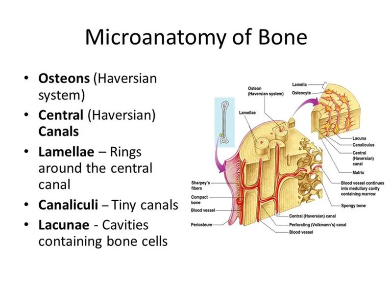
Bone remodelling and new bone formation : Bone formation takes place to support mechanical forces, growth and reparative process. Bone remodeling is a process of constantly resorbing bone by osteoclasts and replace it by osteoblast. Osteocytes act as mechanosensors and instruct the osteoclast where to resorb bone, and osteoblast when and where to form new bone.1,14 Bone remodeling consists of four phases. First the activation phase, this phase involves detection of a remodeling signal due to changes in physical forces recognized by the osteocytes or damage to the bone matrix due to osteocyte apoptosis, these are signals for the osteoclast. The second phase is the resorption phase, osteoclast breaks down the bone matrix. Third is the reversal phase, the mononuclear cell removes the undigested demineralized collagen matrix in the lacunae and prepare the bone surface for bone formation
Anatomical basis of bone implant- A computer numerically controlled (CNC) milling machine can also be used to manufacture a mold from which an implant could be cast. Cranial implants do not require extra strength, so plastic materials are usable for this point of view. Furthermore, it has lower thermal conductivity, which is very important because, this way, the brain is not exposed to extreme temperatures so easily.
Furthermore, magnetic resonance imaging examinations induce electrical currents in these medical implants. These eddy currents may heat the implants and thus may be capable of causing localized tissue heating. Some implants can be made from transparent material, which is an additional advantage of plastics. This way, the organ can be seen without severe operation. In this case, ultra-high molecule-weight polyethylene (UHMW-PE) can be chosen as implant material. This material is a very rigid, noncorrosive, nontoxic thermoplastic. It is relatively cheap and has good radiological transparency. In general, standard implants are not suitable in these surgeries of skull defects compared to joint prostheses because of the complexity and the difference in the anatomy of the skulls. The cranial implant is fabricated on a customized and individual basis based on case differences. This study aims to introduce a new methodology to design a patientspecific implant (PSI), a uniquely shaped implant for every unique patient based on different medical imaging data such as CT or Cone Beam Computer Tomography (CBCT).
Enumerate the indications and contraindications for the bone implant-Craniofacial reconstruction is a highly complex surgery because the operated body parts contain the brain, eyes, and other sensory organs. Sometimes a bone defect has a particular shape with different curvatures in various planes, so the implant has to be individually adjusted by a neurosurgeon during the surgery, which increases blood loss, infection risk, and the operating time. The best way of treating cranial defects would be autogenous bone transplantation because this will have fewer complications of infections when compared to implants from other materials. Currently, titanium is the commonly used material for cranial reconstruction due to its excellent biocompatibility, customization, and mechanical performance. However, several non-carcinogenic biocompatible materials are lighter such as polymethylmethacrylate (PMMA), Polyetheretherketone (PEEK), and hydroxyapatite (HA), which would be better as an implant.