Project 1
Part A
Digestive System - Cholelithiasis
1. Describe one common clinical condition/ disorder associated with the system involved
Gallbladder
- Small, thin-walled green sac
- Pear shaped
- Attached to the posterior surface of the liver by connective tissue
- Upto 10 cm in length
- Hold up to 50 cc of fluid
- Parts of gall bladder
- Fundus - expanded end
- Body - main part
- Neck - continuous with cystic duct
- Peritoneum covers the inferior surface
- Upper surface is in direct contact with the liver
- Held in place by the visceral peritoneum that covers the liver
- Cystic duct arises from the common bile duct and inserts at the neck of the gallbladder
- Cystic artery supplies blood to the gall bladder
- Cystic vein drains the blood from the gall bladder
- When there is no digestion of food, bile is stored in the gall bladder
- Acts as reservoir of bile
- In the gall bladder, bile is concentrated by the removal of water
- When fatty food enters the duodenum, a hormonal stimulus prompts the gall bladder to contract and release bile to the duodenum
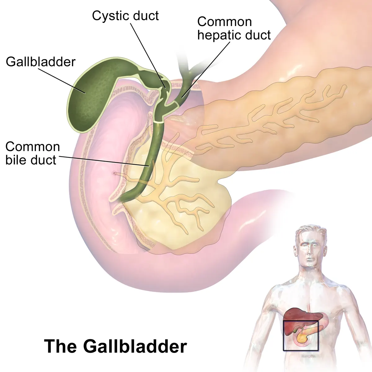
Fig. Gall Bladder
2. Describe in common disorders in association with the organ
Cholelithiasis
- Hardened deposits of digestive fluid
- Cholelithiasis - Temporary obstruction of the cystic duct resulting in biliary pain and is short lived
- Acute cholecystitis - Persistent stone dislodged the cystic duct causes the gallbladder to become distended and inflamed
- Choledocholithiasis - Gallstones obstruct the common bile duct and impedes the flow of bile from liver to intestine
- Cholangitis - Triggered by colonization of bacteria and overgrowth in static bile above an obstructing common duct stone
- 3 types of gallstones - Cholesterol, Black pigment and Brown pigment gallstones
- Cholesterol gallstones - Formed due to over secretion of cholesterol by liver cells and hypomotility or impaired emptying of the gallbladder
- Pigmented gallstones - High heme, Bilirubin may be present in bile at higher rate than normal concentration
- Risk factors - Obesity, Age, Female gender, Pregnancy, Genetics, Total parenteral nutrition, Rapid weight loss and certain medications
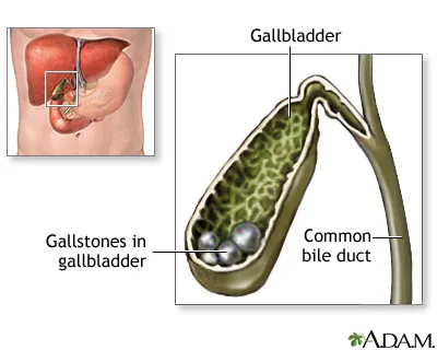
Fig. Cholelithiasis
Diagnostic Techniques
- CBC - Complete Blood Count Test
- CMP - Comprehensive Metabolic Panel Test
- PT/PTT - Prothrombin Time/ Partial Thromboplastin Time Test
- Lipase, Amylase, Alk Phos, Total bilirubin Test
- Urine Analysis
- Ultrasound
- CT Imaging
- Endoscopic Retrograde Cholangiopancreatography
- Magnetic Retrograde Cholangiopancreatography
Endoscopic Retrograde Cholangiopancreatography
- Procedure used to diagnose and treat problems in the liver, gallbladder, bile ducts, and pancreas
- Combines X-ray and endoscope
- The endoscope is guided through the mouth down to the duodenum
- It helps to view the inner part of these organs
- A dye is injected into the bile duct that highlights the organs on X-ray image
- Various X-ray images are then obtained
- After the procedure is done, the endoscope is withdrawn
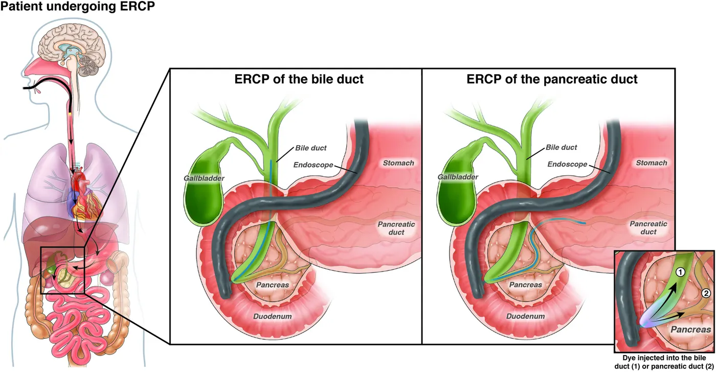
Fig. ERCP - Endoscopic Retrograde Cholangiopancreatography
3. Describe the management and treatment plan utilized in the disorder
Medical Management
- Cholelithiasis without complications
- Can be treated with oral or parenteral analgesia
- Offered dietary advice to reduce the chance of recurrent episodes
- Referred to general surgeon for laparoscopic cholecystectomy
- Acute cholecystitis
- Must be admitted at the hospital
- Requires surgical consult and intravenous antibiotics
- Choledocholithiasis/ Gallstone pancreatitis
- Must be admitted at the hospital
- Requires gastrointestinal consultation & ERCP or MRCP
- Acute Ascending Cholangitis
- Require aggressive resuscitation and ICU care
- Require surgical intervention to drain infection in the biliary tract
Laparoscopic Cholecystectomy - Surgical Procedure
- Preoperative antibiotics is given 30 minutes of incision per protocol
- Anaesthesia and intubation is performed
- Insufflation of the abdomen is achieved to 15 mmHg using carbon dioxide
- Four small incisions are made in the abdomen for trocar placement
- Utilizing a laparoscope and long instruments the gallbladder retracted over the liver
- This allows for exposure of the proposed region of the hepatocystic triangle.
- Careful dissection is carried out to achieve the critical view of safety.
- This view is defined as
- Clearance of fibrous and fatty tissue from the hepatocystic triangle,
- The presence of only two tubular structures entering into the base of the gallbladder
- The separation of the lower third of the gallbladder from the liver to visualize the cystic plate.
- Once this view is adequately achieved, the operating surgeon can proceed with confidence that he/she has isolated the cystic duct and cystic artery.
- Both structures are carefully clipped and transected.
- Electrocautery or harmonic scalpel is then used to separate the gallbladder from the liver bed completely.
- Hemostasis should be achieved after the abdomen is allowed to deflate to 8 mmHg for 2 minutes.
- This technique is employed to avoid missing potential venous bleeding that can be tamponaded by elevated intra-abdominal pressure (15 mmHg).
- The gallbladder is removed from the abdomen in a specimen pouch.
- All trocars should be removed under direct visualization.
- Closure of port sites is surgeon specific
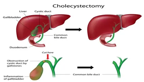
Fig. Choleccystectomy
4. Describe the surgical management of the disorder
Laparoscopic Cholecystectomy
- Minimally invasive procedure
- Removal of diseased gallbladder
- Replaced the open cholecystectomies
- Used to treat acute/chronic cholecystitis, symptomatic cholelithiasis, biliary dyskinesia, acalculous cholecystitis, gallstone pancreatitis, and gallbladder masses/polyps
- Anatomical Basis - Hepatocystic triangle
- Triangle of Calot
- Surgical anatomical landmark created by the cystic duct laterally, common hepatic duct medially and the liver edge superiorly
- Is of surgical importance because this is the location for the most common path of the cystic artery to the gallbladder
- Sentinel lymph node (aka Lund’s node) of the gallbladder resides within the hepatocystic triangle
- Instruments and Equipments
- Two laparoscopic monitors
- One laparoscope (5/10mm, 0/30 degrees) including camera cord and light source
- Carbon dioxide source and tubing for insufflation
- 5 mm to 12 mm trocars (average three 5 mm working trocars and one 10 mm to 12 mm trocar)
- Laparoscopic instruments: Atraumatic graspers, Maryland grasper, clip applier, electrocautery (eg: hook, spatula) and a retrieval bag
- Scalpel (11/15 blade), forceps, needle driver and absorbable sutures
- Major open tray for possible conversion
Post Operative Care
- Patient is admitted at the hospital
- Undergoes ERCP on the same day
Fig. Laparoscopic Chloeccystectomy
5. Self study on the recent advances in the field
Extracorporeal Shock Wave Lithotripsy
- Nonsurgical alternative to manage gallstones
- This treatment is provided to the patients whose gallbladder is functioning normally and the size of the stones are small
- Anesthesia is not required
- Uses high energy sound waves to produce shock waves
- These shock waves disintegrate the gallstones
- Patients who cannot undergo surgery can be treated with ESWL
Percutaneous Therapy
- In a case where the surgery is too dangerous for the patient, percutaneous treatment may be used
- In one procedure, an instrument called cholecytoscope is used for the treatment
- Another percutaneous method is called as dissolution therapy
- In this therapy, a catheter is inserted into the gallbladder and a solvent is injected directly to the gallbladder
- This solvent dissolves the cholesterol stones
Endoscopic Gallbladder Stenting
- An ERCP is performed
- A stent is inserted from the gallbladder to the duodenum
- Stent relieves the biliary symptoms and complications
Fig. Extracorporeal Shock Wave Lithotripsy
Reproductive System - Adenomyosis of Uterus
1. Describe one common clinical condition/ disorder associated with the system involved
Uterus
- Hollow, pear-shaped organ
- Located in the pelvis, posterior to the bladder and anterior to the rectum
- Divides into 4 parts
- Fundus - broad curved area in which fallopian tubes connects to the uterus
- Corpus (Body) - Main part of the uterus that starts directly below the level of fallopian tubes and continues downward
- Isthmus - lower neck region of the uterus
- Cervix - extends downwards from the isthmus and opens in vagina
- Arterial supply - Uterine and ovarian arteries
- Nerve supply - Autonomic - T11, T12; Sympathetic - Hypogastric plexus;
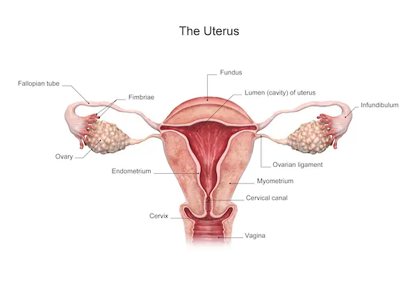
Fig. Uterus
2. Describe in common disorders in association with the organ
Adenomyosis of Uterus
- Endometrial tissue lining the inner wall grows into muscular wall of the uterus
- The tissue performs normal function to thicken, break down and bleed during each menstrual cycle
- Uterus enlarges
- Results in painful and heavy periods
- Resolves after menopause
Fig. Adenomyosis of Uterus
Diagnostic Techniques
- Vaginal Examination
- Transvaginal Ultrasound
- Magnetic Resonance Imaging
Transvaginal Ultrasound
- Also called endovaginal ultrasound
- Helps to examine female reproductive system
- Consists of transvaginal probe inserted through the vagina
- Checks for the shape, position and size of the ovaries and uterus
- Measures the thickness and length of the cervix
- Blood flow through the organs in the pelvis can be monitored
- Thickness and presence of fluids near the bladder or in the fallopian tubes, myometrium and endometrium
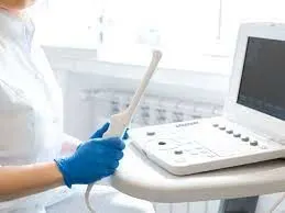
Fig. Transvaginal Ultrasound
3. Describe the management and treatment plan utilized in the disorder
Medical Management
- Exercise
- Meditation
- Acupuncture
- Anti-inflammatory pain killers
- Antifibrinolytic medication
- Intrauterine Device
- Contraceptive Pill
- Surgery - Endometrial Ablation, Hysterectomy
Surgical Procedure
- Endometrial Ablation
- Uses hysteroscope guidance
- Uses monopolar or bipolar loop electrode
- Endometrium is dessicated to the level of the basalis layer using thermal energy
- Does not use resectoscope
- Devices used are global and treats the whole endometrial cavity
- Include thermal fluid, microwave or bipolar radiofrequency electrical energy, laser thermotherapy, and cryoablation
- Minimally invasive surgical procedure to alleviate abnormal uterine bleeding
- Destroys functioning layer of the endometrium to prevent regrowth and suppress menstruation
- 2 Techniques - Resectoscopic Endometrial Ablation & Nonresectoscopic systems
- Resectoscopic Endometrial Ablation
- Nonresectoscopic Systems
- Hysterectomy - Surgical Procedure
- A large incision is made in the abdomen
- Incision may be vertical or horizontal
- Uterus is then removed
- A small incision is made in the vagina
- The uterus is then removed through this incision
- Laparoscope is used
- 3 or 4 small incision is made instead of one large incision
- The instrument is then inserted through this incision
- The uterus is then cut into small pieces and then removed
- 3 types - Supracervical or subtotal, total, and radical
- 2 approaches - open & minimally invasive
- Performed under general anesthesia.
- The procedure lasts for about one to two hour
- To begin the procedure, a urinary catheter is passed through your urethra to empty the bladder.
- The catheter remains in place during surgery and for a short time afterward.
- The abdomen and vagina are cleaned with a sterile solution before surgery.
- A general or local anesthetic is given to the patient
- Hysterectomy can be performed in 3 ways - Abdominal, Vaginal & Laparoscopic
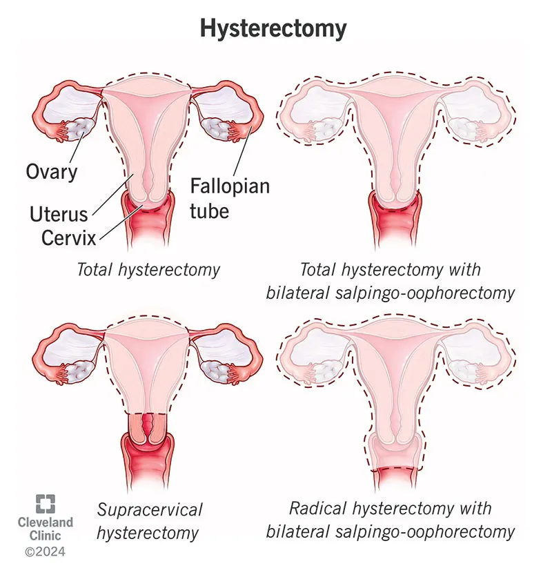
Fig. Hysterectomy
4. Describe the surgical management of the disorder
- Anatomical basis of the surgery
- Organ structure and anatomical relation of the organ to be noted while doing the procedure
- Detailed description of the instruments and equipments used for the surgery
- Brief note on postoperative care
- Elaborate on the equipments/medical devices used to monitor in post operative care
Anatomical Basis
- The surgeon then makes an incision in the lower abdomen, using one of two approaches:
- A vertical incision, which starts in the middle of your abdomen and extends from just below your navel to just above your pubic bone
- A horizontal bikini-line incision, which lies about an inch above your pubic bone
Instruments and Equipments
- Long, heavy Mayo scissors
- Short and long weighted vaginal speculums with an extra-long blade
- Heaney right-angle retractors
- Jorgenson scissors
- Long Allis clamps
- Deaver retractors
- A long needle holder
- Heany clamps
- Single tooth tenaculum
- Bovie extender
- Suction apparatus
- Laparoscope
Post Operative Care
- Mental and Emotional Support
- Pain relieving medication
- Hospital stay for 2-4 days
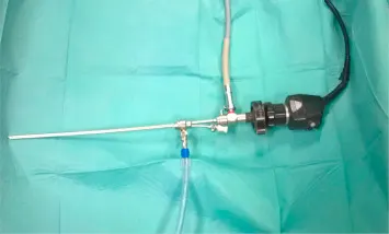
Fig. Hysteroscope
5. Self study on the recent advances in the field
- Transvaginal Ultrasound
- Magnetic Resonance Imaging
- High Intensity Focus Ultrasound
- Uterine Artery Embolization
Part B
A 25 year old biker indulged in illegal racing met with a road traffic accident and several head injury and skull fracture. After resuscitation and operation done for the hematoma it was resorted to do skull implant for his fracture by the orthopaedician
Skull - Type of Bone
- Skull belongs to flat bones
- The outer surface of the bone is called periosteum. It contains blood vessels and nerves that help provide nutrients to the bone
- Flat bones are made up of two layers
- Spongy/Cancellous Bone
- Inner part of the bone.
- Also known as cancellous bone
- Lightweight
- Helps absorb sudden stress
- Compact/Cortical Bone
- Spongy bone is then covered by a layer of compact bone.
- Also known as cortical bone.
- Very hard, dense type of bony tissue
Fig. Skull - Flat Bone
Microscopic anatomy of Bone
- Microstructural unit of compact bone - Osteon or Harvesian system
- Osteon
- Composed of concentric rings of calcified matrix - Lamellae
- The centre of each osteon consists of central canal Harvesian canal
- Harvesian canal contains blood vessels, nerves, and lymphatic vessels
- The vessels and nerves branch off at right angles through a perforating canal also known as Volkman’s canal
- Osteocytes are located inside spaces called lacunae
- Lacunae is found at the borders of adjacent lamellae
- Canaliculi of one lamellae connects with the canaliculi of other lamellae and then connects eventually with the central canal
- This allows nutrient transport and waste removal from them
- Microscopic anatomy of spongy bone
- Osteocytes are housed in the lacunae
- Not arranged in concentric circles
- But the lacunae and osteocytes are found in a lattice like network of matrix spikes called trabeculae
- Trabeculae appear to be a random network
- Each trabeculae forms along the lines of stress to provide strength to the bone
- Spaces in some spongy bones contain red marrow protected by the trabeculae, where hematopoiesis occur
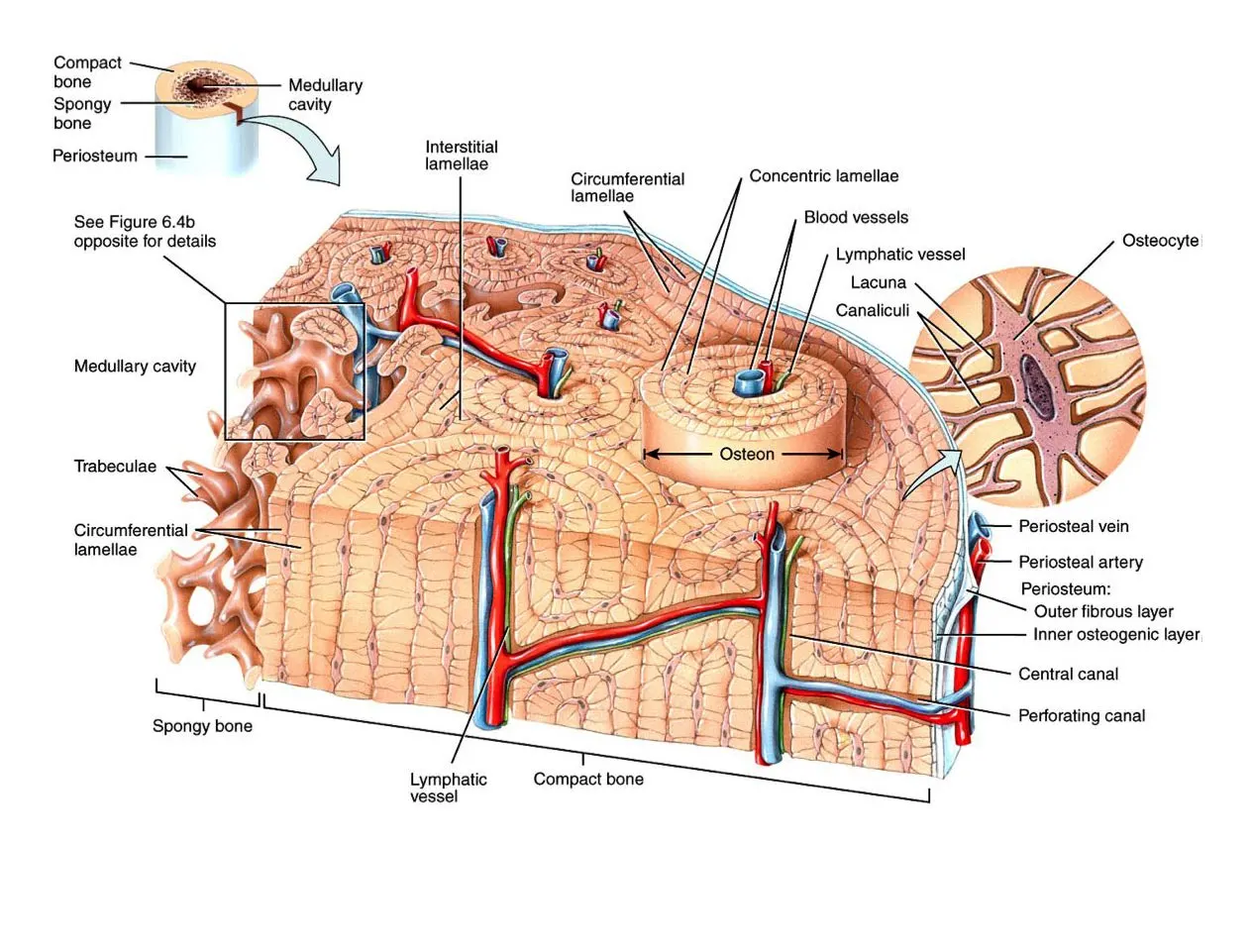
Fig. Microscopic Anatomy of Bone
Blood supply of Skull Bone
- Artery
- External carotid artery - main blood supply to the skull bone and meninges, divides into eight main branches
- Maxillary artery - most prominent and clinically relevant
- Internal carotid - supplies intracranial structures
- Internal carotid & vertebral arteries - large anastomosis - Circle of Wilis
- Veins
- Dural venous sinuses - superior sagittal sinuses, straight sinuses & transverse sinuses
- Superficial and deep veins of the head - Cerebral veins, great vein of Galen, Cerebellar & Facial veins
- Dural venous sinuses, superficial and deep veins of the head drain into the internal and external jugular veins bilaterally and then to the superior venacava
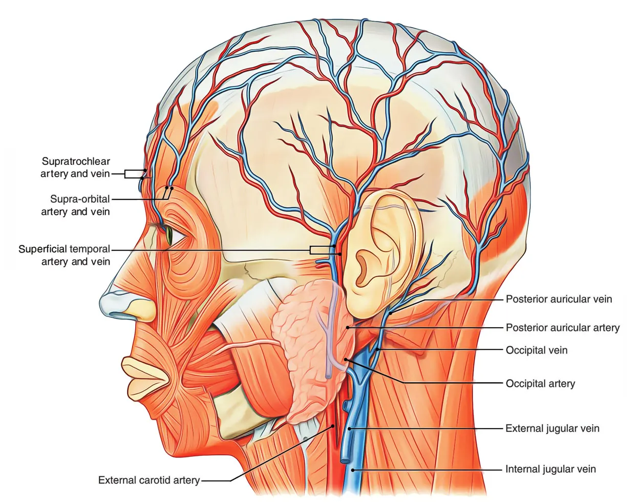
Fig. Blood Supply to Skull
Bone modeling and Bone remodeling
Bone Modeling
- Either the formation of bones by osteoblasts or resorption of bone by osteoclasts
- Formation modeling - Modeling by osteoblasts
- Resorptive modeling - Modeling by osteoclasts
- Bone modeling increases bone mass and maintain or alter bone shape
- Both process occur simultaneously throughout the skeleton
- They both must be coordinated to shape bone
- Occurs on a preexisting bone surface
- Can occur on periosteal, endocortical and trabecular bone surfaces or envelopes
Bone Remodeling
- Involves sequential osteoclast mediated bone resorption and osteoblast mediated bone formation at the same location
- Discrete locations of the skeleton are replaced by quantum packets of bone through the coupled activity of osteoclasts and osteoblasts
- Mechanism for replacement of the bone matrix and repair of small defects
- Renews skeleton over time
- Occur on surfaces such as periosteal, endocortical, trabecular and intracortical
- Process
- Osteoblasts - secrete bone matrix
- Chondroblasts - produce cartilage
- The inner cellular layer of the bone consists of progenitor stem cells
- The progenitor stem cells develop into
- When osteoblasts senses microcrack at any location, they produce a substance called RANKL
- RANKL binds with RANK receptors on the surface of nearby monocytes
- RANKL induces the monocytes to fuse together to form a multinucleated osteoclast cell
- RANKL also helps osteoclast mature and get activated
- Osteoclast secrete lysosomal enzymes like collagenase that digests collagen in the organic matrix
- This forms a hole on the bone surface known as Howship’s lacunae
- Osteoclasts starts producing hydrochloric acid HCl that dissolves hydroxyapatite into soluble calcium and phosphate ions
- These ions gets released into the blood stream
- The osteocytes are scattered within the bony matrix
- These osteocytes are phagocytosed by the osteoclasts
- Then the osteoblasts secrete osteoprotegerin that binds to RANKL and prevents it from activating RANK receptors
- This slows down the activation of osteoclasts
- Once phagocytosis of osteocytes are done the osteoclasts undergo apoptosis
- Following bone resorption, osteoblasts starts secreting osteoid seam
- Osteoid seam is a substance made up of collagen to fill in the lacunae created by the osteoclasts
- Calcium and phosphate begin to deposit on the seam forming hydroxyapatite
- Also many of the osteoblasts gets trapped within the bony matrix and turn into osteocytes
Fig. Bone remodeling cycle
Skull Implant
- Cranioplasty is the surgical procedure performed for cranial defects
- In this procedure, the injured area of the skull is replaced with original skull piece or with skull implants
- Autografts - Cranium, Tibia, Rib, Scapula, Fascia, Sternum, Ilium,
- Allograft - Cadaver cartilage
- Xenograft - Animal Bones
- Non-metal Allografts - Celluloids, Methyl-methacrylate, Hydroxyapatite, Polyethylene and silicon, Chorale, Ceramic, Cortoss
- Metal Allografts - Aluminium, Gold, Silver, Tantalum, Stainless steel, Titanium, Lead, Platinum, Vitallium, Ticonium
- Current Materials used - Titanium (plate or mesh), synthetic bone substitute in liquid form, solid biomaterial
- Indications - Cranial defects, Neurological function improvement, aesthetics, headaches
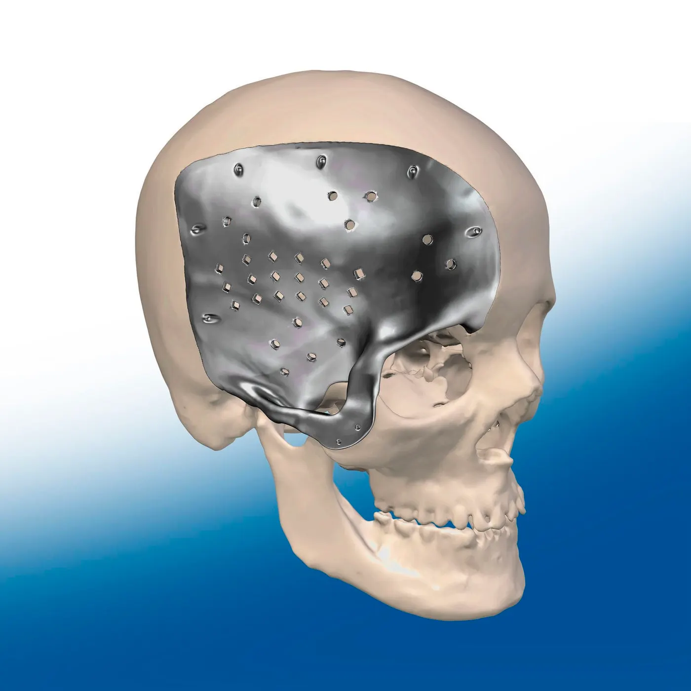
Fig. Skull Implant