Project 1
PART A
1.
1.1
Clinical condition - Stroke (The Brain)
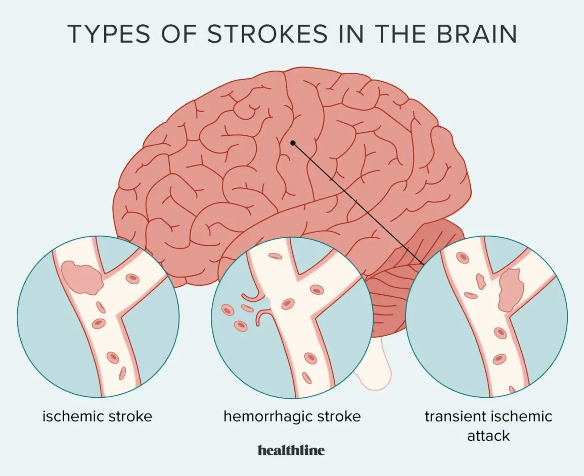

Organ Involved - The Brain
Gross Anatomy:
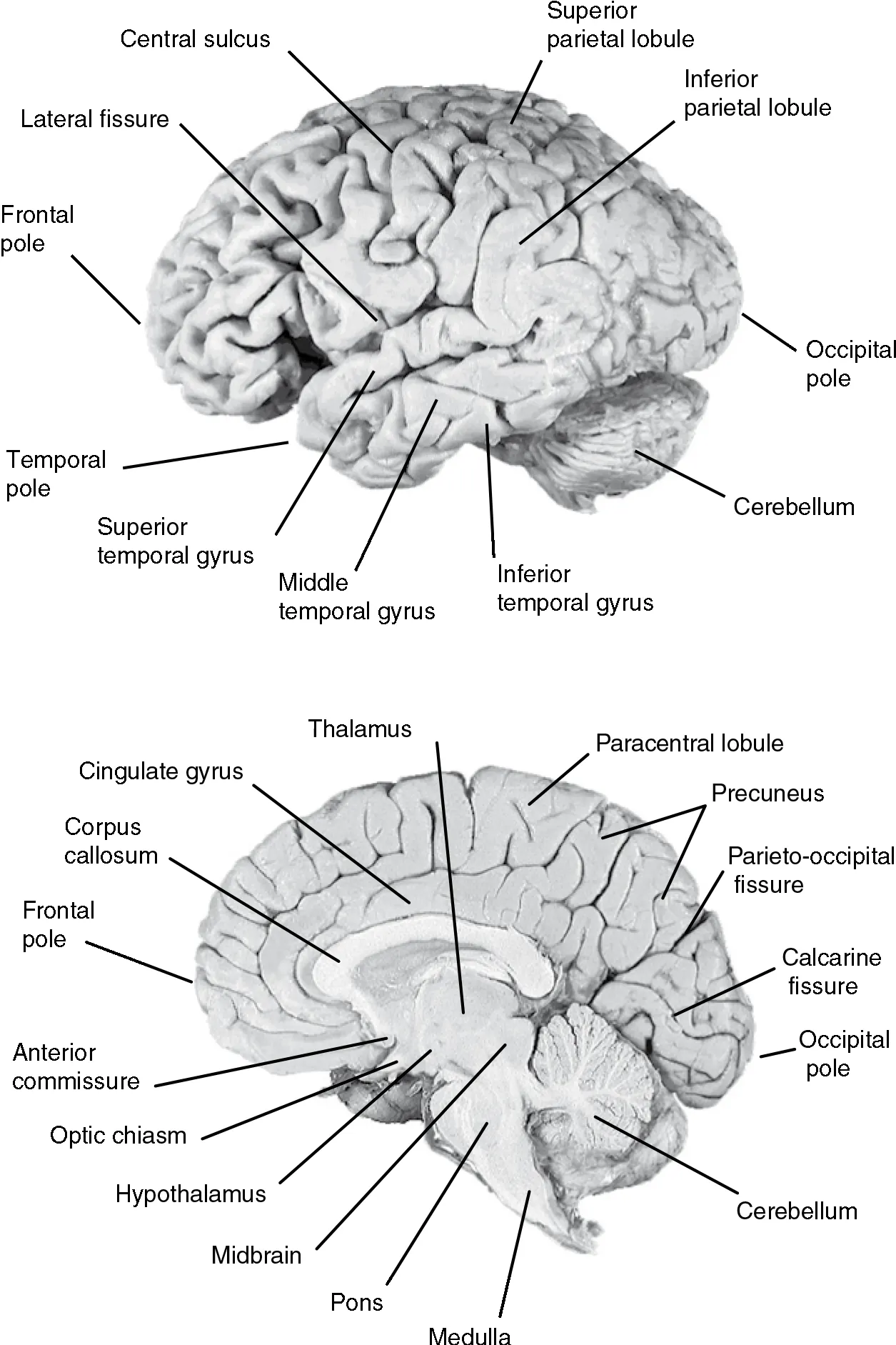
Figure: David L. Clark, Nash N. Boutros and Mario F. Mendez,"The Brain and Behavior An Introduction to Behavioral Neuroanatomy", pp. 4 - 13, Cambridge University Press, 2010. DOI: https://doi.org/10.1017/CBO9780511776915.004. https://static.cambridge.org/binary/version/id/urn:cambridge.org:id:binary:15852:20160618061252183-0516:14229fig2_2.gif?pub-status=live
Blood Supply:
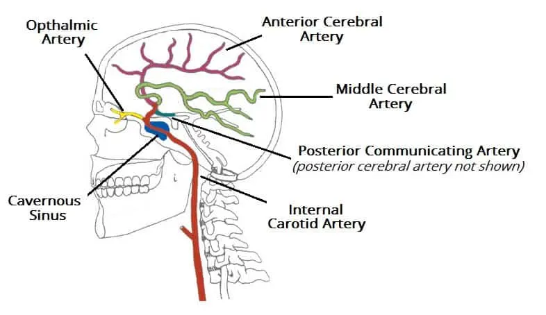
Figure: By Frank Gaillard and Patrick J. Lynch [CC-BY-SA-3.0], via Wikimedia Commons. https://teachmeanatomy.info/wp-content/uploads/Branches-of-the-Internal-Carotid-Artery-Arterial-Supply-to-the-Brain.jpg
Diagnostic technique - MRI
Parts of MRI:
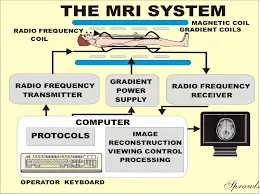
Figure: http://www.sprawls.org/mripmt/MRI02/MRI%202-1.jpg
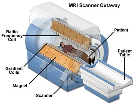
Figure: http://snc2dmri.weebly.com/uploads/2/4/5/6/24569353/7752085.jpg?438
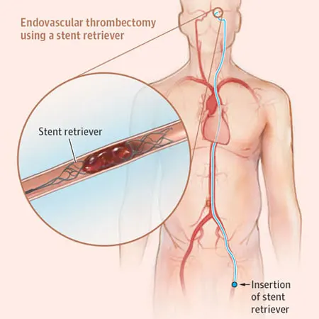
Surgical management - Endovascular thrombectomy
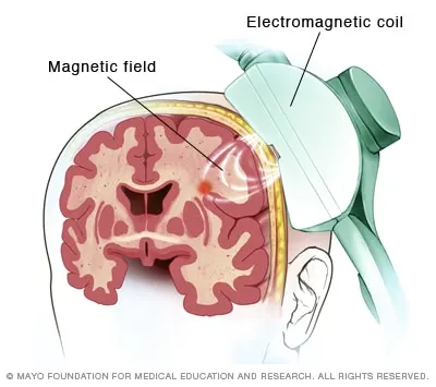
Recent Advancement in Stroke Management - Repetitive transcranial magnetic stimulation
1.2
Clinical condition - Kidney Stones
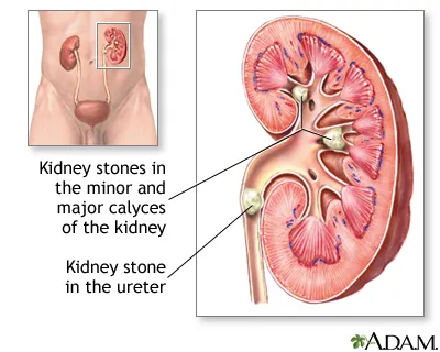
Figure: https://medlineplus.gov/ency/images/ency/fullsize/17091.jpg
Organ Involved - Kidneys
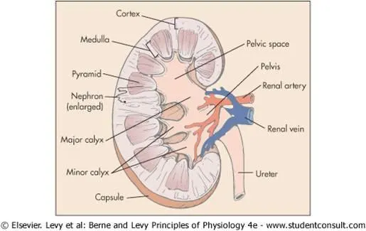
Figure: https://www.onlinebiologynotes.com/wp-content/uploads/2017/05/LS-of-kidney-1.jpg
Blood Supply:
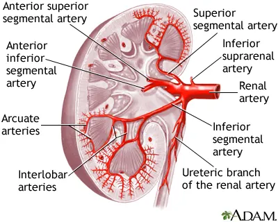
Figure: https://medlineplus.gov/ency/images/ency/fullsize/8817.jpg
Diagnostic technique - X-ray
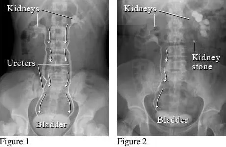
Figure: https://schoolworkhelper.net/wp-content/uploads/2017/03/Kidney-Stones-xray.jpg
Parts of X-ray:
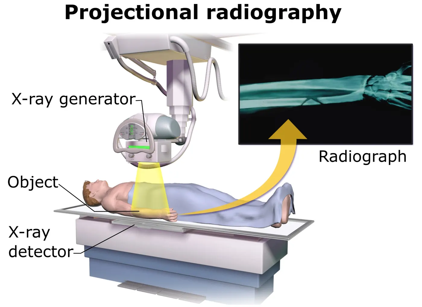
Figure: https://upload.wikimedia.org/wikipedia/commons/d/db/Projectional_radiography_components.jpg
Surgical management - Uteroscopy
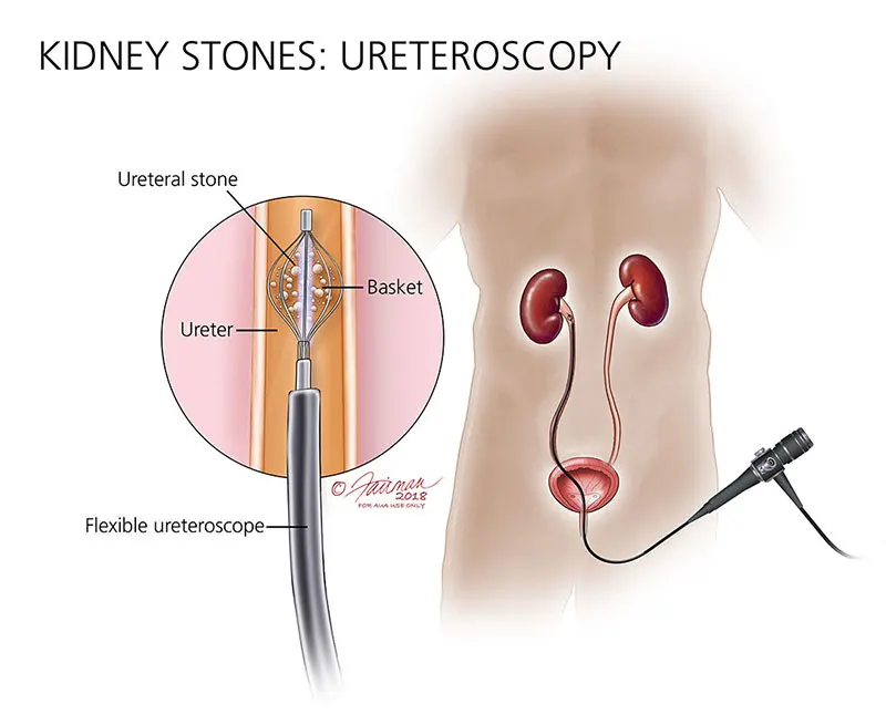
Figure: https://www.urologyhealth.org/images/Conditions/KidneyStones_Ureteroscopy_lightbox.jpg
Recent Advancement in Kidney Stone Removal - High-powered Laser Facilitates Stone Removal
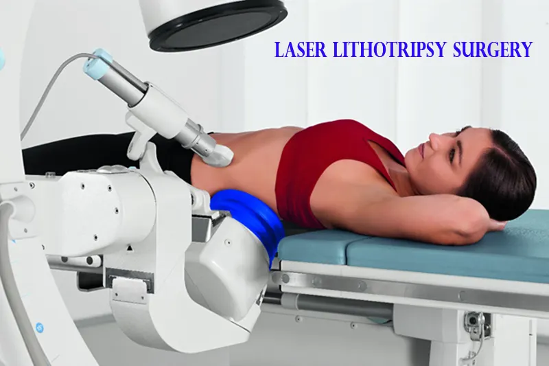
Figure: https://www.urolife.in/wp-content/uploads/2021/04/laser-kidney-stone-surgery-in-pune.png
1.3
Clinical condition - Chronic Obstructive Pulmonary Disease (COPD)
Figure: https://www.chandigarhayurvedcentre.com/wp-content/uploads/2022/12/HealthyvsCOPDlungs960px.jpg
Organ Involved - The lungs
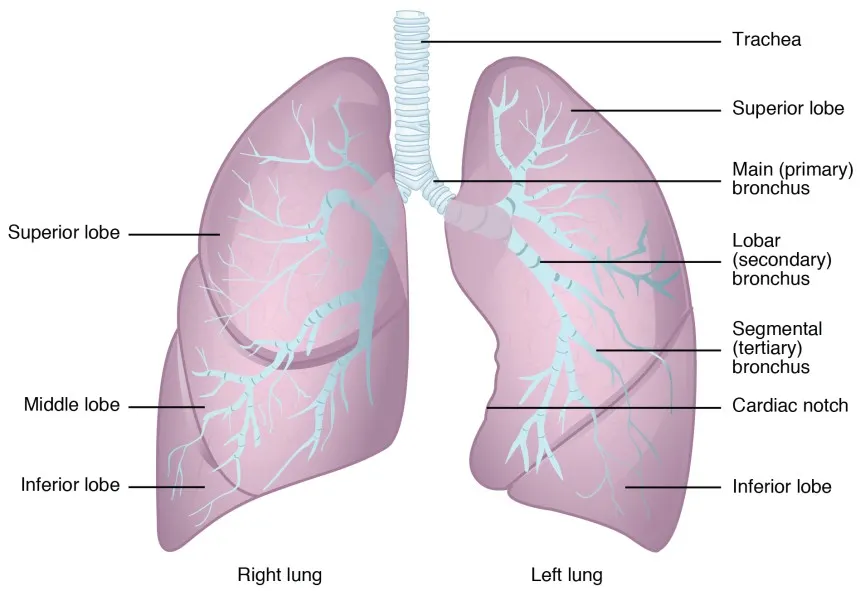
Blood Supply:
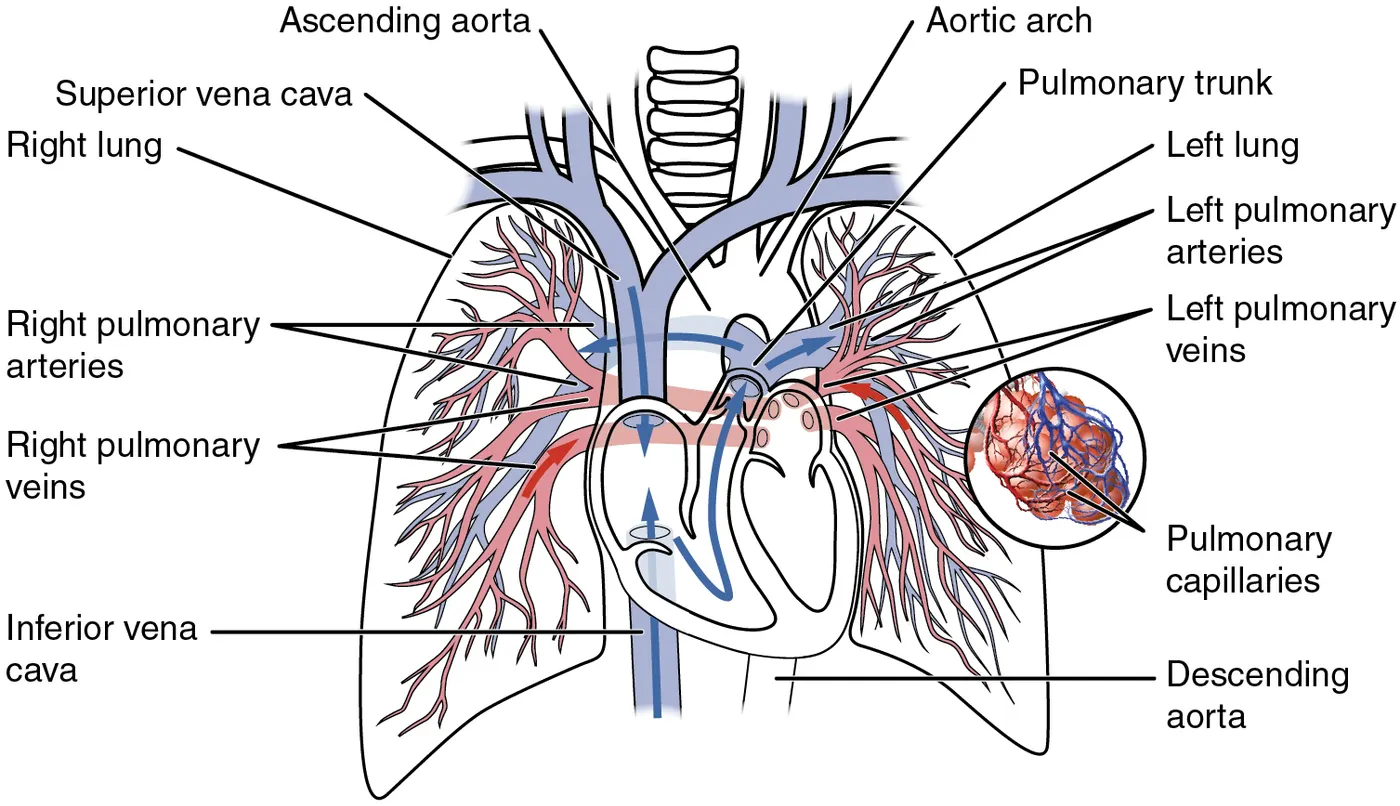
Figure: https://upload.wikimedia.org/wikipedia/commons/3/36/2119_Pulmonary_Circuit.jpg
Diagnostic technique - Spirometry
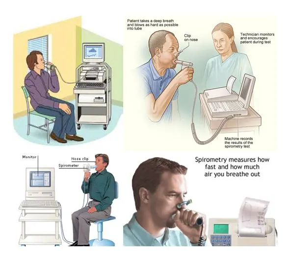
Figure: https://vaishnaviheartcentre.com/wp-content/uploads/2019/05/PFT.jpg
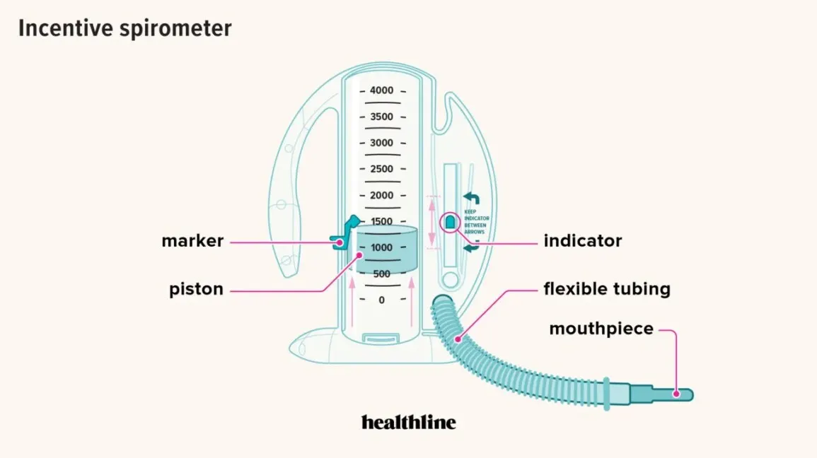
Parts of Spirometer
Surgical Management - Bullectomy
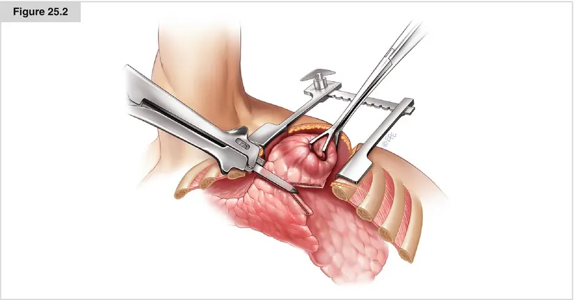
Figure: https://thoracickey.com/wp-content/uploads/2017/04/A194303_1_En_25_Fig2_HTML.jpg
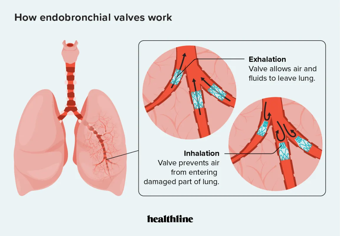
Recent Advancement in Kidney Stone Removal - Endobronchial Valves or Zephyr Valves
PART B
CASE STUDY
- Type of bone the skull belongs to:
The skull is a complex structure that consists of several bones that join together to form the cranium and the face. The bones that make up the cranium are flat bones, which means that they are broad, thin, and usually curved. The bones of the skull are divided into two groups: cranial bones and facial bones. The cranial bones include the frontal, parietal, temporal, occipital, sphenoid, and ethmoid bones. These bones protect the brain and provide attachment sites for muscles and ligaments.
- Microscopic anatomy of bone:
Bone is a living tissue that is composed of cells and extracellular matrix. The cells of bone include osteoblasts, which build new bone, and osteoclasts, which break down bone tissue. The extracellular matrix of bone is made up of collagen fibers and minerals, such as calcium and phosphate. The collagen fibers provide flexibility and tensile strength, while the minerals provide hardness and compressive strength.
- Blood supply of bone:
Bone has an extensive blood supply that provides oxygen and nutrients to the cells. The blood vessels that supply bone include arteries, veins, and capillaries. The blood vessels enter the bone through small channels called foramina, which are located on the surface of the bone. The blood vessels also communicate with the bone cells through small channels called canaliculi, which allow the exchange of nutrients and waste products.
- Bone remodeling and new bone formation:
Bone is constantly being remodeled throughout life in a process called bone remodeling. This process involves the removal of old bone by osteoclasts and the deposition of new bone by osteoblasts. Bone remodeling is essential for maintaining bone health and strength, as well as for repairing bone after injury.
New bone formation occurs through a process called ossification, which involves the deposition of calcium and other minerals onto a matrix of collagen fibers. There are two types of ossification: intramembranous ossification, which occurs in flat bones like the skull, and endochondral ossification, which occurs in long bones.
- Anatomical basis of bone implant:
A bone implant is a device that is used to replace a missing or damaged bone. The implant is usually made of a biocompatible material, such as titanium, that can integrate with the surrounding bone tissue. The implant is secured to the remaining bone with screws or other fixation devices.
- Indications and contraindications for bone implant:
Indications for bone implantation include bone fractures that cannot be treated with conservative methods, bone defects resulting from trauma or disease, and joint replacements. Contraindications for bone implantation include infection, poor bone quality, and inadequate soft tissue coverage. Additionally, patients with certain medical conditions, such as osteoporosis or diabetes, may not be good candidates for bone implantation.